| Physiology |
Functional processes and regulatory mechanisms, including:- Cellular physiology (e.g., membrane potentials)
- Systemic physiology (e.g., cardiovascular dynamics)
- Pathophysiology (disease mechanisms)
- Neurophysiology (signal transmission)
- Endocrine/metabolic regulation
|
- Electrophysiology (e.g., ECG, EEG)
- Biochemical assays (e.g., glucose tolerance tests)
- Genetic sequencing (e.g., CRISPR edits)
- Animal models (e.g., rodent stroke studies)

Structural Organization of the Human Body
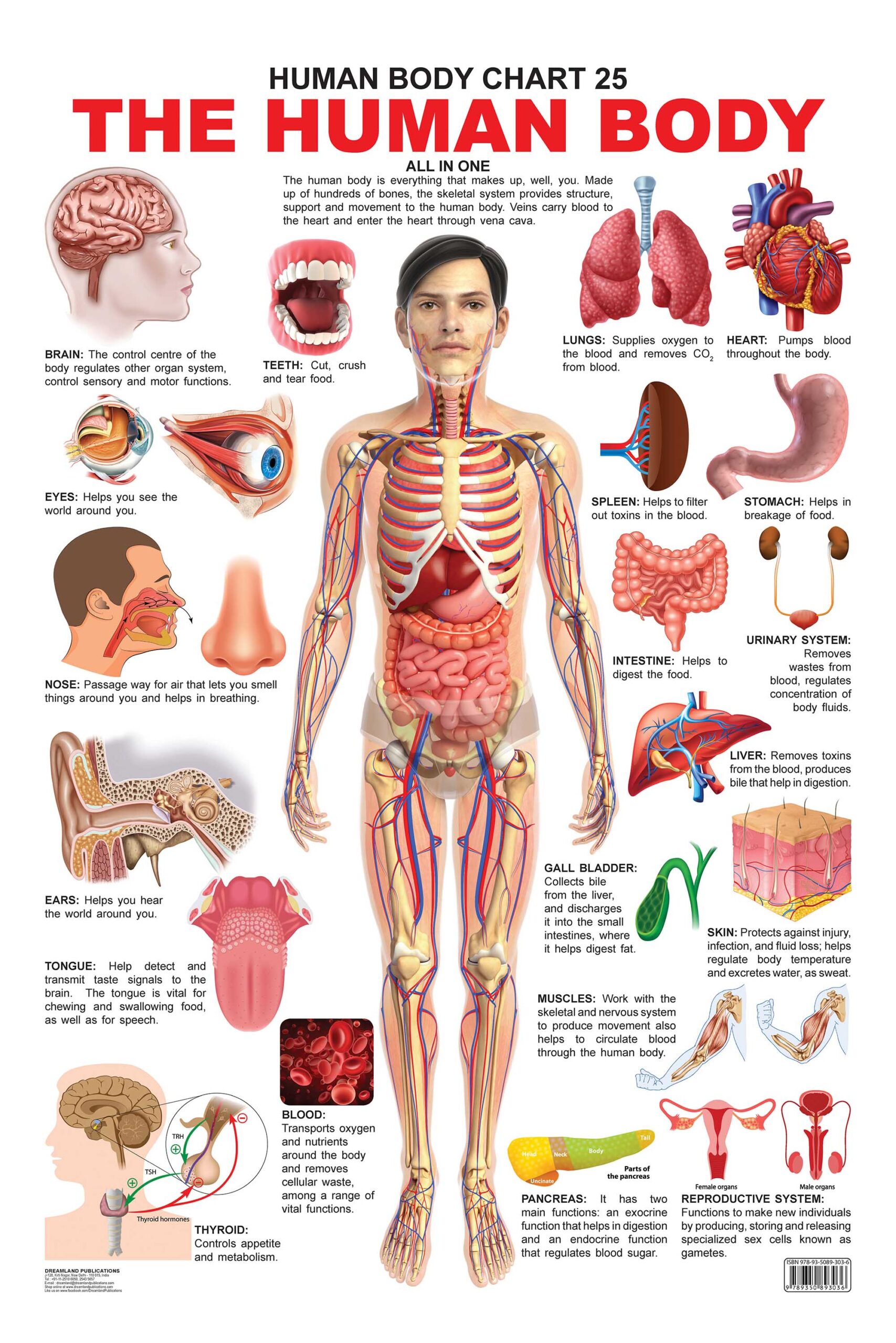
The human body exhibits a highly organized hierarchical structure, where each level builds upon the preceding one to form increasingly complex systems. This organization ensures coordinated function, from the molecular interactions within cells to the integrated operations of multiple organ systems. Understanding this hierarchy is essential for comprehending how anatomical structures support physiological processes, such as nutrient transport, waste elimination, and signal transduction. The following sections explore the levels of organization, anatomical terminology, and system-specific comparisons, alongside a procedural example linking structure to function.
Hierarchical Levels of Organization
The human body is structured across six interconnected levels of organization, each contributing to the overall function of the organism. These levels—chemical, cellular, tissue, organ, system, and organismal—demonstrate how discrete components integrate to maintain homeostasis. Below is a text-based hierarchical diagram illustrating this progression, followed by a description of each level’s role.Text-Based Hierarchy Diagram: Organism (Human Body)
├── Organ Systems (e.g., Circulatory, Nervous)
│ ├── Organs (e.g., Heart, Brain)
│ │ ├── Tissues (e.g., Cardiac Muscle, Nervous Tissue)
│ │ │ ├── Cells (e.g., Cardiomyocytes, Neurons)
│ │ │ │ ├── Organelles (e.g., Mitochondria, Nucleus)
│ │ │ │ │ ├── Macromolecules (e.g., Proteins, DNA)
│ │ │ │ │ └── Atoms/Molecules (e.g., Amino Acids, Water)
│ │ │ └── Extracellular Matrix (e.g., Collagen Fibers)
│ └── Interactions Between Systems (e.g., Nervous System Regulating Heart Rate)
└── Homeostasis (Dynamic Equilibrium) Key Levels Explained:
- Chemical Level: Composed of atoms, molecules, and macromolecules (e.g., carbohydrates, lipids, proteins, nucleic acids). These molecules form the building blocks for cellular structures and energy production.
- Example: Hemoglobin (a protein) transports oxygen in red blood cells; ATP (adenosine triphosphate) provides energy for cellular processes.
- Cellular Level: The basic unit of life, where cells (e.g., epithelial, muscle, nerve) perform specialized functions. Each cell contains organelles like mitochondria (energy production) and the nucleus (genetic control).
- Example: A neuron transmits electrical signals via neurotransmitters, while a hepatocyte (liver cell) processes toxins and synthesizes bile.
- Tissue Level: Groups of similar cells working together for a common function. Four primary tissue types exist:
- Epithelial: Covers surfaces (e.g., skin, lining of intestines).
- Connective: Supports and binds structures (e.g., bone, blood).
- Muscle: Enables movement (e.g., skeletal, cardiac).
- Nervous: Transmits signals (e.g., brain, spinal cord).
- Example: The stratified squamous epithelium of the esophagus resists abrasion during food passage.
- Organ Level: Two or more tissues collaborating to perform complex functions. Organs have distinct structures (e.g., layers, lumens) that reflect their roles.
- Example: The stomach contains:
- Mucous epithelium (protection),
- Smooth muscle (mechanical digestion),
- Gastric glands (enzyme secretion).
- System Level: Organs working together to achieve a physiological goal. Systems interact to maintain homeostasis (e.g., the endocrine system regulates metabolism via hormones, while the immune system defends against pathogens).
- Example: The cardiovascular system (heart + blood vessels) circulates blood to deliver oxygen and nutrients, while the lymphatic system returns excess fluid and filters pathogens.
- Organismal Level: The integrated functioning of all systems, resulting in a living, self-sustaining entity capable of growth, reproduction, and response to stimuli.
- Example: During exercise, the muscular system contracts, the cardiovascular system increases heart rate, and the respiratory system enhances oxygen uptake—all coordinated by the nervous system.
Anatomical Position and Directional Terminology
Precise directional terms and anatomical position (standardized reference posture) are critical for accurately describing locations and relationships within the body. This terminology ensures clarity in medical, research, and clinical contexts. Below are key terms with real-world applications, including descriptions of organ locations using standardized language.Anatomical Position:
- Body erect, facing forward, arms at sides, palms forward, and feet parallel.
- Serves as the reference point for all directional descriptors.
Directional Terms: | Term | Definition | Example Application |
|---|
| Superior | Above; toward the head. | The heart is superior to the diaphragm. |
| Inferior | Below; toward the feet. | The stomach is inferior to the lungs. |
| Anterior | Front; toward the belly. | The nose is anterior to the brain. |
| Posterior | Back; toward the spine. | The heart is posterior to the sternum. |
| Medial | Nearer to the midline of the body. | The trachea is medial to the lungs. |
| Lateral | Farther from the midline. | The arms are lateral to the chest. |
| Proximal | Closer to the origin of a limb or the body’s center. | The elbow is proximal to the wrist. |
| Distal | Farther from the origin. | The fingers are distal to the palm. |
| Superficial | Nearer to the surface. | The skin is superficial to the muscles. |
| Deep | Farther from the surface. | The lungs are deep to the rib cage. |
Practical Examples:
1. Heart Location:
- Anterior to the vertebral column,
- Posterior to the sternum,
- Superior to the diaphragm,
- Medial to the lungs,
- Located in the thoracic cavity, between the 2nd and 6th ribs.
2. Liver Location:
- Superior to the stomach,
- Inferior to the diaphragm,
- Right hypochondrium (upper-right quadrant of the abdomen),
- Anterior to the right kidney,
- Medial to the right lung.
3. Knee Joint:
- Distal to the femur (thigh bone),
- Proximal to the tibia (shin bone),
- Posterior to the patella (kneecap),
- Contains articular cartilage (superficial to bone surfaces).
Comparison of Four Major Body Systems
The following table compares four critical body systems—circulatory, nervous, digestive, and muscular—highlighting their primary organs, physiological functions, anatomical landmarks, and common disorders. This comparison underscores the interdependence of systems and the consequences of dysfunction.
| System |
Primary Organs |
Key Physiological Functions |
Anatomical Landmarks |
Common Disorders Linked to Dysfunction |
|---|
| Circulatory System |
Heart |
- Pumps blood through arteries (oxygenated) and veins (deoxygenated).
- Regulates blood pressure via cardiac output (stroke volume × heart rate).
- Distributes hormones, nutrients, and immune cells.
|
- Located in the mediastinum, between the lungs.
- Apex points toward the left hip; base attaches to major vessels.
- Coronary arteries supply the myocardium.
|
- Coronary Artery Disease (CAD): Plaque buildup in coronary arteries, leading to myocardial infarction (heart attack).
Cellular and Molecular Foundations of Anatomy and Physiology
The cellular and molecular frameworks underpinning human anatomy and physiology establish the functional unity of living systems. Eukaryotic cells, the building blocks of multicellular organisms, integrate specialized structures—such as organelles and biomolecules—to execute processes like energy conversion, signal transduction, and genetic inheritance. These components operate in a hierarchical manner, where molecular interactions (e.g., protein folding, DNA replication) directly influence macroscopic physiological functions, from muscle contraction to neural signaling. Understanding these foundations elucidates how cellular architecture enables physiological homeostasis and disease pathology.
Structure and Function of Eukaryotic Cells
Eukaryotic cells exhibit a compartmentalized organization, where each membrane-bound structure (organelle) performs distinct yet interdependent roles. The plasma membrane, a phospholipid bilayer embedded with proteins and cholesterol, regulates selective permeability, facilitates cell signaling, and maintains ionic gradients critical for processes like action potentials in neurons. Below, the key organelles and their physiological contributions are detailed:
-
Nucleus: The genetic control center housing DNA in chromatin form. The nuclear envelope, perforated by nuclear pores, permits bidirectional transport of RNA and proteins. Within the nucleus, the nucleolus assembles ribosomal subunits, while transcription factors regulate gene expression in response to extracellular cues (e.g., steroid hormones binding to nuclear receptors).
-
Mitochondria: Double-membrane organelles where oxidative phosphorylation occurs, generating ATP via the electron transport chain. Their inner membrane folds (cristae) increase surface area for enzymatic complexes, while mitochondrial DNA encodes select proteins essential for energy metabolism. Dysfunction in mitochondria underlies neurodegenerative diseases (e.g., Parkinson’s) and metabolic disorders.
-
Endoplasmic Reticulum (ER): A continuous membrane network divided into rough (studded with ribosomes) and smooth regions. The rough ER synthesizes and folds secretory/transmembrane proteins, while the smooth ER metabolizes lipids (e.g., steroid synthesis in adrenal cells) and detoxifies drugs via cytochrome P450 enzymes. ER stress, triggered by misfolded proteins, activates the unfolded protein response (UPR), a pathway linked to diabetes and Alzheimer’s.
-
Golgi Apparatus: Modifies, sorts, and packages proteins/lipids into vesicles for secretion or lysosomal degradation. Its cis-to-trans orientation ensures sequential glycosylation and phosphorylation, critical for protein functionality (e.g., lysosomal enzymes require mannose-6-phosphate tags for targeting).
-
Lysosomes and Peroxisomes: Lysosomes degrade macromolecules via hydrolytic enzymes (acidic pH), while peroxisomes oxidize fatty acids and detoxify hydrogen peroxide. Lysosomal storage diseases (e.g., Tay-Sachs) arise from enzyme deficiencies, whereas peroxisomal disorders (e.g., Zellweger syndrome) impair lipid metabolism.
-
Cytoskeleton: A dynamic network of microtubules, microfilaments, and intermediate filaments that maintains cell shape, enables motility (e.g., cilia in respiratory epithelium), and facilitates intracellular transport via motor proteins (kinesin/dynein). Microtubule disassembly during mitosis ensures chromosomal segregation; defects cause cancer (e.g., taxol stabilizes microtubules, arresting mitosis in tumor cells).
Prokaryotic vs. Eukaryotic Cells: Structural and Physiological Comparisons
Prokaryotic cells (bacteria/archaea) lack membrane-bound organelles and a defined nucleus, yet their structural simplicity supports rapid replication and metabolic versatility. Below is a comparative summary of anatomical and physiological distinctions:
Anatomical Differences:
- Nucleoid Region: Prokaryotes possess a single circular chromosome in an unbound nucleoid; eukaryotes have linear chromosomes within a nuclear envelope.
- Organelles: Eukaryotes contain mitochondria, ER, Golgi, and lysosomes; prokaryotes rely on the plasma membrane and mesosomes for compartmentalization.
- Cell Size: Prokaryotes (0.1–5 µm) are smaller; eukaryotic cells (10–100 µm) accommodate larger organelles.
- Cell Wall Composition: Prokaryotes have peptidoglycan (bacteria) or pseudopeptidoglycan (archaea); eukaryotes use cellulose (plants) or chitin (fungi).
Physiological Implications:
- Energy Production: Prokaryotes use plasma membrane invaginations or thylakoids (photosynthetic bacteria); eukaryotes localize respiration to mitochondria.
- Protein Synthesis: Prokaryotic ribosomes (70S) lack a nuclear membrane, enabling coupled transcription-translation; eukaryotic ribosomes (80S) require mRNA export.
- Genetic Exchange: Prokaryotes use plasmids and horizontal gene transfer (conjugation/transduction); eukaryotes rely on meiosis and sexual reproduction.
- Antibiotic Targets: Prokaryotic-specific structures (e.g., 70S ribosomes, peptidoglycan) are exploited by drugs (e.g., tetracycline, penicillin).
Biomolecules and Their Role in Cellular Anatomy and Physiology
Biomolecules—carbohydrates, lipids, proteins, and nucleic acids—serve as structural scaffolds, energy reserves, and information carriers. Their three-dimensional conformations dictate function through non-covalent interactions (e.g., hydrogen bonds, hydrophobic effects). Key examples include:
-
Carbohydrates: Monosaccharides (glucose, fructose) provide immediate energy via glycolysis; polysaccharides (glycogen, starch) store energy, while glycolipids and glycoproteins mediate cell recognition (e.g., ABO blood groups). The branched structure of glycogen allows rapid glucose release during exercise.
-
Lipids: Phospholipids form bilayers for membranes; cholesterol modulates fluidity. Triacylglycerides store energy, while eicosanoids (e.g., prostaglandins) act as local signaling molecules. The amphipathic nature of phospholipids enables compartmentalization in organelles.
-
Proteins: Primary structure (amino acid sequence) folds into secondary (α-helices/β-sheets) and tertiary structures stabilized by disulfide bonds. Quaternary structure (e.g., hemoglobin’s tetramer) enables cooperative binding. Misfolding (e.g., prions in Creutzfeldt-Jakob disease) disrupts cellular function.
-
Nucleic Acids: DNA’s double helix (Watson-Crick model) protects genetic information; RNA’s secondary structures (e.g., tRNA’s cloverleaf) facilitate translation. Base pairing (A-T/U, G-C) ensures accurate replication and transcription.
Structural-Functional Relationships:
- Protein Folding: The hydrophobic collapse of polypeptide chains buries nonpolar residues, forming active sites (e.g., enzymes like lysozyme). Chaperone proteins (e.g., HSP70) assist folding to prevent aggregation.
- DNA Supercoiling: Topoisomerases introduce supercoils to compact DNA (e.g., nucleosomes in eukaryotes), regulating gene accessibility during transcription.
- Lipid Rafts: Cholesterol-rich microdomains in membranes concentrate signaling proteins (e.g., G-protein-coupled receptors), enabling efficient signal transduction.
Central Dogma of Molecular Biology: Anatomical and Physiological Flowchart
The central dogma describes the flow of genetic information from DNA to RNA to protein, with each step anchored in cellular anatomy. Below is a text-based flowchart with physiological annotations:
Step 1: Replication (DNA → DNA)
- Location: Nucleus (eukaryotes); nucleoid (prokaryotes).
- Process: DNA polymerase synthesizes complementary strands (5’→3’), with helicase unwinding the helix and topoisomerase relieving supercoiling.
- Physiological Relevance: Ensures genetic fidelity during cell division; errors (mutations) may cause cancer or hereditary diseases (e.g., sickle cell anemia).
Step 2: Transcription (DNA → RNA)
- Location: Nucleus (eukaryotes); cytoplasm (prokaryotes).
- Process: RNA polymerase II synthesizes mRNA from a DNA template, with introns spliced out by the spliceosome (eukaryotes). Promoters (e.g., TATA box) and enhancers regulate initiation.
- Physiological Relevance: Produces mRNA for protein synthesis; alternative splicing generates protein diversity (e.g., troponin isoforms in muscle).
Step 3: RNA Processing (Eukaryotes)
- Location: Nucleus.
- Process: 5’ cap and poly
Physiological Processes and Homeostasis
Homeostasis represents the dynamic equilibrium maintained by the human body to sustain optimal cellular function despite internal or external perturbations. This equilibrium relies on intricate physiological processes, including feedback mechanisms, cell signaling pathways, and coordinated system interactions. Negative feedback loops serve as the primary regulatory framework, ensuring deviations from set points are corrected efficiently. Below, the mechanisms of homeostasis are explored through systemic examples, followed by an analysis of cell signaling pathways and a case study of homeostatic imbalance.
Mechanisms of Homeostasis and Negative Feedback Loops
Negative feedback loops are self-regulating mechanisms where the output of a process inhibits its further progression, restoring balance. These loops operate across multiple body systems, often involving sensors, control centers, and effectors. Three key examples illustrate their function:
-
Thermoregulation via the Integumentary System
The hypothalamus acts as the control center, detecting temperature deviations through peripheral thermoreceptors. When core temperature rises, sweat glands (effectors) activate, releasing sweat to evaporate and cool the body. Conversely, vasoconstriction and shivering conserve or generate heat. This loop ensures temperature remains within the narrow range of 36.5–37.5°C (97.7–99.5°F).
-
Blood Glucose Regulation via the Endocrine System
The pancreas monitors glucose levels via beta and alpha cells in the islets of Langerhans. Elevated glucose triggers insulin secretion, promoting glucose uptake by cells and storage as glycogen. Hypoglycemia stimulates glucagon release, converting glycogen to glucose. This dual mechanism maintains glucose levels at 70–99 mg/dL (3.9–5.5 mmol/L) fasting.
-
pH Balance in the Respiratory System
Central and peripheral chemoreceptors detect changes in blood pH (via CO₂ levels). Hypercapnia (elevated CO₂) increases respiratory rate, expelling CO₂ and reducing acidity (raising pH). Conversely, hypocapnia slows breathing, retaining CO₂ to restore acid-base balance. This system preserves arterial pH within 7.35–7.45.
Negative feedback loops are characterized by the formula:
Stimulus → Sensor → Control Center → Effector → Response → Correction of Stimulus.
Cell Signaling Pathways Bridging Anatomy and Physiology
Cell signaling pathways transmit information between cells, linking anatomical structures (e.g., endocrine glands) to physiological responses. Three primary pathways—endocrine, paracrine, and autocrine—mediate these interactions:
-
Endocrine Signaling
Hormones released into the bloodstream (e.g., adrenaline from the adrenal medulla) travel systemically to target distant organs. During the fight-or-flight response, adrenaline binds to beta-adrenergic receptors in the heart, liver, and skeletal muscles, triggering:
- Increased cardiac output (heart rate and stroke volume).
- Glycogenolysis in the liver (glucose release).
- Vasodilation in muscles, vasoconstriction in skin/gut.
Anatomical basis: Adrenal medulla (chromaffin cells) secretes adrenaline into capillaries, transported via blood to effector tissues.
-
Paracrine Signaling
Local mediators (e.g., histamine from mast cells) act on nearby cells without entering circulation. In inflammation, histamine released at injury sites binds to H₁ receptors on endothelial cells, causing:
- Vasodilation and increased permeability.
- Recruitment of immune cells (neutrophils).
Anatomical basis: Mast cells in connective tissue release histamine into the extracellular matrix.
-
Autocrine Signaling
Cells secrete signals that bind to their own receptors, regulating growth or function. For example, interleukin-2 (IL-2) produced by activated T-cells stimulates their own proliferation during immune responses.
Key distinction: Endocrine = systemic; Paracrine = local; Autocrine = self-regulation.
Case Study: Homeostatic Imbalance in Diabetes Mellitus Type 1
Diabetes mellitus Type 1 (T1D) arises from autoimmune destruction of pancreatic beta-cells, disrupting insulin secretion and glucose homeostasis. The following steps outline the anatomical and physiological consequences:
-
Anatomical Changes
- Destruction of beta-cells in the islets of Langerhans (pancreas) via T-cell-mediated cytotoxicity.
- Reduced insulin production (<10% of normal levels).
- Secondary glucagon excess due to unopposed alpha-cell activity.
-
Physiological Dysfunctions
- Hyperglycemia: Unopposed glucagon promotes gluconeogenesis and glycogenolysis, elevating blood glucose to >250 mg/dL (13.9 mmol/L).
- Osmotic Diuresis: Excess glucose in urine draws water, leading to dehydration and polyuria (excessive urination).
- Metabolic Acidosis: Fat metabolism (lipolysis) produces ketone bodies, lowering blood pH (<7.35, diabetic ketoacidosis).
- Neuropathy/Vascular Damage: Chronic hyperglycemia glycates proteins (e.g., hemoglobin → HbA1c), impairing nerve and blood vessel function.
-
Compensatory Mechanisms (Failed)
- Initial counterregulatory hormone release (glucagon, cortisol, growth hormone) worsens hyperglycemia.
- Renal threshold for glucose reabsorption (~180 mg/dL) is exceeded, exacerbating glycosuria.
-
Clinical Manifestations
- Polyuria, polydipsia (excessive thirst), and weight loss despite increased appetite.
- Fatigue, blurred vision (osmotic shifts in lenses), and risk of hypoglycemic unawareness if treated with insulin.
Comparison of Major Control Systems
The nervous, endocrine, and immune systems regulate homeostasis through distinct yet overlapping mechanisms. Below is a comparative analysis of their primary messengers, targets, pathways, and dysregulation examples:
| Control System |
Primary Messengers |
Target Organs/Tissues |
Anatomical Pathways |
Example of Dysregulation |
|---|
| Nervous System |
Neurotransmitters (e.g., acetylcholine, norepinephrine) |
Muscles, glands, other neurons |
Nerve impulses via axons; synapses |
Parkinson’s Disease: Dopamine deficiency in basal ganglia disrupts motor control. |
| Neuropeptides (e.g., oxytocin, vasopressin) |
Hypothalamus → posterior pituitary → systemic circulation |
Diabetes Insipidus: Vasopressin (ADH) deficiency causes polyuria. |
| Neuromodulators (e.g., serotonin, GABA) |
CNS regions (e.g., limbic system, spinal cord) |
Anxiety Disorders: Dysregulated serotonin signaling in the amygdala. |
| Endocrine System |
Hormones (e.g., insulin, thyroid hormone) |
Metabolic tissues (liver, adipose), reproductive organs |
Ductless glands → bloodstream → target receptors |
Hyperthyroidism (Graves’ Disease): Autoantibodies stimulate TSH receptor, overproducing thyroid hormones. |
Anatomy and physiology reveal the human body as a masterpiece of complexity, where every structure has a purpose and every function relies on precise anatomical arrangements. From the historical breakthroughs of Vesalius to modern advancements in cellular imaging, these disciplines continue to redefine our understanding of health and disease. By mastering their principles—whether through dissections, molecular pathways, or homeostatic mechanisms—we gain the tools to address medical challenges, innovate therapeutic solutions, and appreciate the extraordinary harmony between form and function that defines life.
The study of anatomy and physiology is not merely academic; it is the foundation upon which clinical practice, biomedical research, and public health strategies are built. As we explore their depths, we recognize that the body’s efficiency lies in its integration of structure and process, offering insights that extend beyond textbooks into real-world applications—from diagnosing disorders to designing life-saving interventions. This interplay remains the key to unlocking the mysteries of human biology and advancing the frontiers of medicine.
FAQ
What exactly is the difference between anatomy and physiology, and why do they matter together?
Anatomy is the study of the structure of the body—its organs, tissues, and cells—while physiology examines how those structures function and work together. They matter together because understanding both reveals how form enables function (e.g., the shape of a heart’s chambers directly affects blood flow). Together, they form the foundation for medicine, biology, and health sciences.
How is anatomy and physiology specifically applied in nursing practice?
In nursing, anatomy and physiology help professionals understand how diseases affect the body, interpret symptoms, administer treatments, and educate patients. For example, knowing lung anatomy/physiology lets nurses assess breathing issues or teach asthma management. It’s also critical for procedures like IV insertion (vein anatomy) or monitoring vital signs (heart/lung function).
What topics are covered in a typical anatomy and physiology course labeled as "Anatomy and Physiology 2"?
"Anatomy and Physiology 2" usually focuses on systems beyond the basics: the nervous system (brain, spinal cord, reflexes), endocrine system (hormones/glands), and often includes reproduction, blood, and immunity in depth. Labs may involve dissection (e.g., sheep hearts) or microscopy of tissues. Prerequisites often require completion of "AP1" (covering cells, tissues, and organ systems like cardiovascular or respiratory).
Can you give a simple definition of anatomy and physiology?
Anatomy and physiology are the science of the human body’s structure and function. Anatomy studies parts (e.g., bones, neurons) and their relationships, while physiology explains processes (e.g., digestion, muscle contraction). Together, they describe how the body maintains life—from cellular actions to whole-organism systems.
What are the main themes or key ideas that anatomy and physiology covers overall?
The core themes include homeostasis (body’s stable internal environment), cellular processes (e.g., mitosis, diffusion), organ system interactions (e.g., how the digestive and circulatory systems work together), and how structure enables function (e.g., alveoli’s thin walls for gas exchange). It also covers growth, reproduction, and responses to stress/disease.
What can I expect to learn in an anatomy and physiology class, and how is it taught?
An anatomy and physiology class typically covers cells → tissues → organs → systems (e.g., muscular, lymphatic) through lectures, labs (dissections, models), and often digital tools like 3D animations. Teaching methods include memorization (e.g., muscle origins/insertions), problem-solving (e.g., "Why does diabetes affect nerves?"), and clinical applications (e.g., how drugs target organs). Labs may require microscopes, cadavers (in some programs), or simulations.
|
|

Leave a Comment
Comments are moderated before appearing. The data you submit is processed according to the Privacy Policy of Voltefac.