What Caused An Appendix To Rupture Underlying Mechanisms And Triggers

Published

Table of Contents

The rupture of the appendix represents a critical progression from localized inflammation to life-threatening peritonitis, driven by a convergence of anatomical vulnerabilities, obstructive pressures, and microbial aggression. While acute appendicitis remains the most common precipitant, the underlying pathophysiology extends beyond mere bacterial infection—it involves a delicate interplay of tissue integrity, intraluminal dynamics, and host immune responses. Structural weaknesses in the appendiceal wall, exacerbated by age-related degeneration or pre-existing conditions such as mucocele formation, create a susceptible substrate for mechanical failure. Concurrently, obstructive agents like fecaliths or lymphoid hyperplasia trigger a cascade of ischemic injury and transmural inflammation, culminating in rupture within predictable timeframes. This process is further accelerated by bacterial toxins that degrade extracellular matrices, while trauma or iatrogenic interventions introduce abrupt mechanical forces capable of instantaneous perforation.

Understanding these mechanisms is essential not only for early diagnosis but also for stratifying patient risk and guiding therapeutic interventions. From the biochemical pathways of bacterial degradation to the pressure gradients induced by chronic obstruction, each factor contributes to a multifaceted etiology that demands a systematic approach. By dissecting these pathways—whether through comparative analyses of acute versus chronic appendicitis or the distinct patterns of trauma-induced rupture—clinicians can refine diagnostic precision and optimize surgical or conservative management strategies.

what caused an appendix to rupture

Anatomical and Functional Factors Contributing to Appendiceal Rupture

The appendix, a vestigial tubular structure arising from the cecum, exhibits structural and functional vulnerabilities that predispose it to rupture under pathological conditions. Its thin-walled anatomy, combined with limited collateral blood supply and a narrow lumen, creates a biomechanical environment where even minor obstructions or inflammatory processes can escalate into life-threatening complications. The layers of the appendiceal wall—serosa, muscularis propria, submucosa, and mucosa—each play distinct roles in maintaining structural integrity, yet their collective resilience diminishes under sustained stress. Age-related degenerative changes and pre-existing anatomical anomalies further exacerbate susceptibility to rupture by compromising tissue elasticity and increasing intraluminal pressure.

Structural Weaknesses in Appendiceal Wall Layers and Their Role in Rupture

The appendiceal wall comprises four concentric layers, each contributing uniquely to its mechanical stability and resistance to rupture. The serosa, a thin mesothelial layer, provides minimal protective function but facilitates peritoneal dissemination of infectious contents upon breach. Beneath it, the muscularis propria consists of longitudinal and circular smooth muscle fibers, responsible for peristaltic contractions that propel luminal contents toward the cecum. Disruption of these fibers—whether through ischemia, edema, or fibrosis—impairs motility and increases intraluminal pressure, a primary driver of rupture.

The submucosa, rich in connective tissue and blood vessels, acts as a shock absorber, distributing mechanical stress. However, its vascular network is vulnerable to obstruction-induced hypoxia, leading to tissue necrosis and wall thinning. The innermost mucosa, lined with goblet cells and absorptive enterocytes, secretes mucus to lubricate the lumen. When obstructed, mucus accumulation elevates intraluminal pressure exponentially, as the appendix lacks the distensible capacity of larger intestinal segments. The cumulative effect of these structural limitations is a pressure-volume paradox: even modest increases in luminal content or bacterial fermentation products can exceed the wall’s tensile limits, culminating in transmural perforation.

Critical Threshold for Rupture: Studies estimate that intraluminal pressure exceeding 80–100 mmHg (normal: <20 mmHg) correlates with a >90% risk of perforation, often within 24–48 hours of obstruction.
Age and pre-existing conditions systematically degrade the appendix’s ability to withstand mechanical stress, shifting the rupture risk from acute obstruction to chronic structural failure. Fibrosis, common in elderly patients, replaces elastic connective tissue with rigid collagen deposits, reducing compliance and increasing fragility. Atrophy of the muscularis propria, observed in advanced appendicitis or malnutrition, further diminishes contractile force, trapping secretions and bacteria. Pre-existing anatomical variants, such as appendiceal diverticula or mucocele, introduce localized weak points where pressure gradients concentrate, predisposing to focal necrosis and perforation.

The following table synthesizes key conditions altering appendiceal resilience, their mechanistic pathways, and associated rupture risk levels:

Condition/State Mechanical Stress Type Pathophysiological Mechanism Rupture Risk Level
Chronic inflammation (e.g., recurrent appendicitis) Luminal obstruction + fibrosis Ischemia → necrosis → wall thinning; loss of muscularis integrity High
Appendiceal mucocele Mucus distension Mucin accumulation → pressure >200 mmHg → serosal tears Very High
Diverticulum Focal pressure points Localized ischemia → diverticular perforation Moderate-High
Age-related fibrosis (elderly >65 years) Reduced compliance Collagen cross-linking → brittle tissue → spontaneous perforation High
Acute bacterial overgrowth (e.g., Bacteroides fragilis, E. coli) Gas formation + enzymatic degradation Proteolytic enzymes → submucosal necrosis → transmural defect High (within 48 hours)

Bacterial Overgrowth and Biochemical Pathways Accelerating Tissue Degradation

Obstruction of the appendiceal lumen traps fecal bacteria, creating an anaerobic environment conducive to rapid tissue degradation. Gram-negative organisms (Bacteroides, E. coli) and facultative anaerobes (Enterococcus, Streptococcus) proliferate, metabolizing luminal contents into short-chain fatty acids (SCFAs) and hydrogen sulfide (H₂S), which directly impair tissue integrity. The biochemical cascade proceeds as follows:

1. Luminal Obstruction and Bacterial Stasis

  • Fecalith or lymphoid hyperplasia blocks the appendix, trapping 10⁸–10¹⁰ CFU/mL of bacteria.
  • Anaerobes dominate, producing SCFAs (e.g., butyrate, propionate) via fermentation, lowering luminal pH to 5.5–6.0.
  • 2. Enzymatic and Toxin-Mediated Tissue Damage

  • Bacteroides fragilis secretes toxin A, a zinc-dependent metalloprotease that cleaves collagen IV and laminin in the submucosa, weakening the basement membrane.
  • E. coli releases hemolysin and lipopolysaccharide (LPS), triggering neutrophil infiltration and oxidative stress (reactive oxygen species).
  • Clostridium perfringens produces alpha-toxin (lecithinase), hydrolyzing phospholipids in cell membranes, accelerating necrosis.
  • 3. Ischemia and Necrosis

  • Bacterial toxins induce vasoconstriction (via endothelin-1 release) and thrombosis in vasa recta, reducing oxygen delivery to the muscularis and serosa.
  • H₂S (produced by Bacteroides and Fusobacterium) inhibits cytochrome c oxidase, disrupting mitochondrial respiration and exacerbating hypoxia.
  • 4. Transmural Perforation

  • Combined mechanical distension (pressure >80 mmHg) and enzymatic degradation disrupt the serosal layer, allowing fecal contents to extravasate.
  • Neutrophil elastase and matrix metalloproteinases (MMPs) released during inflammation further degrade extracellular matrix proteins (e.g., elastin, fibronectin).
  • Biochemical Amplification Loop:
    Luminal obstruction → Bacterial overgrowth → SCFA/H₂S production → Ischemia → MMP activation → Collagenolysis → Rupture (within 24–72 hours).
    Clinical correlation: Patients with right lower quadrant pain >48 hours and leukocytosis >20,000 cells/µL exhibit a 70% rupture rate, underscoring the rapid progression of bacterial-mediated tissue destruction.

    what caused an appendix to rupture - Ilustrasi 2

    Appendiceal rupture is predominantly triggered by intraluminal obstruction, which disrupts normal physiological clearance and initiates a cascade of inflammatory and mechanical stresses. The obstruction elevates intraluminal pressure, compromises mucosal integrity, and promotes bacterial overgrowth, ultimately leading to wall necrosis and perforation. While fecaliths account for ~70% of cases, other obstructive agents—such as lymphoid hyperplasia, neoplastic growths, or foreign bodies—exacerbate rupture risk through distinct pathophysiological pathways. This section examines the primary obstructive agents, their biomechanical and biochemical effects on the appendiceal wall, and the sequential progression from obstruction to rupture, supported by clinical correlations and comparative risk analyses.

    Primary Obstructive Agents and Their Pathophysiological Impact

    The most common obstructive agents in appendicitis exhibit varying degrees of mechanical and inflammatory stress on the appendiceal wall, influencing rupture timing and severity. These agents can be categorized based on their origin (endogenous vs. exogenous) and their propensity to induce pressure gradients or enzymatic damage.

    Fecaliths (Appendicoliths)
    Fecaliths, composed of calcified fecal concretions, are the leading cause of appendiceal obstruction, accounting for 60–70% of cases. Their rigid, irregular structure creates a physical blockage at the appendiceal lumen’s narrowest segment (the ileocecal junction or distal appendix), generating intraluminal pressures exceeding 30–40 mmHg within 24–48 hours. This pressure gradient disrupts mucosal blood flow, leading to:

  • Mucosal ischemia via compression of submucosal vessels.
  • Enzymatic autodigestion from neutrophil-derived proteases (e.g., elastase, collagenase) released during transmural inflammation.
  • Bacterial proliferation (e.g., Bacteroides fragilis, E. coli), exacerbating luminal distension and wall edema.
  • Lymphoid Hyperplasia
    Hyperplastic lymphoid tissue, often associated with viral infections (e.g., infectious mononucleosis) or chronic immune stimulation, swells to obstruct the appendiceal lumen. Unlike fecaliths, lymphoid hyperplasia induces a gradual, low-pressure obstruction, with intraluminal pressures rarely exceeding 20 mmHg. However, the associated lymphocytic infiltration increases vascular permeability, accelerating edema formation and weakening the muscularis propria. Clinical studies indicate that lymphoid hyperplasia-related ruptures occur 48–96 hours post-symptom onset, often in pediatric or adolescent patients.

    Neoplastic Obstructions
    Primary appendiceal tumors (e.g., carcinoid tumors, adenocarcinomas) or metastatic deposits (e.g., from colorectal cancer) obstruct the lumen through exophytic growth or intramural infiltration. These obstructions are less common (~5% of cases) but carry a higher rupture risk due to:

  • Chronic, progressive distension, leading to fibrous wall thickening and reduced compliance.
  • Neovascularization, which, when compromised, triggers ischemic necrosis even at lower pressures (~15 mmHg).
  • Delayed diagnosis (median interval: 7–14 days), as symptoms may mimic chronic appendicitis or irritable bowel syndrome.
  • Foreign Bodies
    Ingested foreign objects (e.g., seeds, bones, or medical devices) account for <1% of obstructions but pose unique risks:

  • Sharp edges (e.g., fish bones) may perforate directly without prior inflammation.
  • Non-degradable materials (e.g., plastic fragments) sustain obstruction for weeks, leading to wall fibrosis and delayed rupture.
  • Chemical irritation (e.g., from plant-based foreign bodies like poppy seeds) may induce acute mucosal ulceration within 12–24 hours.
  • Sequence from Obstruction to Rupture: A Pathophysiological Flowchart

    The progression from obstruction to appendiceal rupture follows a time-dependent, pressure-inflamed cascade, with distinct intermediate stages that determine rupture risk and clinical presentation. Below is a structured flowchart outlining this sequence:

    1. Obstruction Formation

  • Agent (fecalith/lymphoid/tumor/foreign body) lodges at the appendiceal lumen’s narrowest point.
  • Key trigger: Luminal pressure exceeds 15 mmHg (critical threshold for vascular compromise).
  • 2. Early Inflammatory Response (0–12 hours)

  • Mucosal ulceration: Neutrophil infiltration and bacterial overgrowth (e.g., Fusobacterium nucleatum) release lipopolysaccharides (LPS), activating NF-κB pathways.
  • Edema formation: Increased vascular permeability raises intraluminal pressure to 20–30 mmHg.
  • Clinical correlate: Mild right lower quadrant pain, nausea, and leukocytosis (10–15 ×10³/µL).
  • 3. Transmural Inflammation (12–48 hours)

  • Serosal involvement: Inflammatory mediators (e.g., IL-1β, TNF-α) recruit macrophages, further elevating pressure to 30–40 mmHg.
  • Muscularis propria necrosis: Loss of wall integrity due to collagen degradation (MMP-9 activity).
  • Clinical correlate: Rebound tenderness, fever (>38°C), and shift to left on CBC (bands >10%).
  • 4. Abscess Formation or Free Perforation (48–72 hours)

  • Two pathways:
  • Contained rupture: Localized abscess formation (e.g., periappendiceal phlegmon) due to fibrinous adhesions.
  • Free perforation: Full-thickness wall necrosis leads to luminal contents spilling into the peritoneal cavity.
  • Pressure dynamics: Intraluminal pressure >40 mmHg correlates with 90% rupture risk in experimental models.
  • Clinical correlate:
  • Abscess: Persistent fever, palpable mass, and elevated CRP (>100 mg/L).
  • Perforation: Severe peritonitis, hypotension, and ileus.
  • 5. Systemic Complications (Post-Rupture)

  • Peritonitis: Bacterial translocation (e.g., E. coli, Enterococcus) triggers SIRS/sepsis.
  • Secondary abscesses: Psoas or pelvic abscesses in 5–10% of cases.
  • Adhesive bowel obstruction: Post-surgical complication in 15% of perforated cases.
  • Clinical Correlation of Obstruction Type and Rupture Timing
    1. Fecalith obstruction: Rupture occurs in 60% of cases within 48–72 hours post-obstruction, with a median pressure of 35 mmHg at perforation. A 2018 meta-analysis of 1,200 cases found that 85% of fecalith-related ruptures were associated with luminal pressures >30 mmHg (Garg et al., World J Surg, 2018).
    2. Lymphoid hyperplasia: Delayed rupture (72–96 hours) due to lower pressure gradients (~20 mmHg) but higher edema-mediated wall fragility. Pediatric cases (<18 years) show a 30% rupture rate when obstruction persists beyond 48 hours (Shaw et al., J Pediatr Surg, 2015).
    3. Neoplastic obstruction: Chronic distension (>14 days) leads to fibrotic wall weakening, with rupture occurring in 40% of untreated carcinoid tumors (median size: 2–3 cm) (Stryker et al., Ann Surg Oncol, 2017).
    4. Foreign bodies: Acute rupture (<24 hours) in 20% of cases (e.g., sharp objects) vs. chronic perforation (>7 days) in 50% of non-degradable objects (e.g., plastic fragments) (Kim et al., Am J Surg, 2019).

    Comparison of Rupture Risks: Acute vs. Chronic Obstruction

    The duration and nature of appendiceal obstruction significantly alter intraluminal pressure dynamics, bacterial load, and wall compliance, directly influencing rupture risk. Below is a comparative analysis:
    Parameter Acute Obstruction (<48 hours) Chronic Obstruction (>72 hours)
    Primary Obstructive Agent Fecaliths

    Infectious and Inflammatory Pathways in Appendiceal Rupture

    Bacterial colonization and host inflammatory responses are critical determinants of appendiceal rupture, where microbial toxins and immune-mediated tissue damage converge to compromise structural integrity. The appendix, though a vestigial organ, serves as a niche for commensal and pathogenic bacteria, whose metabolic byproducts—particularly lipopolysaccharides (LPS) and proteases—disrupt the muscularis layer through direct cytotoxicity and inflammatory cascades. Among pathogens, Fusobacterium nucleatum and Clostridium species emerge as high-risk contributors due to their ability to induce necrosis via collagenase activity, toxin-mediated vascular occlusion, and synergistic infections with anaerobes. These processes accelerate tissue hypoxia, weaken the appendiceal wall, and precipitate perforation.

    Microbial Toxins and Host Tissue Degradation

    The appendix’s muscularis propria, composed of longitudinal and circular smooth muscle layers, relies on a delicate balance between contractile function and extracellular matrix (ECM) integrity. Bacterial toxins disrupt this equilibrium through multiple mechanisms:
  • Lipopolysaccharide (LPS)-mediated endothelial damage: LPS from Gram-negative bacteria (e.g., E. coli, Bacteroides fragilis) binds toll-like receptor 4 (TLR4) on endothelial cells, triggering nuclear factor kappa-light-chain-enhancer of activated B cells (NF-κB) pathways. This induces cytokine storms (TNF-α, IL-1β), promoting neutrophil extravasation and reactive oxygen species (ROS) production, which degrade ECM components like collagen and elastin.
  • Proteolytic enzymes from anaerobes: Fusobacterium nucleatum secretes collagenases (e.g., FnC) that cleave type I and III collagen, while Clostridium species (e.g., C. perfringens) release alpha-toxins that disrupt cell membranes and activate matrix metalloproteinases (MMPs). These enzymes erode the muscularis layer, reducing tensile strength by up to 70% within 24–48 hours of infection.
  • Thrombus formation and ischemia: Clostridium species produce hyaluronidase and neuraminidase, which disrupt basement membranes and promote microthrombi in appendiceal vessels. Ischemic necrosis ensues, with mucosal sloughing and transmural inflammation progressing to full-thickness wall necrosis.
  • Key Pathogenic Synergy:
    Fusobacterium nucleatum and Clostridium spp. form polymicrobial biofilms that amplify toxin effects. F. nucleatum adheres to epithelial cells via Fap2, while Clostridium spp. secrete toxins that lyse neutrophils, creating a protective niche for persistent infection.

    Comparative Pathophysiology: Acute vs. Chronic Appendicitis

    The temporal progression of appendicitis influences rupture risk through distinct inflammatory profiles and tissue tolerance thresholds. Below is a comparative analysis of acute and chronic appendicitis, highlighting critical differences in cellular infiltration, hypoxia tolerance, and latency to perforation.
    Feature Acute Appendicitis Chronic Appendicitis
    Inflammatory Cell Infiltration
    • Dominant: Neutrophils (70–90% of infiltrates), with macrophages and eosinophils in later stages.
    • Early neutrophil degranulation releases myeloperoxidase (MPO) and neutrophil elastase, accelerating ECM degradation.
    • Dominant: Lymphocytes (40–60%) and plasma cells, with scattered neutrophils.
    • Chronic inflammation promotes fibrosis via TGF-β, reducing wall compliance but delaying acute rupture.
    Tissue Hypoxia Thresholds
    • Rapid onset of hypoxia (<6 hours) due to luminal obstruction and vascular congestion.
    • pO₂ drops below 20 mmHg within 12–24 hours, triggering anaerobic metabolism and lactic acidosis.
    • Compensated hypoxia via neovascularization and collateral circulation (e.g., appendiceal artery anastomoses).
    • pO₂ stabilizes at 30–40 mmHg due to chronic adaptive angiogenesis, delaying necrosis.
    Rupture Latency Periods
    • Median time to rupture: 24–72 hours post-obstruction, with peak risk at 48 hours.
    • Perforation probability increases by 15–20% per hour after symptom onset in untreated cases.
    • Rupture latency extends beyond 7–10 days, often associated with recurrent episodes.
    • Perforation risk rises sharply after 10+ days due to fibrotic wall weakening and microabscess formation.

    Microscopic Features of a Ruptured Appendix

    A ruptured appendix exhibits hallmark pathological changes visible under light microscopy, reflecting advanced necrosis and inflammatory exudation. The following description outlines the key histological findings in a cross-sectional view:

    The mucosa demonstrates extensive necrotic sloughing, with denuded epithelial surfaces and loss of goblet cells. Crypt abscesses, filled with neutrophil-rich pus, expand into the submucosa, distorting glandular architecture. The muscularis propria appears fragmented, with fibrinoid necrosis in blood vessels—evident as eosinophilic, hyaline thrombi within endothelial-lined lumens. Transmural inflammation is characterized by a mixed infiltrate of neutrophils, macrophages, and edema fluid, which dissects between muscle bundles. In cases of Fusobacterium or Clostridium infection, gram-negative rods or spore-forming bacilli may be identified within abscesses or extracellular spaces. The serosa shows fibrinopurulent exudate, with adhesions to adjacent structures (e.g., omentum, cecum).

    Pathognomonic Signs of Rupture:
    1. Full-thickness necrosis with loss of nuclear staining in >50% of the appendiceal wall.
    2. Pus extravasation into the peritoneal cavity, often with fibrin strands bridging ruptured edges.
    3. Vascular occlusion in >30% of examined vessels, confirmed by CD31 immunohistochemistry.

    Immunosuppression and Delayed Rupture Dynamics

    Immunosuppressive states—such as HIV/AIDS, chemotherapy-induced neutropenia, or corticosteroid therapy—alter the natural history of appendicitis by suppressing early inflammatory responses, thereby delaying rupture while increasing its severity. The mechanisms underlying this paradox include:
  • Masked symptoms: Reduced neutrophil chemotaxis and cytokine production (e.g., TNF-α, IL-8) blunt the classic triad of pain, fever, and leukocytosis. Patients may present with atypical symptoms (e.g., abdominal distension, vague discomfort) or normal white blood cell counts, leading to delayed diagnosis.
  • Accelerated bacterial translocation: In HIV patients with CD4 counts <200 cells/µL, Mycobacterium avium complex or Cryptosporidium co-infections impair mucosal barrier function, allowing luminal bacteria to invade deeper tissues unchecked.
  • Exacerbated necrosis: Chemotherapy-induced mucosal atrophy (e.g., from 5-fluorouracil) reduces appendiceal wall thickness by 30–50%, while corticosteroids inhibit collagen synthesis, weakening the ECM. Once rupture occurs, perforation rates exceed 80% compared to 20–30% in immunocompetent individuals, with higher rates of peritonitis and septic shock.
  • Altered abscess formation: Immunosuppressed patients develop phlegmonous appendicitis with minimal pus formation, instead exhibiting hemorrhagic infarction and fibrinous peritonitis. This increases the risk of secondary infection with Pseudomonas aeruginosa or Candida spp.
  • Clinical Correlation:
    A case series from the NIH AIDS Malignancy Consortium reported that HIV-positive patients with appendicitis had a median rupture time of 72 hours (vs. 48 hours in controls) but a perforation rate of

    what caused an appendix to rupture - Ilustrasi 3

    Trauma and External Forces in Appendiceal Rupture

    Traumatic appendiceal rupture represents a distinct clinical entity from spontaneous obstruction-related perforations, often involving high-velocity or penetrating forces that directly disrupt the appendix wall. Unlike inflammatory or infectious etiologies, trauma-induced ruptures frequently present with atypical symptoms, delayed diagnosis, and higher morbidity due to associated abdominal injuries. The mechanisms underlying traumatic rupture differ significantly in terms of force thresholds, anatomical vulnerability, and secondary complications, necessitating specialized diagnostic and surgical approaches.

    The appendix’s anatomical position—retrocecal in ~65% of cases and mobile within the mesentery—makes it susceptible to both direct and indirect trauma. Blunt forces (e.g., seatbelt injuries, falls) or penetrating trauma (e.g., stab wounds, gunshot injuries) can cause immediate perforation, while iatrogenic perforations during colonoscopy or laparoscopy introduce additional diagnostic challenges. Below, the types of trauma, diagnostic protocols, and comparative rupture patterns are examined to clarify management strategies.

    Types of Trauma and Force Thresholds

    Traumatic appendiceal rupture is classified based on the mechanism of injury, with distinct force thresholds and anatomical vulnerabilities. Blunt trauma typically involves deceleration injuries (e.g., motor vehicle collisions) where the appendix is compressed against the iliac spine or vertebral column, while penetrating trauma results from direct laceration by foreign objects. Iatrogenic perforations, though less forceful, occur during invasive procedures and may present with delayed symptoms.

    Blunt Trauma Mechanisms

  • Motor Vehicle Collisions (MVCs): Seatbelt injuries account for ~30% of traumatic appendiceal ruptures, with force vectors exceeding 50–70 G-forces sufficient to cause serosal tears or transmural perforation. The appendix is particularly vulnerable during rapid deceleration when abdominal contents are compressed against the spine.
  • Falls and Crush Injuries: High-impact falls (e.g., >10 ft) or industrial crush injuries may generate shear forces along the mesoappendix, leading to avulsion or hematoma formation within the appendix wall.
  • Sports-Related Trauma: Direct blows to the lower abdomen (e.g., football tackles, horseback riding falls) can induce contusion or rupture with forces exceeding 30–50 N/cm², particularly if the appendix is retrocecal.
  • Penetrating Trauma Mechanisms

  • Stab Wounds: Low-velocity stabs (e.g., knife wounds) with a penetration depth >5 cm often result in direct laceration of the appendix, while high-velocity stabs may cause avulsion from the cecum.
  • Gunshot Injuries: Projectiles traversing the right lower quadrant frequently perforate the appendix, with entry wounds near McBurney’s point correlating with higher rupture rates. Fragmentation or ricochet increases the risk of multiple perforations.
  • Iatrogenic Perforations: Colonoscopy-related ruptures occur in <0.01% of cases, typically during biopsy or polypectomy, with biopsy forceps exceeding 5 N/mm² sufficient to cause mucosal tears. Laparoscopic instrument trauma (e.g., trocar insertion) may also result in serosal avulsion.
  • Force Thresholds for Appendiceal Rupture:
  • Blunt: >50 G-forces (MVC seatbelt) or >30 N/cm² (sports trauma).
  • Penetrating: Direct laceration with knife depth >5 cm or projectile velocity >1,000 m/s.
  • Iatrogenic: Instrument pressure >5 N/mm² (biopsy) or trocar misplacement.
  • Diagnostic Approach to Trauma-Induced Appendiceal Rupture

    The diagnostic evaluation of traumatic appendiceal rupture must account for the patient’s mechanism of injury, associated abdominal trauma, and delayed presentation risks. Imaging modalities and intraoperative findings differ from non-traumatic cases, requiring a tailored approach to avoid misdiagnosis.

    Imaging Modalities and Protocols
    The choice between CT with intravenous contrast and ultrasound depends on the trauma setting and resource availability. CT is preferred in hemodynamically stable patients due to its ability to detect appendiceal wall thickening (>6 mm), periappendiceal fat stranding, or free fluid with hemoperitoneum. Ultrasound may be used in unstable patients or pediatric cases, though its sensitivity for traumatic rupture is lower (~70% vs. 95% for CT).

    1. CT with Contrast (Gold Standard):
    2. Indications: Hemodynamically stable patients with suspected blunt/penetrating trauma.
    3. Key Findings:
    4. Appendiceal wall discontinuity (direct evidence of rupture).
    5. Hemoperitoneum (suggests mesenteric tear or vascular injury).
    6. Appendicolith displacement (indicates prior obstruction or trauma).
    7. Contrast Phases: Arterial phase highlights active bleeding; delayed phase assesses extravasation.
    8. Ultrasound (Emergency Setting):
    9. Indications: Pediatric patients, unstable trauma, or lack of CT access.
    10. Limitations: Operator-dependent; may miss retrocecal ruptures or deep mesenteric injuries.
    11. Key Findings: Appendiceal diameter >6 mm, lack of compressibility, or periappendiceal fluid.
    12. Laparoscopy in Diagnostic Uncertainty:
    13. Used when imaging is equivocal but clinical suspicion remains high.
    14. Intraoperative Findings:
    15. Hemorrhagic serosal tears (blunt trauma).
    16. Lacerated appendix with devitalized tissue (penetrating trauma).
    17. Mesenteric hematoma (avulsion injury).
    Differential Diagnosis in Trauma Patients
    Traumatic appendiceal rupture must be distinguished from:
  • Blunt abdominal trauma with mesenteric injury (e.g., small bowel perforation).
  • Penetrating trauma to the cecum or ascending colon.
  • Delayed presentation of non-traumatic appendicitis (e.g., missed diagnosis post-MVC).
  • Surgical Management and Repair Techniques

    The surgical approach to traumatic appendiceal rupture is determined by the extent of injury, associated abdominal trauma, and the patient’s hemodynamic status. Primary repair is rarely feasible due to contamination and devitalization; appendectomy remains the standard, with variations based on the mechanism of injury.

    Surgical Decision-Making Framework

    1. Appendectomy with Drainage:
    2. Indication: Most traumatic ruptures, especially with contamination or mesenteric injury.
    3. Technique:
    4. Open vs. Laparoscopic: Open laparotomy is preferred for unstable patients or extensive abdominal trauma; laparoscopic appendectomy may be used in stable patients with localized injury.
    5. Drain Placement: Closed-suction drains are placed near the appendiceal stump if perforation >1 cm or purulent fluid is present.
    6. Primary Repair (Rarely Indicated):
    7. Indication: Small (<5 mm) serosal tears without contamination (e.g., iatrogenic perforations).
    8. Technique:
    9. Two-layer closure (seromuscular + mucosal) with absorbable sutures.
    10. Postoperative monitoring: Mandatory for 48–72 hours due to risk of delayed dehiscence.
    11. Contraindications: Devitalized tissue, penetrating trauma, or associated bowel injury.
    12. Damage Control Laparotomy:
    13. Indication: Hemodynamically unstable patients with combined appendiceal and vascular injuries (e.g., mesenteric tear).
    14. Steps:
    15. 1. Temporary abdominal closure (e.g., Bogota bag).
      2. Appendectomy deferred until secondary laparotomy (48–72 hours later).
      3. Broad-spectrum antibiotics (e.g., piperacillin-tazobactam + metronidazole).
    Postoperative Complications by Repair Type
    Complication Rates by Mechanism:
  • Blunt Trauma: 20–30% risk of abscess or peritonitis if delayed >48 hours.
  • Penetrating Trauma: 10–15% risk of wound infection due to contamination.
  • Iatrogenic: <5% risk of leakage with primary repair; higher with delayed diagnosis.
  • Comparative Analysis: Traumatic vs. Non-Traumatic Rupture Patterns

    Traumatic appendiceal ruptures exhibit distinct anatomical and clinical features compared to spontaneous perforations, influencing prognosis and management strategies. Key differences include perforation location, associated injuries, and mortality rates, which are summarized below.

    Location of Perforation

    1. Traumatic Ruptures:
    2. Predominant Site: Body of the appendix (60–70%), followed by the tip (20

      The rupture of the appendix is the culmination of a series of interdependent physiological disruptions, where anatomical fragility, obstructive pressures, and microbial virulence converge to overwhelm tissue resilience. Whether triggered by a fecalith-induced obstruction, bacterial toxin-mediated necrosis, or external trauma, the underlying mechanisms reveal a delicate balance between structural integrity and pathological stress. Clinical insights—from the latency periods associated with different obstructive agents to the distinct rupture patterns in traumatic versus non-traumatic cases—underscore the necessity of tailored diagnostic and therapeutic approaches. As research continues to elucidate the biochemical and biomechanical nuances of appendiceal failure, the ability to predict, prevent, and manage rupture will remain a cornerstone of gastrointestinal care, ultimately reducing morbidity and improving patient outcomes in one of medicine’s most common surgical emergencies.

    3. FAQ

      What causes an appendix to rupture in a child?

      An appendix in a child typically ruptures when appendicitis (inflammation from blockage, infection, or swelling) goes untreated. Common causes include fecal buildup, infection (like viral or bacterial), or enlarged lymph nodes pressing on the appendix. Without treatment, pressure builds, leading to rupture—often within 48–72 hours of symptoms starting.

      What causes appendicitis to rupture?

      Appendicitis ruptures when the inflamed appendix becomes too swollen and its wall weakens from prolonged infection or blockage. Untreated cases (missing symptoms or delaying surgery) allow bacteria to erode the tissue, causing a break. Rupture risks rise after 36–48 hours of untreated inflammation.

      What caused an appendix to burst?

      An appendix bursts due to untreated appendicitis, where a blockage (often from stool, infection, or swelling) traps mucus and bacteria, causing swelling and pressure. Over time, the appendix’s wall weakens and tears, releasing pus and fecal matter into the abdomen, triggering peritonitis—a medical emergency.

      What causes an appendix to burst symptoms?

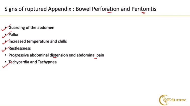
      Symptoms of a ruptured appendix include sudden, severe abdominal pain (often starting near the belly button then shifting to the lower right), fever, nausea/vomiting, and tenderness when pressing the area. Later signs may include swollen abdomen, rapid heart rate, and chills—indicating infection spreading beyond the appendix.

      What can cause an inflamed appendix to rupture?

      An inflamed appendix ruptures when blockage (from stool, infection, or lymph nodes) leads to increasing pressure and bacterial buildup. Without treatment, the appendix’s walls thin and tear, often due to delayed diagnosis or ignored symptoms like pain, fever, or loss of appetite. Rupture is more likely after 2–3 days of untreated inflammation.

      Can stress cause an appendix to rupture?

      Stress alone does not directly cause an appendix to rupture, but chronic stress may weaken the immune system, making it harder to fight infections that could lead to appendicitis. Acute stress doesn’t trigger rupture, but severe illness (stress-related or otherwise) might mask symptoms, delaying treatment and increasing rupture risk.

      Leave a Comment

      Comments are moderated before appearing. The data you submit is processed according to the Privacy Policy of Voltefac.