A Holter monitor represents a cornerstone in modern cardiology, offering continuous and precise heart rhythm assessment beyond the limitations of traditional electrocardiograms (ECGs). Unlike standard ECG tests confined to brief, snapshot recordings, this portable device enables 24-to-48-hour—or even longer—monitoring of cardiac electrical activity in real-world settings. By capturing arrhythmias that may evade detection during clinical visits, Holter monitors bridge the gap between symptomatic episodes and diagnostic certainty, empowering clinicians to tailor interventions with greater accuracy. Their integration into routine practice has transformed the evaluation of conditions ranging from cryptogenic strokes to exercise-induced palpitations, underscoring their indispensable role in both acute and chronic cardiac care.
The device’s functionality hinges on a seamless fusion of hardware and software, where adhesive electrodes record electrical signals transmitted to a compact recorder worn by the patient. Advances in digital storage and telemetry have further enhanced its utility, allowing for remote data transmission and immediate analysis in critical scenarios. Beyond its technical prowess, the Holter monitor’s value lies in its ability to correlate cardiac events with daily activities, sleep patterns, or stress triggers—providing a holistic view of a patient’s physiological state that static tests cannot achieve.

Definition and Core Functionality of Holter Monitors in Cardiac Diagnostics
A Holter monitor is a portable, wearable electrocardiogram (ECG) device designed for continuous, prolonged recording of the heart’s electrical activity over 24 to 48 hours, or in some cases, up to several weeks. Unlike standard ECG tests, which capture heart rhythms for only a few seconds to minutes, Holter monitors provide a comprehensive view of cardiac function during daily activities, sleep, and stress, enabling clinicians to diagnose intermittent arrhythmias that may not manifest during brief clinical evaluations.The primary purpose of a Holter monitor is to detect abnormal heart rhythms (arrhythmias) that could otherwise go undetected due to their sporadic or asymptomatic nature. These devices are instrumental in identifying conditions such as atrial fibrillation, supraventricular tachycardia, ventricular tachycardia, and conduction abnormalities like heart block. By correlating patient symptoms with real-time ECG data, Holter monitors facilitate targeted treatment plans, including medication adjustments, lifestyle modifications, or interventions like catheter ablation.
Comparison of Holter Monitors with Standard ECG in Diagnostic Capabilities
Standard ECG tests record heart activity for 10–15 seconds, offering a snapshot of cardiac function under controlled conditions. In contrast, Holter monitors provide continuous, real-time data over extended periods, capturing variations in heart rate and rhythm that occur during routine physical activities, emotional stress, or sleep. This prolonged monitoring is critical for diagnosing paroxysmal arrhythmias, which may last only seconds or minutes and are unlikely to be detected in a brief ECG.Key differences between Holter monitors and standard ECGs include:
Duration: Standard ECG (seconds to minutes) vs. Holter monitor (24–72 hours or longer).
Portability: Holter monitors are wearable and designed for daily use, while standard ECGs require stationary equipment.
Data Capture: Holter monitors record continuous ECG tracings, whereas standard ECGs provide static images of heart activity.
Clinical Utility: Holter monitors are superior for detecting intermittent or asymptomatic arrhythmias, while standard ECGs are sufficient for initial screening or evaluating known cardiac conditions.
A Holter monitor’s ability to correlate symptoms with ECG changes is its defining advantage, enabling precise diagnosis of conditions such as palpitations, syncope, or chest discomfort that may not be evident during a brief clinical ECG.
Components of a Holter Monitor and Their Functional Roles
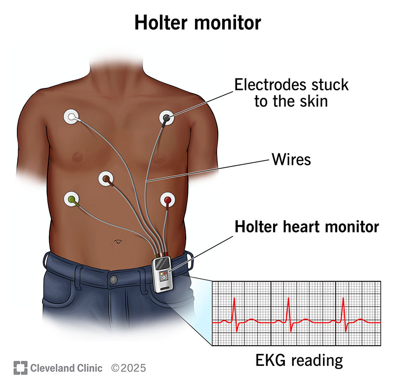
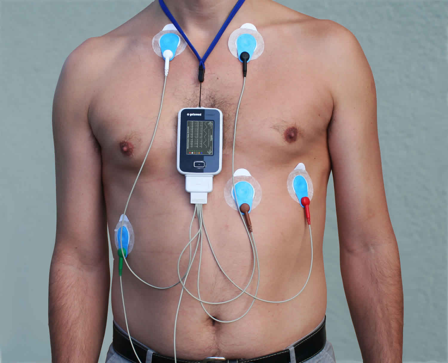
A Holter monitor consists of four primary components, each contributing to its portability, accuracy, and ease of use. Understanding these components clarifies how the device operates in clinical and home settings.The electrodes are adhesive patches applied to the patient’s chest, typically at standardized positions (e.g., V1, V5, and aVL leads) to mirror a 12-lead ECG configuration. These electrodes detect electrical impulses generated by the heart and transmit them to the recorder. High-quality electrodes ensure minimal signal interference and consistent contact, which is critical for accurate rhythm analysis over extended periods.
The recorder unit processes the electrical signals from the electrodes, digitizes them, and stores the data. Modern Holter monitors use digital storage (flash memory or cloud-based systems) to record continuous ECG tracings. Some advanced models incorporate real-time transmission capabilities, allowing clinicians to remotely review data without waiting for the patient to return the device.
The battery powers the recorder, with most devices offering 24–72 hours of continuous operation. Battery life is a critical consideration, as prolonged monitoring requires reliable energy supply. Some monitors feature low-power modes or rechargeable batteries to extend usage duration.
Data storage mechanisms vary by device. Traditional Holter monitors use internal memory cards, while newer models employ wireless transmission to store data on secure servers. This evolution has improved diagnostic workflows by enabling immediate access to recordings for cardiologists, reducing turnaround time for analysis.
The integration of lightweight, high-capacity batteries and miniaturized digital recorders has made Holter monitors more patient-friendly, reducing discomfort and improving compliance during prolonged wear.
Comparison of Holter Monitors with Other Cardiac Monitoring Devices
Cardiac monitoring devices vary in duration, portability, and clinical application. Below is a comparative analysis of Holter monitors against other commonly used devices, highlighting their respective strengths and limitations.
| Feature | Holter Monitor | Event Monitor (External Loop Recorder) | Implantable Loop Recorder (ILR) | Patch Monitor (e.g., Zio Patch) |
|---|
| Monitoring Duration | 24–72 hours (or up to 30 days) | Days to weeks (patient-activated) | Months to years (continuous) | 1–14 days (continuous) |
| Battery Life | 24–72 hours (disposable/rechargeable) | Weeks to months (longer than Holter) | Years (internal battery) | 1–14 days (single-use) |
| Patient Mobility | High (wearable during daily activities) | Moderate (portable but may limit movement) | Very High (internal, no restrictions) | High (adhesive patch) |
| Data Capture | Continuous ECG recording | Intermittent (symptom-triggered) | Continuous, with remote transmission | Continuous, with wireless sync |
| Clinical Use Case | Intermittent arrhythmias, palpitations | Rare or infrequent symptoms | Cryptogenic stroke, unexplained syncope | Post-hospitalization follow-up |
| Patient Compliance | Moderate (24–48 hours is manageable) | High (only activated when symptomatic) | Excellent (internal, no maintenance) | High (easy to apply/remove) |
| Cost | Moderate ($200–$500 per study) | Moderate ($300–$800) | High ($1,500–$3,000) | Moderate ($200–$400) |
While Holter monitors excel in short-to-medium-term continuous monitoring, devices like implantable loop recorders (ILRs) are preferred for long-term, asymptomatic patients with suspected arrhythmias, such as those at risk of cryptogenic stroke. Patch monitors (e.g., Zio) bridge the gap between Holter and ILR by offering extended wear time with wireless data transmission, ideal for post-discharge monitoring.
Cardiac Arrhythmias Detectable via Holter Monitoring and Their Diagnostic Significance
Holter monitors are particularly effective in identifying intermittent or paroxysmal arrhythmias, which may not be captured during a standard ECG or even during a clinical examination. The following arrhythmias are commonly diagnosed using Holter monitoring, along with their clinical implications:Atrial Fibrillation (AFib)
AFib is characterized by rapid, irregular atrial contractions (300–600 bpm) with subsequent irregular ventricular response. Holter monitors detect paroxysmal AFib (episodes lasting <7 days), which may resolve spontaneously and thus go undiagnosed without prolonged monitoring. AFib increases the risk of stroke, heart failure, and thromboembolic events, making early detection critical for initiating anticoagulation therapy (e.g., warfarin or DOACs).
Supraventricular Tachycardia (SVT)
SVT originates above the ventricles (e.g., AV nodal reentry tachycardia, atrial tachycardia) and presents as sudden-onset palpitations with heart rates exceeding 150 bpm. Holter monitors can capture brief SVT episodes, which may terminate spontaneously, confirming the diagnosis and guiding treatment options such as catheter ablation, beta-blockers, or calcium channel blockers.
Ventricular Tachycardia (VT)
VT involves rapid ventricular contractions (>100 bpm) and is often associated with structural heart disease (e.g., prior myocardial infarction, cardiomyopathy). Holter monitoring is essential for asymptomatic VT, which may predispose patients to sudden cardiac death. Detection of VT prompts implantable cardioverter-defibrillator (ICD) placement or antiarrhythmic drug therapy.
Bradyarrhythmias and Heart Block
Conditions such as second-degree AV block (Mobitz type I/II) or complete heart block may manifest as symptomatic bradycardia (heart rate <60 bpm). Holter monitors reveal intermittent conduction delays, which are critical for determining the need for pacemaker implantation in high-risk patients.
Premature Atrial/VENTRICULAR Contractions (PACs/PVCs)
Frequent or multifocal PACs/PVCs may indicate underlying cardiac pathology (e.g., ischemia, cardiomyopathy) or electrolyte imbalances. Holter monitoring quantifies the burden of
Procedure and Patient Experience in Holter Monitoring
Holter monitoring is a non-invasive yet meticulously executed diagnostic procedure that requires precise preparation, patient cooperation, and careful attention to detail. The success of the test hinges on proper electrode placement, accurate symptom documentation, and adherence to protocol to ensure uninterrupted recording of cardiac activity. Patient experience during monitoring may vary, influenced by factors such as electrode sensitivity, device weight, and psychological comfort. Understanding the procedural steps, patient instructions, and strategies to mitigate discomfort ensures optimal diagnostic yield while maintaining patient compliance and satisfaction.
Preparation and Placement of the Holter Monitor
Preparation for Holter monitor placement begins with thorough skin and device preparation to ensure optimal signal quality and patient comfort. The process involves multiple steps, including skin cleansing, electrode positioning, and device calibration to minimize artifacts and maximize recording accuracy.
Skin Preparation and Electrode Application
The patient’s skin must be clean, dry, and free of lotions or oils to prevent poor electrode contact or irritation. A clinician typically follows these steps:
1. Skin Cleansing: The chest area is cleaned with an alcohol wipe or mild abrasive pad to remove dead skin cells and enhance conductivity. Excessive scrubbing should be avoided to prevent micro-tears.
2. Hair Removal: If excessive chest hair interferes with electrode adhesion, it may be clipped or trimmed gently to ensure proper contact.
3. Electrode Placement: Electrodes are positioned according to standard ECG lead placements (e.g., V1–V6, lead II, or modified chest leads). Common placements include:
Standard Limb Leads: Right arm (RA), left arm (LA), right leg (RL), and left leg (LL) electrodes.
Chest Leads: Typically V1–V6, aligned horizontally across the chest to capture anterior, lateral, and inferior myocardial activity.
Alternative Configurations: Some monitors use fewer electrodes (e.g., 3–5 leads) for simplified recording, particularly in pediatric or portable Holter variants.
4. Electrode Attachment: Electrodes are secured using adhesive pads or gel-filled patches. Conductive gel ensures low-impedance contact, while adhesive strength prevents displacement during movement. Hypoallergenic adhesives are preferred to reduce skin irritation, especially for prolonged wear (24–48 hours).Device Calibration and Connection
Once electrodes are applied, the Holter monitor undergoes calibration to verify signal integrity:
Impedance Check: Each electrode’s impedance is measured (ideal range: <5 kΩ) to confirm proper contact. High impedance may indicate poor adhesion or skin contamination.
Baseline ECG Verification: A preliminary ECG trace is recorded to assess for artifacts (e.g., muscle noise, power-line interference) or signal dropout.
Device Programming: The monitor is configured for the recording duration (typically 24–48 hours), sampling rate (usually 128–500 Hz), and memory capacity. Patient-specific details (e.g., age, medical history) are logged for reference.
Patient Symptom Diary and Activity Logging
Accurate symptom documentation is critical for correlating ECG abnormalities with patient experiences. A well-maintained diary enhances diagnostic precision by linking arrhythmias, ischemia, or other events to specific triggers such as exertion, sleep, or emotional stress.Components of an Effective Symptom Diary
Patients are instructed to record the following details in a structured diary provided by the healthcare provider:
Timestamp: Exact time of symptom onset and resolution, formatted as HH:MM (24-hour clock).
Symptom Description: Specific characteristics of the event, including:
Type: Palpitations, dizziness, chest pain, shortness of breath, or syncope.
Severity: Mild, moderate, or severe (e.g., "palpitations rated 8/10").
Duration: How long the symptom lasted (e.g., "30 seconds of rapid heartbeat").
Associated Activities:
Physical: Exercise intensity (e.g., "walking uphill," "lifting 10 lbs"), posture (sitting, standing), or sleep stages (REM vs. non-REM).
Emotional: Stressors (e.g., "argument with family," "work deadline"), anxiety levels, or relaxation techniques used.
Environmental: Temperature changes, caffeine/alcohol consumption, or recent meals.
ECG Correlation: Patients are advised to note if they felt a pulse irregularity or chest discomfort simultaneously with the symptom.Example Diary Entry Format
Date: [DD/MM/YYYY]
Time: 14:30
Symptom: Sudden onset of rapid, pounding heartbeat (palpitations)
Severity: 7/10
Duration: 2 minutes
Activity: Climbing three flights of stairs (moderate exertion)
Additional Notes: Felt lightheaded but no chest pain. Ate a high-caffeine coffee 1 hour prior.
ECG Event: Noted irregular rhythm during episode (patient pressed event marker).
Instructions for Event Marking
Patients are trained to use the Holter monitor’s event marker (a button or switch) to log symptoms in real time. This creates a timestamped annotation on the ECG trace, aiding clinicians in identifying the exact cardiac event. Key reminders include:
Pressing the marker immediately upon symptom onset, not after resolution.
Avoiding repeated presses, which may create false annotations.
Noting the marker number (if applicable) in the diary for cross-reference.
Patient Instructions Checklist for Holter Monitoring
Clear, concise instructions minimize complications and ensure data integrity. Medical professionals should provide patients with a printed or digital checklist covering essential guidelines. Below is a structured checklist with explanatory context for each item.General Device Care
The Holter monitor must remain functional and undisturbed throughout the recording period. Patients should:
Keep the device dry: Avoid swimming, bathing, or showering without a waterproof cover (if provided). Sweat may not require avoidance but should be blotted gently.
Avoid magnets and electronic interference: Stay clear of MRI machines, arc welders, or large transformers, which can corrupt recordings. Keep the device at least 6 inches away from electronic devices (e.g., microwaves, cell phones).
Wear loose clothing: Tight garments may dislodge electrodes or cause discomfort. Avoid restrictive necklaces or belts over the monitor.
Sleep position: Lie on the back or side; avoid sleeping on the device to prevent pressure-related artifacts.Electrode and Skin Maintenance
Proper electrode care prevents signal loss and skin reactions:
Do not remove or adjust electrodes: Self-adjustment risks poor contact or disconnection. If an electrode loosens, gently press it back into place.
Avoid applying lotions or oils: These can degrade adhesive strength or create conductive barriers.
Monitor for skin irritation: Redness, itching, or blistering should be reported immediately. Hypoallergenic electrodes reduce but do not eliminate this risk.Activity and Lifestyle Guidelines
Patients should maintain their usual daily routine but log deviations that may affect heart rhythm:
Exercise as tolerated: Engage in normal physical activity, including walking, climbing stairs, or light sports. Avoid extreme exertion (e.g., marathon running) unless cleared by a physician.
Record sleep patterns: Note bedtime, wake time, and any nighttime symptoms (e.g., sleep apnea-related bradycardia).
Avoid alcohol and caffeine: These substances can induce arrhythmias or mask symptoms. If consumed, the amount and timing should be documented.
Stress management: Patients prone to anxiety should practice relaxation techniques (e.g., deep breathing) and log emotional triggers.Emergency and Technical Issues
Patients must recognize when to seek medical attention or contact the monitoring facility:
Chest pain or severe symptoms: Immediate cessation of monitoring and emergency care (e.g., 911 or local emergency services).
Device malfunction: If the monitor stops recording, beeps erratically, or displays errors, contact the provider for troubleshooting.
Lost or damaged device: Return the monitor as soon as possible, even if incomplete data was recorded.
Minimizing Patient Discomfort During Holter Monitoring
Patient discomfort during Holter monitoring often stems from electrode irritation, device bulk, or psychological anxiety. Addressing these factors through proper technique, material selection, and patient education enhances compliance and data quality.Electrode Placement Techniques for Comfort
Strategic electrode positioning reduces irritation and movement-related artifacts:
Avoid sensitive areas: Place electrodes away from bony prominences (e.g., clavicles, sternum) or highly innervated regions (e.g., axillae). The mid-chest or upper abdomen often provides a balance of signal quality and comfort.
Use hypoallergenic adhesives: Hydrocolloid or silicone-based adhesives minimize allergic reactions and reduce tape residue upon removal. For sensitive skin, consider 24-hour wear patches (e.g., Medtronic’s ProSensor electrodes).
Secure electrodes with minimal tension: Over-tightening can cause skin trauma, while loose electrodes may shift. A gentle press to ensure adhesion is sufficient.
Technical Specifications and Data Interpretation in Holter Monitoring
Holter monitors vary significantly in technical specifications, influencing their operational efficiency, patient compliance, and diagnostic accuracy. Battery life, data storage mechanisms, and real-time transmission capabilities define their functionality, while artifacts and signal integrity challenges require careful interpretation by clinicians. Understanding these technical nuances ensures optimal use in cardiac diagnostics, balancing convenience with precision in rhythm analysis.
Battery Life and Monitoring Duration Across Holter Monitor Models
Battery life is a critical factor in Holter monitors, directly impacting the duration of continuous ECG recording and patient convenience. Modern Holter monitors typically operate for 24 to 48 hours, with some advanced models extending up to 72 hours or longer using low-power designs. For example:
Standard 24-hour monitors (e.g., GE Healthcare’s CardioSoft Holter) use lithium or rechargeable batteries, ensuring uninterrupted recording while minimizing patient burden.
Extended-duration monitors (e.g., Medtronic’s Reveal LINQ Insertable Loop Recorder) leverage miniaturized, long-life batteries (up to 3 years for implantable versions) but are not traditional Holter devices.
Portable event monitors (e.g., KardiaMobile) prioritize short-term use (minutes to hours) with rapid recharging capabilities.Longer battery life reduces the need for frequent recharging but may compromise recording quality due to prolonged electrode-skin contact or patient movement. Conversely, shorter battery life (e.g., 12–24 hours) improves signal fidelity but limits detection of infrequent arrhythmias. Patient convenience is further enhanced by wireless charging in some models, eliminating the need for manual battery replacements.
Data Storage and Transmission in Holter Monitoring
Holter monitor data storage has evolved from analog tape recordings to digital memory systems and cloud-based platforms, improving accessibility and analysis efficiency. Key storage and transmission methods include:- Digital Memory Storage:
High-capacity flash memory (typically 1GB–4GB) stores ECG traces at 128–512 samples per second, preserving high-resolution data for offline analysis. Memory cards (e.g., SD cards) or internal storage in wearable devices (e.g., patch-based monitors) ensure data integrity until download.
- Cloud-Based Systems:
Telemetric Holter monitors (e.g., BioTelemetry’s CardioNet) transmit ECG data in real time via cellular networks (4G/LTE) or Wi-Fi, enabling immediate review by cardiologists. This is critical for urgent cases, such as suspected ST-segment elevation myocardial infarction (STEMI) or ventricular tachycardia, where delays in diagnosis can be life-threatening.
- Hybrid Models:
Some monitors combine local storage with optional telemetry, allowing clinicians to prioritize urgent transmissions while retaining full recordings for retrospective analysis.
Telemetry in Emergency Scenarios:
Real-time transmission reduces the time-to-intervention for high-risk patients. For instance, a patient experiencing syncope during monitoring can trigger an automatic alert to the monitoring center, where a cardiologist reviews the ECG and advises immediate medical response. However, telemetry requires robust cybersecurity measures to protect patient data and reliable network connectivity to prevent data loss.
Key Technical Limitations of Holter Monitors
Holter monitors, despite their utility, are constrained by motion artifacts, electrode dislodgment, and environmental interference, which can degrade diagnostic accuracy. These limitations necessitate careful patient instruction, equipment calibration, and clinician expertise in distinguishing true cardiac events from false signals.
Common technical challenges include:
Motion Artifacts:
Patient movement (e.g., walking, lying down) introduces high-frequency noise that obscures ECG signals, particularly in leads with poor electrode contact. Artifacts often appear as irregular, high-amplitude spikes or baseline wander.- Electrode Dislodgment:
Improper electrode placement or detachment during monitoring leads to signal loss or intermittent recordings, reducing diagnostic yield. Gel-based electrodes are more stable than dry electrodes but require meticulous skin preparation.
- Electromagnetic Interference (EMI):
Electronic devices (e.g., smartphones, microwaves, MRI machines) generate 50/60Hz interference, manifesting as sine-wave patterns superimposed on the ECG. Shielded cables and proper grounding mitigate this issue.
- Battery Drain and Heat Generation:
Prolonged use may cause thermal artifacts or premature battery depletion, truncating recordings. Some monitors include automatic shutdown features to prevent data loss.
Downloading and Analyzing Holter Monitor Data
The process of downloading and interpreting Holter monitor data involves specialized software platforms and cardiologist-led analysis, ensuring accurate diagnosis of arrhythmias and other cardiac events. Key steps include:- Data Extraction:
ECG recordings are downloaded from the monitor’s memory via USB, Wi-Fi, or proprietary docking stations into analysis software (e.g., MUSE Cardio, GE’s CardioLab, or Mortara’s ELI 350). This step requires compatible firmware and patient-specific calibration to maintain signal integrity.
- Software Tools for ECG Analysis:
Automated algorithms in these platforms perform initial rhythm classification, flagging potential abnormalities such as:
Atrial fibrillation (irregularly irregular rhythm).
Supraventricular tachycardia (narrow QRS complexes >100 bpm).
Ventricular ectopy (premature beats with widened QRS).
Clinicians then overread the data, adjusting automated interpretations for false positives/negatives.- Role of Cardiologists in Interpretation:
Holter analysis is not fully automated; cardiologists assess:
Correlation between symptoms and ECG findings (e.g., palpitations during documented tachycardia).
Subtle arrhythmias (e.g., paroxysmal atrial flutter) that may be missed by algorithms.
Artifact differentiation (e.g., distinguishing muscle tremor from ventricular ectopy).Example Workflow:
1. Patient wears the Holter monitor for 24 hours.
2. Data is downloaded into MUSE Cardio, which generates a summary report with annotated rhythms.
3. The cardiologist reviews specific time stamps where symptoms occurred, comparing them to the ECG trace.
4. Final diagnosis is documented, guiding treatment (e.g., antiarrhythmic medication, catheter ablation).
Common Artifacts in Holter Monitor Recordings and Their Distinction from Cardiac Events
Artifacts in Holter recordings can mimic or obscure cardiac pathology, requiring clinicians to apply pattern recognition and contextual analysis. The following table outlines common artifacts and their distinguishing features:
| Artifact Type |
Appearance on ECG |
Distinguishing Features |
Clinical Context |
|---|
| Baseline Wander |
Slow, undulating baseline shifts |
- Caused by respiratory movements or poor electrode contact.
- Does not affect QRS morphology.
- Often seen in lead II or V5 due to diaphragmatic motion.
|
Common in supine patients; may obscure ST-segment changes. |
| Muscle Tremor |
High-frequency, irregular oscillations |
- Associated with patient shivering, anxiety, or movement.
- Appears as fine, rapid spikes superimposed on the ECG.
- Most prominent in limb leads (I, II, III).
|
May be confused with ventricular fibrillation; context (e.g., patient activity) is key. |
| Electrode Pop |
Sudden, transient spikes or notching |
- Caused by electrode displacement or skin contact loss.
- Appears as sharp, isolated deflections without P-QRS-T sequence.
- Often seen during patient repositioning.
|
Can mimic premature beats; verify with adjacent leads. |
| Power Line Interference (50/60Hz) |
Regular, sine-wave oscillations |
- Result of electromagnetic fields from nearby devices.
- Appears as horizontal, repetitive waves at 50/60Hz.
-
Clinical Applications and Patient Groups in Holter Monitoring
Holter monitoring plays a pivotal role in cardiac diagnostics by providing continuous, ambulatory ECG recordings that capture transient arrhythmias often missed by shorter diagnostic tools. Its utility extends beyond routine evaluations, serving as the gold standard for conditions where symptoms are episodic, unpredictable, or poorly correlated with conventional testing. This section explores the specific medical indications where Holter monitoring is indispensable, the patient populations that derive the most benefit, and its comparative advantages in outpatient versus inpatient settings. Emerging applications in post-surgical and long-term arrhythmia management further highlight its evolving role in modern cardiology.
Conditions Where Holter Monitoring Is the Gold Standard
Holter monitoring is the preferred diagnostic tool for several cardiac conditions where symptoms are intermittent, symptoms lack clear triggers, or alternative tests fail to provide definitive answers. Its ability to record prolonged ECG data increases the likelihood of capturing rare or paroxysmal arrhythmias that standard 12-lead ECGs or event monitors may miss.Syncope and Near-Syncope
Syncope, or fainting, often stems from arrhythmias such as sinus node dysfunction, atrioventricular (AV) block, or supraventricular tachycardia (SVT). Standard ECG may appear normal between episodes, while exercise stress tests or tilt-table testing may not reproduce symptoms. A 24–48-hour Holter monitor detects bradyarrhythmias, tachyarrhythmias, or asystolic pauses that correlate with syncopal events. For example, a patient with recurrent syncope and no structural heart disease may present with 2:1 AV block or paroxysmal atrial fibrillation (AF) only captured during Holter monitoring. Palpitations and Cryptogenic Stroke
Patients with palpitations often describe irregular heartbeats that resolve before medical evaluation. Holter monitoring identifies atrial fibrillation (AF), atrial flutter, or ventricular tachycardia (VT) that may not be evident on a single ECG. In cryptogenic stroke—where no clear cause is identified—Holter monitoring detects paroxysmal AF in up to 30% of cases, guiding anticoagulation therapy. A study in The New England Journal of Medicine demonstrated that 30-day cardiac monitoring (including Holter) detected AF in 12.5% of cryptogenic stroke patients, significantly higher than shorter monitoring durations. Unexplained Dyspnea or Chest Pain
Patients with exertional dyspnea or angina-like chest pain may have silent ischemia or arrhythmias not detected by stress testing. Holter monitoring reveals ST-segment changes, ventricular ectopy, or non-sustained VT during daily activities. For instance, a middle-aged athlete with exertional dyspnea may exhibit asymptomatic ventricular premature contractions (VPCs) or supraventricular ectopy only detectable during prolonged monitoring. Alternative Tests and Their Limitations
- Standard 12-lead ECG: Captures only a snapshot; transient arrhythmias may not be present.
- Exercise Stress Test: Limited to provoked arrhythmias; silent or post-exercise arrhythmias may be missed.
- Event Monitors: Patient-activated, reducing capture rates for infrequent or asymptomatic episodes.
- Implantable Loop Recorders (ILRs): More invasive; reserved for high-risk patients with rare symptoms.
Patient Populations Benefiting Most from Holter Monitoring
Holter monitoring is particularly valuable for patient groups where arrhythmias are prevalent, symptoms are vague, or traditional diagnostics are insufficient. These populations include elderly individuals, athletes, post-surgical patients, and those with pacemakers or implantable devices.Elderly Patients with Multimorbidities
Elderly patients often present with atrial fibrillation, heart block, or bradyarrhythmias that contribute to falls, confusion, or fatigue. A 48-hour Holter monitor in this group may reveal:
- Nocturnal bradycardia or pause-dependent syncope.
- Paroxysmal AF with rapid ventricular response, explaining recurrent hospitalizations for heart failure.
- Drug-induced arrhythmias (e.g., QT prolongation from diuretics or antiarrhythmics).
Athletes with Suspected Arrhythmias
Athletes undergoing pre-participation screening may have asymptomatic arrhythmias that could pose risks during competition. Holter monitoring identifies:
- Long QT syndrome or Brugada syndrome in high-risk sports (e.g., swimming, long-distance running).
- Atrial or ventricular ectopy that may warrant further evaluation (e.g., cardiac MRI for structural abnormalities).
- Exercise-induced arrhythmias (e.g., atrial fibrillation in endurance athletes).
Patients with Pacemakers or Implantable Cardioverter-Defibrillators (ICDs)
Holter monitoring ensures device functionality and detects:
- Pacemaker-mediated tachycardia (PMT) or oversensing leading to inappropriate pacing.
- ICD lead fractures or insulation defects causing missed ventricular tachycardia.
- Atrial or ventricular arrhythmias that may require programming adjustments.
Post-Surgical Cardiac Patients
After coronary artery bypass grafting (CABG), valve repair, or catheter ablation, patients may develop new-onset arrhythmias due to surgical trauma or scar-related reentry circuits. Holter monitoring in this context:
- Detects post-ablation atrial flutter or ventricular tachycardia requiring additional interventions.
- Monitors atrial fibrillation recurrence after pulmonary vein isolation, guiding antiarrhythmic therapy.
- Evaluates post-CABG ischemia via ST-segment analysis during daily activities.
Case Studies Highlighting Diagnostic Value
Holter monitoring has resolved diagnostic dilemmas in patients with vague, non-specific symptoms where other tests were inconclusive. The following cases illustrate its impact:Case 1: Cryptogenic Syncope in an Elderly Woman
A 78-year-old female presented with three episodes of unexplained falls over six months. Standard ECG showed left bundle branch block (LBBB), but no arrhythmias. A 48-hour Holter monitor revealed intermittent 3:1 AV block with pauses up to 3.2 seconds, correlating with syncopal events. Pacemaker implantation resolved symptoms. Case 2: Palpitations and Cryptogenic Stroke in a Middle-Aged Man
A 55-year-old male with hypertension suffered a lacunar stroke with no identifiable cause. 24-hour Holter monitoring detected paroxysmal AF with a maximum heart rate of 180 bpm, explaining the embolic event. Direct oral anticoagulant (DOAC) therapy prevented recurrent strokes. Case 3: Silent Ventricular Tachycardia in an Athlete
A 22-year-old marathon runner reported fatigue and near-syncope during training. Exercise stress test was normal, but 72-hour Holter monitoring revealed non-sustained VT (LBBB morphology, rate 160 bpm) during sleep. Electrophysiology study (EPS) confirmed idiopathic VT, leading to successful catheter ablation. Case 4: Post-Ablation Atrial Flutter in a Patient with Paroxysmal AF
A 60-year-old male underwent pulmonary vein isolation (PVI) for AF but continued experiencing palpitations. 7-day Holter monitoring identified atrial flutter (cycle length 220 ms) originating from the caval-tricuspid isthmus, not detected during the initial procedure. Additional ablation restored sinus rhythm.
Outpatient vs. Inpatient Holter Monitoring: Comparative Analysis
The choice between outpatient and inpatient Holter monitoring depends on patient stability, symptom frequency, and resource availability. Each setting has distinct advantages in terms of cost, compliance, and diagnostic yield.Outpatient Holter Monitoring
- Advantages:
- Cost-effective: Reduces hospital stay costs and minimizes patient burden.
- High compliance: Patients in familiar environments are more likely to adhere to monitoring instructions.
- Real-world data: Captures arrhythmias during daily activities, sleep, and stress, which may differ from inpatient settings.
- Limitations:
- Missed episodes: Infrequent or unpredictable symptoms may not coincide with monitoring periods.
- Technical issues: Electrode displacement or signal artifacts can occur without immediate technical support.
- Best suited for:
- Stable patients with intermittent symptoms (e.g., palpitations, syncope).
- Long-term monitoring (e.g., 7-day Holter or event monitors for rare arrhythmias).
Inpatient Holter Monitoring
- Advantages:
- Continuous supervision: Immediate intervention for hemodynamically significant arrhythmias (e.g., VT, complete heart block).
- Controlled environment: Reduces artifact risk and ensures proper electrode placement.
- Higher diagnostic yield: Patients with frequent or severe symptoms (e.g., electrical storms in VT) benefit from round-the-clock monitoring.
- Limitations:
- H
Advanced Features and Innovations in Holter Monitoring
Holter monitors have evolved beyond traditional ambulatory ECG recording, integrating cutting-edge technologies to enhance diagnostic precision, patient convenience, and clinical workflow efficiency. Recent advancements leverage wireless connectivity, artificial intelligence (AI), and hybrid device ecosystems to transform Holter monitoring into a dynamic, data-driven tool for cardiac care. These innovations address limitations in conventional Holter systems, such as delayed data transmission, subjective rhythm interpretation, and fragmented patient monitoring, while expanding applications in both clinical and research settings.The integration of these technologies not only improves arrhythmia detection but also enables proactive cardiac management, remote patient monitoring, and large-scale epidemiological studies. Below, the key advancements—ranging from AI-driven analytics to mHealth integration—are examined in technical and clinical contexts, alongside their implications for modern cardiology.
Wireless Transmission and Real-Time Data Streaming
Modern Holter monitors eliminate the dependency on physical data storage by adopting wireless transmission protocols, including Bluetooth Low Energy (BLE), Wi-Fi, and cellular networks (4G/5G). These systems enable real-time or near-real-time ECG data streaming to centralized platforms, such as electronic health records (EHRs) or cloud-based analytics engines. The transition from batch-processing to instantaneous data transfer allows clinicians to:
- Intervene promptly in cases of acute arrhythmias (e.g., ventricular tachycardia or atrial fibrillation) without waiting for post-procedure analysis.
- Reduce patient anxiety by providing immediate feedback, particularly in high-risk groups such as post-MI or post-ablation patients.
- Enhance compliance through automated reminders and progress updates via companion mobile applications.
Technical Implementation:
Wireless Holter monitors employ secure, encrypted data tunnels to transmit ECG signals to cloud servers or local gateways. Devices like the KardiaMobile (AliveCor) or Zio XT (iRhythm Technologies) utilize proprietary firmware to compress and prioritize data, ensuring minimal latency. Some systems incorporate edge computing—processing raw ECG signals locally before transmitting summarized findings—to conserve bandwidth and reduce power consumption.
Key Advantage: Real-time monitoring bridges the gap between outpatient Holter studies and inpatient telemetry, enabling proactive cardiac care rather than reactive diagnosis.
AI and Machine Learning in Arrhythmia Classification
The integration of machine learning (ML) algorithms into Holter monitor analysis has revolutionized arrhythmia detection by automating pattern recognition and reducing inter-observer variability. Traditional Holter readings rely on manual review by cardiologists or technicians, which is time-consuming and prone to human error. AI-driven systems now classify rhythms with >95% accuracy for common arrhythmias, including:
- Atrial fibrillation (AFib) and flutter
- Supraventricular tachycardia (SVT)
- Ventricular arrhythmias (e.g., PVCs, VT)
- Bradyarrhythmias (e.g., heart block)
Technical Overview of AI Workflows:
1. Preprocessing: Raw ECG signals are filtered to remove noise (e.g., muscle artifact, baseline wander) using adaptive bandpass filters and wavelet transforms.
2. Feature Extraction: Algorithms identify morphological and temporal features, such as:
- P-wave duration and variability (for AFib)
- QRS complex width and morphology (for ventricular arrhythmias)
- RR interval trends (for sinus node dysfunction)
3. Classification: Supervised learning models (e.g., convolutional neural networks (CNNs) or random forests) compare extracted features against labeled datasets (e.g., MIT-BIH Arrhythmia Database). Deep learning architectures, such as 1D-CNNs, excel at detecting subtle ECG patterns without manual feature engineering.
4. Post-Analysis: AI flags high-confidence arrhythmic events for clinician review while suppressing false positives through ensemble methods or confidence scoring.Clinical Impact:
- Reduced reporting time from days to hours, with some systems (e.g., CardioSec’s AI platform) providing preliminary reports within minutes of data upload.
- Improved diagnostic yield in patients with paroxysmal arrhythmias, where traditional Holter may miss intermittent episodes.
- Standardization of reporting across institutions, mitigating variability in interpretation.
Example: The Zio Patch (iRhythm) uses AI to classify 12+ arrhythmia types, including rare variants like atrial tachycardia with block, with sensitivity comparable to expert cardiologists.
Mobile Health (mHealth) Integration and Patient Engagement
The convergence of Holter monitors with mobile health (mHealth) platforms has democratized cardiac monitoring, enabling patients to actively participate in their care. These systems typically include:
- Dedicated smartphone applications for device setup, data synchronization, and educational content.
- Cloud-based dashboards for clinicians to access trends, symptoms logs, and AI-generated summaries.
- Automated alerts for abnormal rhythms or missed recordings.
Key mHealth Features:
- Patient-Driven Data Collection:
- Symptoms correlation (e.g., linking palpitations to detected arrhythmias).
- Activity logs (e.g., syncing with Apple Health or Google Fit to assess rhythm triggers like caffeine or stress).
- Secure Data Sharing:
- HIPAA-compliant APIs allow seamless integration with EHRs (e.g., Epic, Cerner).
- Patient portals enable individuals to review reports and share findings with specialists.
- Remote Patient Monitoring (RPM):
- Chronic AFib patients can track burden metrics (e.g., % time in AFib) over weeks, facilitating shared decision-making with providers.
Case Study: KardiaMobile 6L
This FDA-cleared device pairs with an app to:
1. Record 6-lead ECGs via smartphone.
2. Upload data to a cloud platform for AI analysis.
3. Generate real-time alerts for AFib or bradycardia.
4. Sync with Epic EHR via HL7/FHIR standards.
Patient Benefit: mHealth integration improves adherence rates by ~30% in long-term monitoring studies, as seen in the LOOP Study (2021), where patients preferred digital follow-up over traditional clinic visits.
Hybrid Monitoring Systems for Comprehensive Cardiac Assessment
To address the limitations of standalone Holter monitors—such as isolated ECG data—modern systems integrate with wearable sensors, blood pressure cuffs, and activity trackers to provide a holistic view of cardiovascular health. These hybrid ecosystems are particularly valuable in:
- Hypertension management (e.g., linking arrhythmias to blood pressure spikes).
- Heart failure monitoring (e.g., detecting fluid overload via weight scales + ECG changes).
- Athlete or occupational health (e.g., assessing exertional arrhythmias during exercise).
Examples of Hybrid Systems: | Device/System | Integrated Sensors | Clinical Application |
|---|
| Apple Watch + ECG App | Photoplethysmography (PPG), ECG | AFib screening + activity correlation |
| BioTelemetry’s Zio XT | ECG + accelerometer | Fall detection + arrhythmia correlation |
| Withings ScanWatch | ECG, PPG, blood pressure, SpO₂ | Comprehensive cardiac risk stratification |
| Medtronic CareLink | Implantable loop recorder + remote | Post-ablation arrhythmia surveillance |
Technical Integration:
- Data Fusion Algorithms: Combine ECG with PPG-derived heart rate variability (HRV) or step-count data to assess autonomic dysfunction.
- Predictive Analytics: ML models correlate nocturnal hypertension (from BP cuffs) with symptomatic bradycardia (from Holter).
- Interoperability Standards: Adoption of IEEE 11073 or Continuity of Care (CCD) formats ensures seamless data exchange between devices.
Research Application: The European SOCRATES study (2022) used hybrid monitors (ECG + BP + activity) to identify silent atrial high-rate episodes (AHRE) in 22% of patients initially deemed "normal" by Holter alone.
Role in Large-Scale Cardiac Research and Epidemiology
Holter monitors have become indispensable in population-based studies investigating arrhythmia prevalence, risk factors, and therapeutic efficacy. Their scalability, non-invasive nature, and long-term wearability make them ideal for:
- Epidemiological Surveys: Assessing AFib burden in aging populations (e.g., ARIC Study, Framingham Heart Study).
- Drug Trials: Evaluating proarrhythmic effects of new medications (e.g., SGLT2 inhibitors in T2DM patients).
- Device Validation: Testing the accuracy of wearable AFib
From its foundational role in diagnosing elusive arrhythmias to its evolving integration with wearable technology and AI-driven analytics, the Holter monitor exemplifies how innovation in medical devices can redefine diagnostic precision. Its ability to transform vague symptoms into actionable insights has made it a staple in cardiology, particularly for patients whose conditions defy conventional testing. As advancements in machine learning and hybrid monitoring systems expand its capabilities, the Holter monitor continues to set benchmarks for non-invasive cardiac assessment, ensuring that even the most subtle irregularities are identified with clarity and efficiency. For clinicians and patients alike, its enduring relevance lies in its promise: to illuminate the hidden complexities of the heart with unparalleled accuracy.
FAQ
what is a holter monitor used for?
Q: What medical conditions is a Holter monitor used for?
what is a holter monitor test?
Q: What exactly is a Holter monitor test?
what is a holter monitor and how does it work?
Q: What is a Holter monitor, and how does it work?
what is a holter monitor used to diagnose?
Q: What conditions can a Holter monitor help diagnose?
what is a holter monitor test used for?
Q: What is a Holter monitor test used for?
what is a holter monitor and what does it do?
Q: What is a Holter monitor, and what does it do?
|
Leave a Comment
Comments are moderated before appearing. The data you submit is processed according to the Privacy Policy of Voltefac.