What Is A Heart Stent And Its Critical Role In Cardiovascular Health
Table of Contents
- Definition and Basic Function of a Heart Stent
- Mechanism of Stent Function and Material Properties
- Comparison of Bare-Metal Stents (BMS) and Drug-Eluting Stents (DES)
- Anatomical Locations and Clinical Significance of Stent Implantation
- Medical Procedures Involving Heart Stents: Angioplasty and Beyond
- Procedural Steps in Percutaneous Coronary Intervention (PCI) with Stent Placement
- Elective vs. Emergency Stent Placement
- Contraindications to Stent Placement
- Types of Heart Stents and Their Clinical Applications
- Comparative Analysis of Drug-Eluting Stents (DES) and Bare-Metal Stents (BMS)
- Niche Applications of Stents Beyond Coronary Arteries
- Bioresorbable Stents: Technical Overview and Clinical Potential
- Pharmacological Coatings and Mechanisms of Restenosis Prevention
- Patient Experience and Post-Procedure Care Following Heart Stent Placement
- Recovery Timeline and Immediate Post-Procedure Guidelines
- Monitoring for Post-Stent Complications
- Lifestyle Adjustments for Long-Term Stent Success
- Psychological Impact and Coping Strategies
- Technological Advancements and Future Directions in Stent Design
- Smart Stents with Embedded Sensors for Real-Time Monitoring
- 3D Printing and Customization in Stent Manufacturing
- Fully Biodegradable Stents: Balancing Degradation and Arterial Healing
- Robotic-Assisted Stent Procedures vs. Traditional Manual Techniques
- FAQ
- What medical conditions is a heart stent used to treat?
- What materials are heart stents typically made from?
- How is a heart stent procedure performed step by step?
- What exactly is a heart stent, and how does it physically work inside the body?
- What happens during a heart stent operation, and is surgery required?
- How do you say "heart stent" in Spanish?
A heart stent is a life-saving medical device designed to restore and maintain optimal blood flow within narrowed or blocked arteries, playing a pivotal role in treating cardiovascular diseases that affect millions globally. By acting as a scaffold within the arterial walls, stents prevent collapse and ensure uninterrupted circulation, reducing the risk of heart attacks, strokes, and other life-threatening complications. Advances in biomaterials and minimally invasive techniques have transformed stent technology into a cornerstone of modern interventional cardiology, offering tailored solutions for diverse patient needs—from emergency blockages to chronic arterial conditions.
The functionality of a heart stent extends beyond its mechanical support, integrating innovative designs such as drug-eluting coatings and biodegradable frameworks to enhance efficacy and minimize long-term risks. Procedures like percutaneous coronary intervention (PCI) leverage real-time imaging and precision engineering to deploy stents with minimal invasiveness, while post-operative care emphasizes patient education, lifestyle modifications, and vigilant monitoring to prevent complications. As technology evolves, smart stents and 3D-printed customizations are redefining treatment paradigms, promising safer, more sustainable interventions for an aging global population.
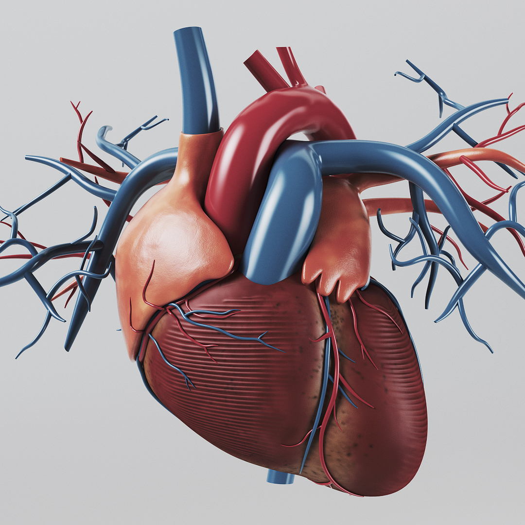
Definition and Basic Function of a Heart Stent
A heart stent is a small, mesh-like tube inserted into narrowed or weakened arteries to restore and maintain adequate blood flow. Its primary function is to provide structural support, preventing arterial collapse and ensuring uninterrupted perfusion to vital organs, particularly the heart muscle. Stents are integral in treating conditions such as coronary artery disease (CAD), peripheral artery disease (PAD), and carotid artery stenosis, where plaque buildup or structural weaknesses compromise vascular integrity.
The deployment of a stent involves a precise interplay between mechanical intervention and biological adaptation. The device expands within the artery, compressing atherosclerotic plaques or structural defects while promoting endothelialization—the natural process by which the inner arterial lining regenerates over the stent’s surface. This dual mechanism of mechanical scaffolding and biological integration ensures long-term patency, reducing the risk of ischemia and associated complications such as angina, heart attack, or stroke.
Mechanism of Stent Function and Material Properties
The physical operation of a stent relies on its radial force—the outward pressure exerted to counteract arterial elastic recoil and plaque compression. During implantation, a stent is delivered via a catheter-based system, typically using balloon angioplasty or self-expanding mechanisms. Once positioned at the target lesion, the stent expands to match the artery’s diameter, creating a stable lumen. The materials used in stent construction are engineered to balance biocompatibility, hemocompatibility, and mechanical resilience.Key material categories include:
The ideal stent material must resist thrombosis, minimize inflammatory response, and maintain structural integrity under physiological stresses (e.g., pulsatile blood flow, bending in dynamic arteries).Stents achieve their function through:
1. Mechanical scaffolding: Immediate restoration of luminal diameter to alleviate flow-limiting stenosis.
2. Endothelialization: Promotion of a continuous endothelial layer to prevent clot formation.
3. Hemodynamic normalization: Reduction of turbulent flow, which can exacerbate plaque progression.
Comparison of Bare-Metal Stents (BMS) and Drug-Eluting Stents (DES)
The choice between stent types depends on patient-specific factors, lesion complexity, and risk profiles. Below is a comparative analysis of bare-metal stents (BMS) and drug-eluting stents (DES), highlighting their material composition, clinical performance, and applications.| Feature | Bare-Metal Stent (BMS) | Drug-Eluting Stent (DES) |
|---|---|---|
| Material Composition | 316L stainless steel, cobalt-chromium alloys, or platinum-chromium alloys; uncoated or coated with heparin or phosphorylcholine. | Same base metals as BMS, with a polymer coating (e.g., durable or biodegradable) impregnated with antiproliferative drugs (e.g., sirolimus, everolimus). |
| Durability | Permanent implantation; no degradation over time. | Durable DES: Permanent polymer coating; bioabsorbable DES: Polymer/drug layer degrades within 12–36 months. |
| Risk of Clotting (Thrombosis) | Higher early risk (3–6 months) due to lack of antiproliferative protection; requires dual antiplatelet therapy (DAPT) for 1–3 months. | Lower early risk but delayed endothelialization may increase late thrombosis if DAPT is discontinued prematurely; typical DAPT duration: 6–12 months (longer for high-risk lesions). |
| Typical Use Cases |
|
|
| Restenosis Rate | 15–30% within 6–12 months (higher in diabetic or complex lesions). | 5–10% with durable DES; bioabsorbable DES may reduce very late restenosis but carry higher early thrombosis risk. |
DES significantly reduced restenosis rates by ~50% compared to BMS in clinical trials (e.g., SIRIUS, TAXUS), though their use requires stricter adherence to DAPT protocols to mitigate late stent thrombosis.
Anatomical Locations and Clinical Significance of Stent Implantation
Stents are deployed in arteries where luminal narrowing or structural weakness threatens organ perfusion or risks acute occlusion. The most critical implantation sites include:1. Coronary Arteries
Coronary stents are the most frequently implanted, with ~1.2 million procedures annually in the U.S. alone, primarily for obstructive CAD or acute coronary syndromes.2. Peripheral Arteries
3. Carotid Arteries
Criticality of These Locations:
Stent placement in these regions is guided by angiographic assessment, fractional flow reserve (FFR), or intravascular ultrasound (IVUS) to ensure optimal lesion coverage and minimize procedural complications.
Medical Procedures Involving Heart Stents: Angioplasty and Beyond
Percutaneous coronary intervention (PCI) with stent placement represents a cornerstone of interventional cardiology, offering a minimally invasive solution to restore blood flow in obstructed coronary arteries. The procedure combines catheter-based techniques with advanced imaging to achieve immediate mechanical revascularization, often as a lifesaving measure in acute ischemic events or an elective strategy for chronic obstructive disease. Beyond traditional angioplasty, modern stent technologies—such as drug-eluting stents (DES) and bioresorbable scaffolds—have expanded therapeutic possibilities while refining patient-specific treatment pathways.
The success of stent-based interventions hinges on precise procedural execution, real-time diagnostic feedback, and careful patient selection. Elective and emergency placements differ significantly in urgency, risk stratification, and clinical outcomes, with emergency cases often dictated by time-sensitive hemodynamic instability. Contraindications to stenting, though rare, necessitate thorough preoperative evaluation to mitigate procedural risks, particularly in patients with comorbid conditions that may compromise procedural safety or long-term stent patency.
Procedural Steps in Percutaneous Coronary Intervention (PCI) with Stent Placement
PCI with stent deployment follows a standardized sequence of steps, integrating vascular access, coronary angiography, lesion preparation, and stent optimization. The procedure is performed under sterile conditions in a cardiac catheterization laboratory, with the patient under conscious sedation or general anesthesia. Key phases include:1. Pre-procedural Preparation
2. Catheter Insertion and Coronary Angiography
3. Balloon Angioplasty (Pre-dilatation)
4. Stent Deployment
5. Procedure Completion
Elective vs. Emergency Stent Placement
The indication for stent placement spans a spectrum from elective, planned revascularization to emergency intervention during acute coronary syndromes (ACS). Key distinctions include:- Elective Stent Placement
- Emergency Stent Placement
Primary PCI within 12 hours of symptom onset in STEMI reduces 30-day mortality by ~50% compared to thrombolysis, with benefits sustained at 1-year follow-up (STEMI-3 Trial, 2017).
Contraindications to Stent Placement
Absolute or relative contraindications to PCI with stenting are rare but critical to identify pre-procedurally to avoid complications. The following conditions may preclude safe stent deployment or necessitate alternative revascularization strategies:-
Severe Renal Dysfunction (eGFR <30 mL/min/1.73 m² or on dialysis)
- Explanation: Contrast-induced nephropathy (CIN) risk exceeds 20% in high-risk patients. Pre-procedural hydration, low-osmolar contrast, and minimal contrast volume (<100 mL) are mandatory. Alternative imaging (e.g., intravascular ultrasound [IVUS]) may reduce contrast use.
-
Active Systemic Infection or Endocarditis
- Explanation: Stent implantation carries a risk of systemic embolization or infection (e.g., stent-endocarditis), particularly in patients with prosthetic valves or intravenous drug use. Delayed revascularization or medical management is preferred until infection resolves.
-
Uncontrolled Coagulopathy (INR >3.0, Platelet Count <50,000/µL)
- Explanation: Increased bleeding risk during vascular access and stent deployment. Correction of coagulopathy (e.g., vitamin K, platelet transfusions) or alternative revascularization (e.g., coronary artery bypass grafting [CABG]) may be required.
-
Structural Heart Defects with High Surgical Risk
- Example: Severe aortic stenosis with critical left ventricular outflow tract obstruction or complex congenital heart disease (e.g., univentricular physiology).
- Explanation: PCI may exacerbate valvular dysfunction or hemodynamic instability. Hybrid approaches (e.g., transcatheter aortic valve replacement [TAVR] + PCI) or CABG are considered.
-
Allergy to Contrast Media or Antiplatelet Agents
- Explanation: Life-threatening anaphylaxis or treatment failure. Pre-medication (e.g., corticosteroids, antihistamines) or alternative imaging (e.g., IVUS) may mitigate contrast reactions, while drug-eluting stents require strict antiplatelet compliance.
-
Severe Peripheral Arterial Disease (PAD) with Critical Limb Ischemia
- Explanation: Vascular access complications (e.g., femoral artery occlusion) or distal embolization risk may outweigh benefits. Endovascular treatment of PAD or CABG may be prioritized.
-
Recent Stroke (≤30 Days) or Intracranial Hemorrhage
- Explanation: Antiplatelet therapy increases hemorrhagic
- Complex lesions (long segments, bifurcations, chronic total occlusions).
- Diabetic patients or those with high-risk features (e.g., small vessels, left main disease).
- Elective procedures where long-term outcomes are prioritized.
- Acute myocardial infarction (STEMI) with short lesion lengths.
- Patients with contraindications to DAPT (e.g., high bleeding risk).
- Emergency revascularization where immediate stent apposition is critical.
- Material: Nitinol or stainless steel alloys for flexibility and radial strength.
- Design: Modular or bifurcated configurations to adapt to aortic geometry.
- Coatings: Hydrophobic polymers (e.g., polyethylene terephthalate) to prevent thrombus formation.
- Example: Medtronic’s Valiant Thoracic Stent Graft combines self-expanding nitinol with a low-profile delivery system for complex aortic pathologies.
- Radial Force: Higher than coronary stents to penetrate calcified plaques (e.g., Boston Scientific’s Lifestent).
- Hemocompatibility: Phosphorylcholine or heparin coatings to reduce thrombosis in high-flow environments.
- Drug-Eluting Variants: Paclitaxel-coated stents (e.g., Zilver PTX) demonstrate reduced restenosis in femoropopliteal arteries (up to 70% reduction at 2 years per ZILVER PTX trial).
- Low Profile: Compatibility with microcatheters (e.g., Covidien’s Neuroform Atlas).
- Radiopacity: Platinum or gold markers for fluoroscopic visibility.
- Thromboresistance: Polyethylene oxide coatings to minimize embolic risk.
- Example: Enterprise VRD stent (Codman) combines self-expanding nitinol with a closed-cell design to prevent distal migration.
- Degradation Process: Hydrolytic cleavage of ester bonds in PLLA/PCL releases lactic/caproic acid, metabolized via the Krebs cycle.
- Timeline:
- 6–12 months: Scaffold maintains structural integrity.
- 2–3 years: Complete resorption, with arterial remodeling and vasomotion recovery.
- Biocompatibility: FDA-approved PLLA is used in resorbable sutures (e.g., Vicryl), validating its safety profile.
- Younger Patients: Ideal for pediatric congenital heart disease or adults with expected long lifespans, avoiding chronic foreign-body risks.
- Late-Course Benefits: Potential reduction in very late stent thrombosis (beyond 1 year) and improved long-term endothelial function.
- Example: The ABSORB II trial demonstrated non-inferiority in major adverse cardiac events (MACE) at 5 years, though higher rates of scaffold thrombosis (ST) necessitated rigorous patient selection.
- Procedural Complexity: Requires precise deployment to avoid malapposition or edge dissection.
- Cost: ~5× higher than DES, limiting widespread adoption.
- Data Gaps: Long-term (>5 years) outcomes remain under investigation, particularly for complex lesions.
- Resume light activities (e.g., short walks, household chores) within 3–5 days, provided no complications arise.
- Avoid contact sports or high-impact exercises (e.g., running, weightlifting) for 4–6 weeks, as these increase the risk of stent displacement or bleeding.
- Limit stair climbing to 1–2 flights at a time for the first 2 weeks, progressing as tolerated.
- Attend cardiac rehabilitation programs (if prescribed), which combine supervised exercise, education on heart-healthy living, and psychological support.
- Hydration: Increased fluid intake (unless restricted by kidney function) to aid circulation and medication absorption.
- Low-sodium intake (<1,500–2,000 mg/day) to reduce blood pressure and strain on the heart.
- Heart-healthy fats: Emphasis on omega-3 fatty acids (e.g., fatty fish, flaxseeds) and avoidance of trans fats and excessive saturated fats.
- Fiber-rich foods: Whole grains, fruits, and vegetables to support cholesterol management and digestive health.
- Avoidance of alcohol and caffeine for at least 48 hours post-procedure, as these can interact with antiplatelet medications (e.g., clopidogrel) and increase bleeding risk.
- Echocardiogram or stress test (4–6 weeks post-procedure) to assess heart function and stent efficacy.
- Bloodwork (e.g., lipid panel, complete blood count, kidney function tests) to monitor medication side effects and metabolic health.
- Coronary angiography repeat (rare, but may be indicated if symptoms persist or restenosis is suspected).
- Chest discomfort: Persistent or worsening pressure, squeezing, or heaviness (may indicate restenosis or poor blood flow).
- Fatigue or weakness: Unusual tiredness lasting >24 hours, particularly with minimal exertion.
- Swelling or bruising: Excessive swelling at the catheter insertion site (e.g., groin, wrist) or new bruising beyond the expected area.
- Fever or chills: Temperature >100.4°F (38°C), suggesting infection at the insertion site or systemic inflammation.
- Shortness of breath: Mild dyspnea during routine activities (e.g., walking up stairs), possibly indicating fluid retention or heart failure.
- Nausea or vomiting: Unrelieved by antiemetics, which may signal heart strain or medication side effects.
- Headache or dizziness: Severe or persistent headaches, especially if accompanied by vision changes or confusion.
- Crushing chest pain radiating to arms, jaw, or back: Classic angina or myocardial infarction (heart attack) symptoms.
- Severe shortness of breath: Sudden onset, at rest, or with minimal activity (possible stent thrombosis or pulmonary edema).
- Rapid or irregular heartbeat: Palpitations, fluttering, or a pulse >100 bpm (tachycardia) without exertion.
- Cold, clammy skin: Pale or sweaty skin, indicating shock or severe hypoxia.
- Fainting or near-fainting episodes: Syncope or presyncope, suggesting poor cardiac output or arrhythmias.
- Severe allergic reaction: Rash, itching, swelling of lips/tongue, or difficulty breathing (anaphylaxis to contrast dye or medications).
- Risk reduction: Smoking accelerates atherosclerosis and impairs endothelial function, increasing restenosis risk by 30–50% within 12 months.
- Support strategies:
- Nicotine replacement therapy (patches, gum) under medical supervision.
- Behavioral counseling or support groups (e.g., American Lung Association programs).
- Avoidance of secondhand smoke and vaping, which also damages blood vessels.
- Antiplatelet therapy: Dual antiplatelet therapy (DAPT) (e.g., aspirin + clopidogrel/ticagrelor) is mandatory for at least 6–12 months (longer for DES). Skipping doses increases thrombosis risk by up to 70%.
- Statins: High-intensity statins (e.g., atorvastatin) reduce LDL cholesterol by 50%+, lowering restenosis risk by 25–30%.
- Blood pressure and diabetes management: ACE inhibitors (e.g., lisinopril) or ARBs (e.g., losartan) may be prescribed to protect heart function.
- Tracking side effects: Document any adverse reactions (e.g., gastrointestinal bleeding with aspirin) and report to the cardiologist.
- Chronic stress elevates cortisol levels, promoting inflammation and plaque instability. Techniques to mitigate stress include:
- Mindfulness-based stress reduction (MBSR): Structured programs (e.g., 8-week courses) reduce stress hormones by 20–30%.
- Physical activity: Low-impact exercises (e.g., yoga, tai chi, swimming) improve mental health and cardiovascular fitness.
- Cognitive behavioral therapy (CBT): Addresses anxiety about recurrence or medication dependency, with studies showing 40% reduction in depressive symptoms post-intervention.
- Support resources:
- Cardiac psychology services offered through rehabilitation programs.
- Peer support groups (e.g., American Heart Association’s "Heart to Heart" forums).
- Online tools (e.g., apps like "MoodTools" for tracking emotional well-being).
- Mediterranean diet: Associated with a 25% lower risk of major cardiovascular events post-stent, emphasizing olive oil, nuts, and leafy greens.
- Gradual exercise progression: Transition from cardiac rehab to 150 minutes/week of moderate activity (e.g., brisk walking, cycling) within 3 months.
- Weight management: Achieving and maintaining a BMI <25 reduces restenosis risk by 15–20% through improved endothelial function.
- Anxiety about stent durability: Concerns over restenosis or stent failure, particularly in younger patients or those with complex coronary anatomy.
- Medication burden: Fatigue from
- Pressure-sensitive microelectromechanical systems (MEMS): Embedded within stent struts to measure translesional pressure gradients, enabling early detection of restenosis or graft failure. Trials such as the WISE trial (Wireless Instantaneous Stent Evaluation) demonstrated feasibility in porcine models, with human applications pending regulatory approval.
- Flow-resistant impedance sensors: Detect turbulent flow patterns indicative of stent edge dissection or incomplete expansion, reducing the need for routine angiographic follow-up. Early prototypes (e.g., Boston Scientific’s "Smart Stent" concept) have shown promise in identifying hemodynamically significant lesions within 24 hours of implantation.
- Bioabsorbable sensor arrays: Combining temporary monitoring with biodegradable scaffolds to eliminate long-term foreign-body risks. Research at ETH Zurich has yielded prototypes using piezoelectric polymers that degrade synchronously with arterial healing, avoiding chronic inflammation.
- Congenital heart disease (CHD) interventions: Stents for pulmonary valve stenosis or coarctation of the aorta are now printed with adjustable diameters to accommodate pediatric growth, as demonstrated by Medtronic’s "3D-printed stent graft" used in the CONCERT trial. These implants reduce the need for multiple reinterventions.
- Bifurcation and complex coronary lesions: Hybrid stents combining sirolimus-eluting segments with biodegradable scaffolds are printed to match lesion-specific drug elution profiles, as seen in Abbott’s "Custom Stent" platform, which achieved 92% procedural success in a 2023 multicenter study.
- Peripheral vascular diseases (PVD): Nitinol-based 3D-printed stents for femoropopliteal arteries incorporate variable porosity to enhance endothelialization and reduce restenosis, with Boston Scientific’s "Supera" stent serving as a precursor to next-gen customizable designs.
- Selective Laser Melting (SLM): Produces stents with nanometer-scale surface textures to promote endothelial proliferation, reducing thrombosis risks.
- Electrospinning for biodegradable scaffolds: Yields fiber-based stents with tunable degradation rates, as explored by MIT’s BioMicroSystems lab for coronary applications.
- AI-driven design optimization: Algorithms like DeepStent (Stanford University) generate stent geometries by analyzing CT angiography data, predicting optimal strut placement to minimize shear stress.
- Smart degradation triggers: Incorporating enzymatically responsive polymers that accelerate resorption in high-shear environments (e.g., left main coronary artery) while preserving structural integrity in low-flow regions.
- Hybrid biodegradable-metallic stents: Combining absorbable scaffolds with permanent struts in high-stress zones (e.g., ostial lesions) to mitigate mechanical failure risks.
- In vivo imaging-guided degradation: Using MRI-compatible markers to monitor scaffold resorption dynamically, as demonstrated by Cardiovascular Research Technologies’ "Degradation Tracking" system.
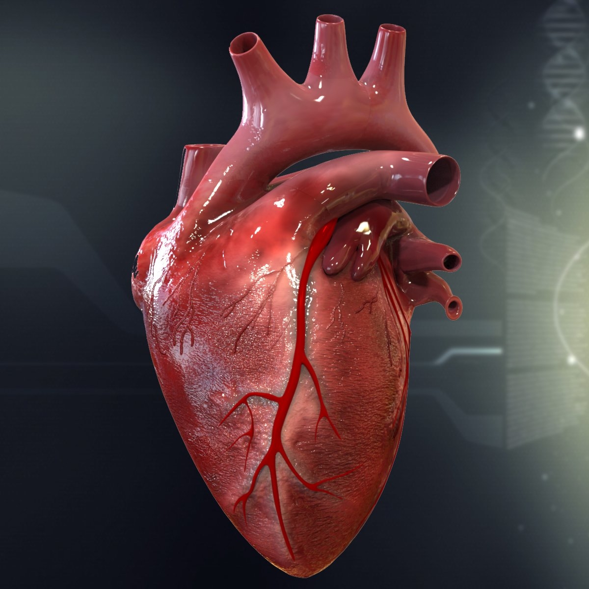
Types of Heart Stents and Their Clinical Applications
Heart stents represent a critical advancement in interventional cardiology, with diverse designs tailored to specific anatomical and pathological requirements. Beyond the fundamental distinction between bare-metal and drug-eluting stents, modern stent technology extends to specialized applications in peripheral, neurovascular, and aortic vasculature. Each variant addresses unique physiological challenges, incorporating material science, pharmacology, and biomechanical engineering to optimize patient outcomes. This section examines the comparative efficacy of stent types, niche vascular applications, and innovative designs such as bioresorbable scaffolds, alongside the role of pharmacological coatings in mitigating restenosis.Comparative Analysis of Drug-Eluting Stents (DES) and Bare-Metal Stents (BMS)
The selection between drug-eluting stents (DES) and bare-metal stents (BMS) hinges on patient risk profiles, procedural urgency, and long-term clinical goals. While BMS offer immediate structural support and shorter healing times, DES provide superior anti-restenotic properties through localized drug delivery. Below is a structured comparison highlighting key clinical and economic considerations:| Parameter | Drug-Eluting Stents (DES) | Bare-Metal Stents (BMS) |
|---|---|---|
| Anti-Restenosis Effectiveness | Highly effective due to sustained release of antiproliferative agents (e.g., sirolimus, paclitaxel). Clinical studies (e.g., RAVEL, SIRIUS) demonstrate 30–50% reduction in restenosis rates at 12 months compared to BMS. Mechanism: Controlled elution of drugs inhibits smooth muscle cell proliferation via mTOR (sirolimus) or microtubule stabilization (paclitaxel). |
Limited efficacy; restenosis rates range from 20–40% at 6–12 months, primarily driven by neointimal hyperplasia. Limitation: Lack of pharmacological intervention leads to higher rates of in-stent restenosis (ISR) in complex lesions. |
| Healing Time and Endothelialization | Delayed re-endothelialization (6–12 months) due to cytotoxic effects of drug coatings, increasing late stent thrombosis risk if dual antiplatelet therapy (DAPT) is prematurely discontinued. Clinical Note: Current guidelines recommend ≥12 months of DAPT for DES to mitigate late thrombosis. |
Faster endothelial coverage (3–6 months), reducing early stent thrombosis risk but not eliminating it entirely. |
| Cost Considerations | Higher upfront cost (2–3× that of BMS), though long-term savings may offset expenses due to reduced target lesion revascularization (TLR). Economic Data: Cost-effectiveness studies (e.g., JAMA 2006) suggest DES are cost-neutral or cost-saving in high-risk populations over 5 years. |
Lower initial cost, but higher procedural costs may arise from repeat revascularizations (e.g., PCI or CABG for ISR). |
| Clinical Indications |
Niche Applications of Stents Beyond Coronary Arteries
Stent technology has expanded into peripheral, neurovascular, and structural heart applications, each requiring specialized designs to accommodate unique anatomical and hemodynamic demands. These include:Aortic Stents (Endovascular Aortic Repair - EVAR/TEVAR)
Stents used in EVAR (abdominal aortic aneurysm) or TEVAR (thoracic aortic aneurysm) must withstand high-pressure environments and resist calcification. Key features include:
Peripheral Artery Disease (PAD) Stents
PAD stents target lower extremity arteries, often in calcified or tortuous vessels. Critical design aspects include:
Neurovascular Stents
Used in carotid artery stenosis or intracranial aneurysms, these stents require:
Bioresorbable Stents: Technical Overview and Clinical Potential
Bioresorbable stents (BRS) represent a paradigm shift by temporarily scaffolding the artery before dissolving, restoring natural vasomotion and reducing long-term complications. The most advanced platforms—Absorb BVS (Abbott Vascular) and Magmaris (Biotronik)—employ poly-L-lactic acid (PLLA) or poly-L-caprolactone (PCL) scaffolds with drug coatings (e.g., everolimus). Key technical and clinical aspects include:Mechanism of Resorption
Clinical Advantages
Limitations and Challenges
Pharmacological Coatings and Mechanisms of Restenosis Prevention
Drug-eluting stents leverage controlled-release pharmacology to inhibit neointimal hyperplasia. The two dominant drugPatient Experience and Post-Procedure Care Following Heart Stent Placement
The recovery phase after coronary stent implantation is critical for ensuring long-term success and minimizing complications. Patients undergo a structured rehabilitation process, combining physical restrictions, dietary adjustments, and vigilant monitoring of symptoms. Understanding the expected timeline, potential warning signs, and necessary lifestyle modifications empowers individuals to actively participate in their recovery while mitigating risks. Psychological support also plays a pivotal role, as patients navigate the emotional and practical challenges of adapting to a new health regimen.Post-procedure care is divided into immediate recovery (first 24–48 hours), short-term rehabilitation (first 30 days), and long-term management (beyond one month). Each phase includes specific guidelines to optimize healing and prevent restenosis or other adverse events. Medical professionals emphasize adherence to follow-up protocols, including diagnostic tests and medication compliance, to monitor vascular health and stent patency.
Recovery Timeline and Immediate Post-Procedure Guidelines
The first 24–48 hours post-stent placement are critical for stabilization. Patients typically remain hospitalized for observation, with nurses monitoring vital signs, pain levels, and signs of bleeding or allergic reactions to contrast dye or antiplatelet medications. Physical activity restrictions begin immediately: patients are advised to avoid heavy lifting (>5 lbs or 2.3 kg), strenuous exercise, or driving for at least 1–2 weeks, depending on the procedure’s complexity and individual recovery.During the first 30 days, the focus shifts to gradual rehabilitation. Patients are encouraged to:
Dietary modifications in the initial phase include:
Follow-up tests within the first 30 days typically include:
Monitoring for Post-Stent Complications
Patients must remain vigilant for signs of complications, which can range from mild discomfort to life-threatening emergencies. The following checklist categorizes symptoms by severity, guiding patients on when to seek immediate medical attention.Mild to Moderate Symptoms (Consult Healthcare Provider Promptly)
Severe or Emergency Symptoms (Seek Immediate Medical Help)
Note: Patients with drug-eluting stents (DES) must be especially cautious, as delayed healing of the arterial lining increases the risk of late stent thrombosis (up to 1 year post-procedure). Symptoms may appear subtler but require urgent evaluation.
Lifestyle Adjustments for Long-Term Stent Success
Adherence to prescribed lifestyle changes is as critical as medication compliance in preventing restenosis and optimizing cardiovascular health. The following adjustments form the cornerstone of post-stent management:Smoking Cessation
Medication Adherence
Stress Management and Mental Health
Dietary and Exercise Sustainability
Psychological Impact and Coping Strategies
The psychological aftermath of a stent procedure often involves a mix of relief, anxiety, and existential adjustments. Patients frequently report fear of recurrence, dependency on medications, and uncertainty about long-term prognosis, which can lead to depression or avoidance behaviors. Addressing these challenges proactively improves quality of life and treatment outcomes.Common Psychological Responses
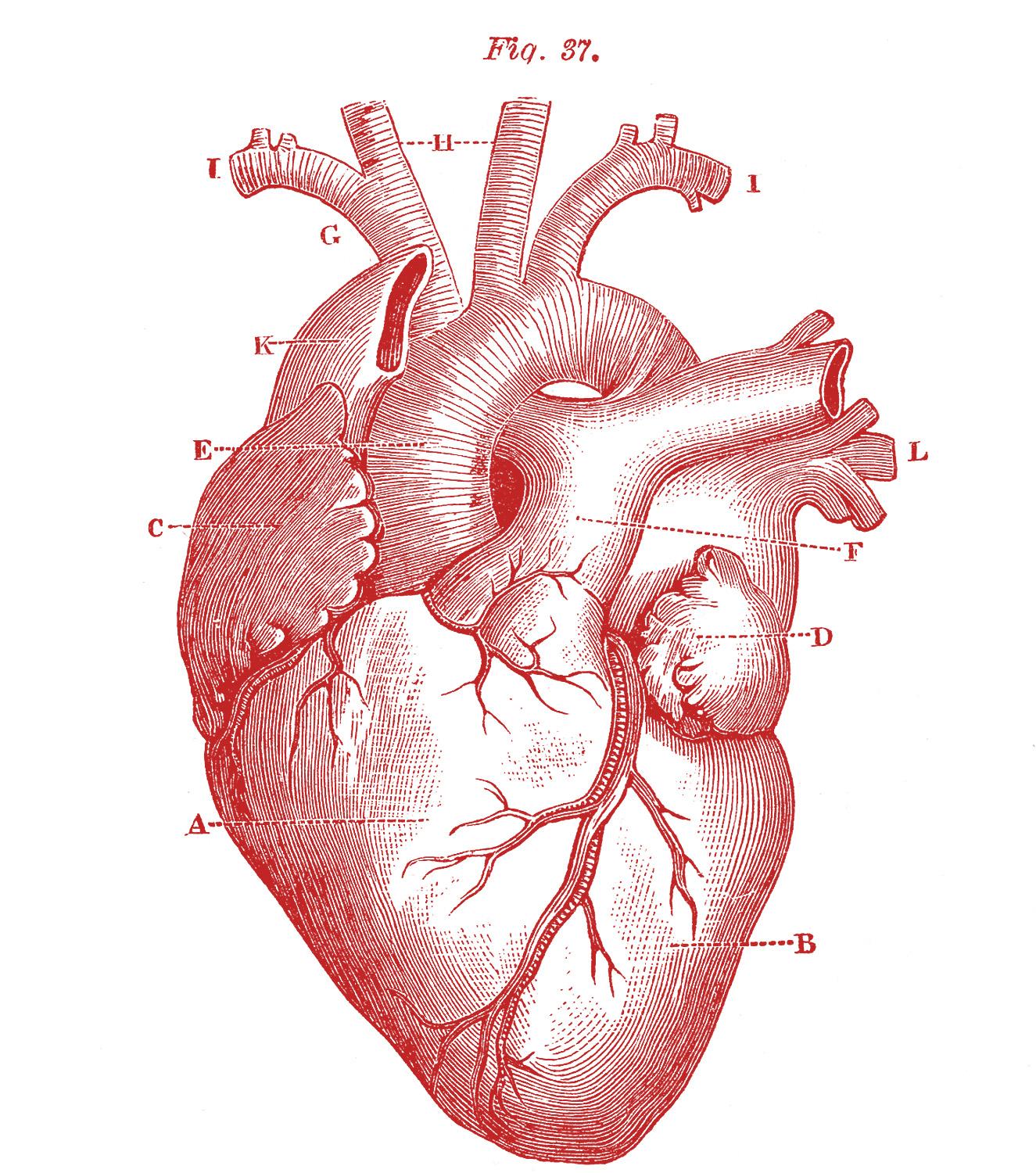
Technological Advancements and Future Directions in Stent Design
Advancements in stent technology continue to redefine interventional cardiology, integrating innovations in biomaterials, digital fabrication, and real-time monitoring to enhance procedural efficacy and patient outcomes. Emerging trends such as smart stents, 3D-printed custom implants, and biodegradable scaffolds address long-standing limitations in durability, biocompatibility, and anatomical adaptability. These developments not only improve immediate procedural success but also optimize long-term vascular health, reducing complications like restenosis and thrombosis. Below, the evolution of stent design is examined through four transformative domains: sensor-embedded smart stents, additive manufacturing for patient-specific solutions, biodegradable materials, and robotic-assisted deployment.Smart Stents with Embedded Sensors for Real-Time Monitoring
The integration of microelectronics and wireless communication into stent structures enables in vivo monitoring of critical hemodynamic and structural parameters, marking a paradigm shift from passive implants to active diagnostic tools. These smart stents incorporate miniature sensors—such as pressure transducers, flow meters, or impedance-based detectors—to transmit real-time data on blood flow dynamics, stent apposition, and potential malappositions or thrombus formation. Clinical applications extend beyond post-procedural surveillance to personalized therapy adjustments, where physicians can remotely adjust medication regimens (e.g., antiplatelet dosing) based on intravascular conditions detected by embedded sensors.Key sensor technologies under development include:
Clinical Impact: Smart stents could reduce unplanned revascularizations by 30–40% through early intervention, while wireless data transmission aligns with the FDA’s Digital Health Innovation Plan, prioritizing remote patient management.
3D Printing and Customization in Stent Manufacturing
Additive manufacturing (3D printing) has revolutionized stent production by enabling patient-specific designs tailored to complex anatomies, congenital defects, or bifurcated lesions that defy off-the-shelf solutions. Traditional stents, fabricated via laser-cutting from metal tubes, are limited to standardized geometries, whereas 3D-printed stents allow for customized strut thickness, porosity, and branching patterns optimized for individual vascular morphologies. This precision reduces procedural risks—such as malapposition in tortuous vessels—and improves long-term patency.Applications of 3D-printed stents include:
Manufacturing Advances:
Fully Biodegradable Stents: Balancing Degradation and Arterial Healing
The development of bioabsorbable stents addresses the foreign-body response associated with permanent metallic implants, which can lead to chronic inflammation, late stent thrombosis, and the need for lifelong dual antiplatelet therapy (DAPT). These stents are engineered from poly-L-lactic acid (PLLA), magnesium alloys, or tyrosine-derived polymers, designed to degrade within 12–36 months, coinciding with arterial remodeling. The primary challenge lies in synchronizing degradation kinetics with endothelialization and vascular healing to prevent early structural failure or delayed restenosis.Key biodegradable stent platforms and their clinical trade-offs:
| Stent Material | Degradation Time | Clinical Applications | Challenges |
|---|---|---|---|
| PLLA (e.g., Abbott’s Absorb BVS) | 12–18 months | Coronary artery disease (CAD) with small vessel diameters (<3.0 mm) | Higher restenosis rates at 3 years (12.4% vs. 8.8% for metallic stents in the ABSORB III trial); risk of late scaffold thrombosis if DAPT is discontinued prematurely. |
| Magnesium alloys (e.g., Biotronik’s Magmaris) | 6–12 months | Peripheral arteries (e.g., femoropopliteal lesions) | Rapid degradation may lead to early loss of radial strength, requiring thicker struts that increase thrombogenicity. |
| Tyrosine-derived polymers (e.g., Reva Medical’s REVA stent) | 24–36 months | Saphenous vein graft lesions, bifurcations | Slower resorption may prolong foreign-body reaction; limited long-term data beyond 5 years. |
Future Directions:
Robotic-Assisted Stent Procedures vs. Traditional Manual Techniques
The adoption of robotic-assisted catheterization systems represents a significant leap in procedural precision, radiation exposure reduction, and ergonomic efficiency compared to conventional manual techniques. Systems such as Corindus’ CorPath GRX and Hansen Medical’s Sensei X leverage haptic feedback, automated contrast injection, and AI-driven lesion assessment to enhance stent deployment accuracy, particularly in complex anatomies like chronic total occlusions (CTOs) or heavily calcified lesions. Below is a comparative analysis of robotic versus manual stent procedures across critical metrics:| Parameter | Robotic-Assisted Procedure | Traditional Manual Procedure Heart stents represent a convergence of medical ingenuity and clinical necessity, bridging the gap between acute cardiovascular emergencies and chronic disease management. From the meticulous selection of materials to the integration of cutting-edge imaging and robotic assistance, each advancement refines the balance between immediate therapeutic outcomes and long-term patient well-being. As research continues to explore biodegradable alternatives and real-time monitoring systems, the future of stent technology holds the potential to further reduce complications, expand accessibility, and redefine recovery trajectories for individuals worldwide. Understanding their mechanics, applications, and post-procedural care is essential for patients, clinicians, and policymakers alike to harness their full transformative potential in global healthcare. FAQWhat medical conditions is a heart stent used to treat?A heart stent is primarily used to treat blocked or narrowed coronary arteries due to plaque buildup (atherosclerosis), improving blood flow to the heart muscle. It can also be used for other arteries (like those in the legs or kidneys) or to treat conditions like heart attacks or chest pain (angina). Stents may also prevent arteries from re-narrowing after angioplasty. What materials are heart stents typically made from?Most heart stents are made from metals like stainless steel, cobalt-chromium alloys, or platinum-chromium, which provide strength and flexibility. Some newer stents use biodegradable polymers that dissolve over time, while drug-eluting stents combine metal with medications (e.g., sirolimus or everolimus) to reduce scarring and re-blockage. How is a heart stent procedure performed step by step?During a stent procedure (angioplasty), a catheter with a deflated balloon is threaded through an artery (often via the wrist or groin) to the blocked area. The balloon is inflated to widen the artery, then a collapsed stent is expanded to hold it open. The catheter is removed, leaving the stent permanently in place. The procedure is usually done under local anesthesia and takes about 1–2 hours. What exactly is a heart stent, and how does it physically work inside the body?A heart stent is a tiny, mesh-like tube that acts as a scaffold to prop open a narrowed or weakened artery, restoring normal blood flow. Once placed, it stays permanently in the artery, preventing collapse or re-blockage. Some stents release drugs (drug-eluting stents) to reduce inflammation and lower the risk of the artery closing again over time. What happens during a heart stent operation, and is surgery required?A heart stent operation is not open-heart surgery—it’s a minimally invasive procedure called percutaneous coronary intervention (PCI). A catheter is inserted through a small incision (often in the wrist or thigh), guided to the blocked artery, and the stent is deployed without cutting open the chest. Recovery is faster than surgery, with patients often leaving the hospital the same day. How do you say "heart stent" in Spanish?In Spanish, a heart stent is called "stent coronario" (for coronary arteries) or simply "stent" in medical contexts. The procedure is known as "angioplastia con stent" or "implante de stent." The term "stent" itself is often used directly, as it’s an international medical term. |
|---|
Leave a Comment
Comments are moderated before appearing. The data you submit is processed according to the Privacy Policy of Voltefac.