What Is Dental Scaling And Its Critical Role In Oral Health
Table of Contents
- Definition and Core Concept of Dental Scaling in Oral Hygiene
- Types of Dental Scaling and Their Clinical Applications
- Procedures and Techniques in Dental Scaling
- Supragingival Scaling Procedure
- Subgingival Scaling Procedure
- Comparison of Manual and Ultrasonic Scaling Methods
- Tools and Equipment in Dental Scaling
- Essential Instruments for Manual and Ultrasonic Scaling
- Patient Experience and Aftercare in Dental Scaling
- Sensory and Physical Experiences During Scaling
- Pain Management and Comfort Techniques
- Post-Scaling Aftercare Checklist
- Recovery Timeline: Supragingival vs. Subgingival Scaling
- Medical and Clinical Significance of Dental Scaling in Periodontal and Systemic Health
- Prevention of Periodontal Diseases and Systemic Health Connections
- High-Risk Patient Groups and Tailored Scaling Protocols
- Integration of Scaling with Comprehensive Periodontal Therapy
- Educational and Preventive Aspects of Dental Scaling
- Patient-Friendly Tips to Minimize Tartar Buildup Between Professional Cleanings
- Debunking Common Myths About Dental Scaling
- FAQ
- What is dental scaling and polishing, and how does it work?
- What is dental scaling and root planing, and when is it needed?
- What is dental scaling for dogs, and why is it important?
- What does the dental scaling procedure involve, and how long does it take?
- What are dental scaling units, and what do they do?
- What is dental scaling for cats, and how often should it be done?
Dental scaling is a cornerstone of preventive dentistry, systematically removing plaque and tartar to safeguard gum health and prevent periodontal disease progression. Unlike superficial polishing, this targeted procedure addresses hardened deposits above and below the gumline, mitigating inflammation and systemic risks like cardiovascular complications. By distinguishing between supragingival and subgingival techniques, dental professionals tailor interventions to patient-specific needs, ensuring optimal outcomes through precision instruments and evidence-based protocols.
The process integrates mechanical and ultrasonic methods, each offering distinct advantages in efficiency, patient comfort, and disease management. From titanium-tipped curettes to high-frequency ultrasonic scalers, modern tools enhance precision while minimizing discomfort, though challenges persist in accessing deep periodontal pockets. Understanding these techniques—not only their execution but also their integration with broader periodontal therapy—empowers patients to engage actively in maintaining oral health between professional visits.
Definition and Core Concept of Dental Scaling in Oral Hygiene
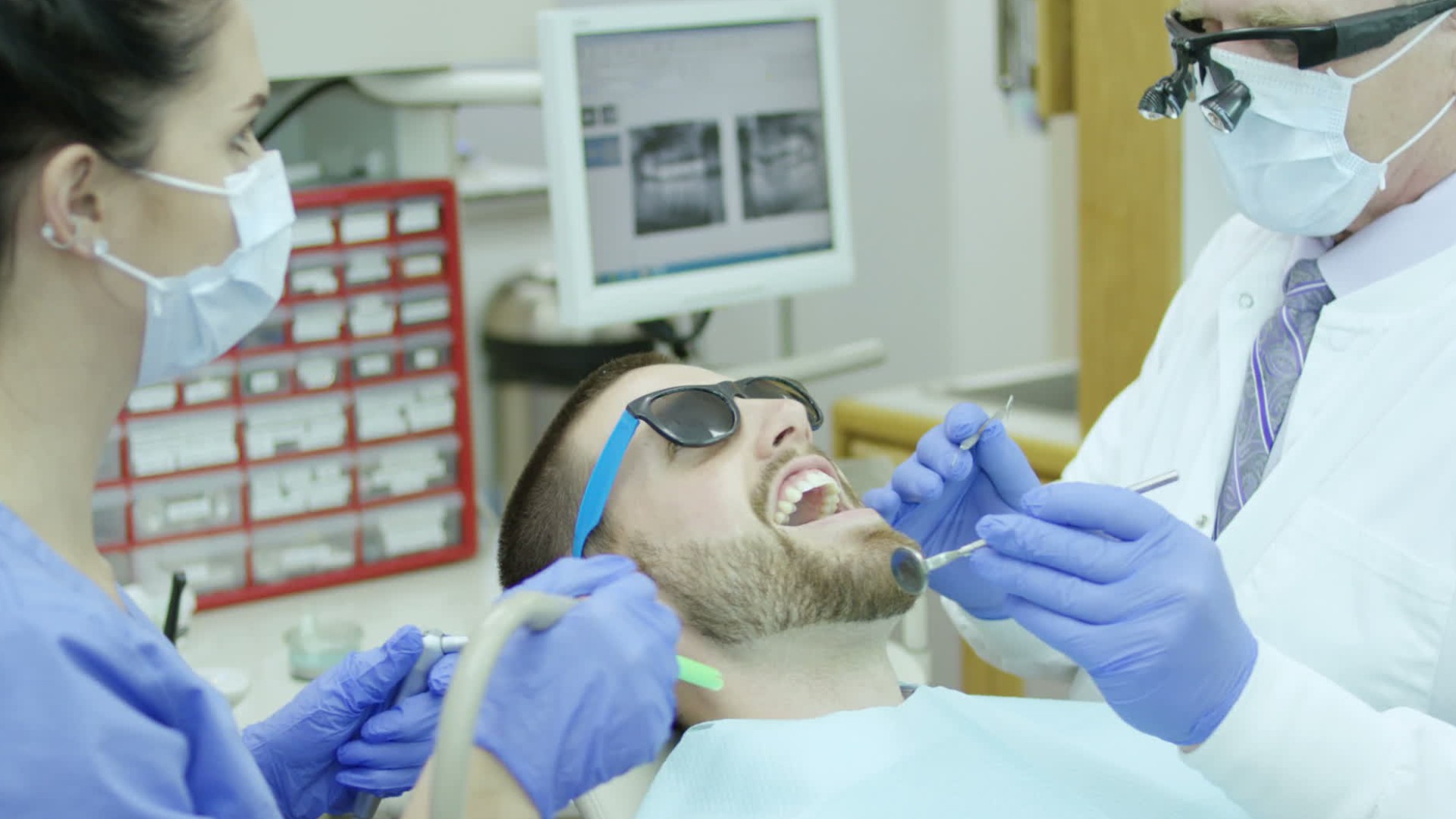
Dental scaling is a non-surgical periodontal procedure designed to remove plaque, tartar (calculus), and bacterial toxins from tooth surfaces and below the gumline. As a foundational component of preventive and therapeutic dentistry, its primary purpose is to eliminate microbial biofilms and hard deposits that contribute to gingivitis, periodontitis, and systemic inflammation. Unlike polishing, which smooths tooth surfaces post-scaling to prevent future plaque adhesion, scaling specifically targets the mechanical removal of mineralized deposits. Root planing, a more invasive extension of scaling, involves smoothing root surfaces to eliminate periodontal pockets, whereas scaling alone may suffice for supragingival (above-gum) plaque control.
The procedure distinguishes itself from other dental interventions by its precision in addressing microbial-induced pathology. While polishing enhances aesthetics and reduces staining, scaling directly targets pathogenic biofilms, which are resistant to brushing and flossing. Its therapeutic role extends beyond cosmetic concerns, as accumulated calculus disrupts the gingival seal, facilitating bacterial invasion and chronic inflammation. Studies indicate that untreated calculus buildup increases the risk of periodontal disease progression by up to 40% (American Academy of Periodontology, 2020), underscoring scaling’s critical role in maintaining periodontal health.
Types of Dental Scaling and Their Clinical Applications
Dental scaling is categorized into two primary techniques based on the anatomical location of plaque and calculus accumulation: supragingival and subgingival. Each method employs distinct instruments and protocols to address specific pathological conditions, ensuring targeted intervention without unnecessary tissue trauma.Supragingival Scaling
Supragingival scaling focuses on the removal of plaque and calculus deposits located coronal to the gingival margin, primarily affecting the visible portions of teeth. This procedure is essential for patients with:
The technique typically utilizes hand instruments (scalers, curettes) or ultrasonic/sonic scalers, with the latter offering advantages in efficiency and reduced patient discomfort. Ultrasonic devices employ vibrations and water irrigation to dislodge calculus, while hand instruments provide tactile precision for delicate areas. Key considerations include:
Subgingival Scaling
Subgingival scaling targets calculus and bacterial biofilms embedded within periodontal pockets (typically ≥4 mm depth) or below the gingival sulcus. This intervention is critical in managing periodontitis, where chronic inflammation leads to attachment loss and alveolar bone resorption. The procedure is indicated for:
Subgingival scaling requires specialized instruments designed to navigate tight pocket spaces without traumatizing the gingival epithelium. Common techniques include:
Comparative Efficacy and Integration with Root Planing
While supragingival scaling addresses visible plaque, subgingival scaling is often combined with root planing to achieve optimal outcomes in periodontitis. Root planing involves smoothing root surfaces to remove embedded calculus and toxic byproducts, creating a glassy, bacteria-resistant surface. Key distinctions:
Supragingival scaling removes coronal deposits and is primarily preventive, whereas subgingival scaling (with or without root planing) is therapeutic, targeting active disease sites.Research demonstrates that scaling and root planing (SRP) reduce pocket depths by 1.0–1.5 mm and achieve clinical attachment gain in 20–30% of treated sites (Papapanou et al., 2018). However, subgingival scaling alone may suffice for patients with shallow pockets (<4 mm) and minimal attachment loss, provided adjunctive therapies (e.g., antimicrobials) are incorporated.
Instrumentation Challenges and Patient Considerations
The depth and morphology of subgingival pockets present unique challenges, including:
Documentation and Periodontal Maintenance
Accurate charting of scaling procedures is critical for monitoring progression and treatment efficacy. Essential documentation includes:
In clinical practice, supragingival scaling is often integrated into prophylaxis visits, while subgingival scaling is reserved for periodontal therapy phases, with maintenance intervals tailored to individual risk factors (e.g., every 3–6 months for high-risk patients).
Procedures and Techniques in Dental Scaling
Dental scaling is a precision-driven procedure that removes plaque, calculus, and bacterial toxins from tooth surfaces and gingival pockets to restore periodontal health. The technique varies depending on the location of deposits—supragingival (above the gumline) or subgingival (below the gumline)—and requires specialized instruments tailored to accessibility, patient comfort, and clinical efficiency. Proper execution minimizes trauma to soft tissues while maximizing plaque biofilm disruption, a critical factor in preventing gingivitis progression and periodontal disease.
The selection of scaling methods—manual or ultrasonic—depends on the deposit’s hardness, patient anatomy, and clinician expertise. Each approach presents distinct advantages, such as reduced patient discomfort or improved calculus removal efficiency, but also challenges, such as aerosol generation or instrument fatigue. Below, the step-by-step protocols for supragingival and subgingival scaling are detailed, alongside a comparative analysis of manual and ultrasonic techniques to guide clinical decision-making.
Supragingival Scaling Procedure
Supragingival scaling targets calculus and plaque deposits visible above the gingival margin, typically accessible during routine prophylaxis. The procedure prioritizes thoroughness while minimizing gingival irritation, as excessive pressure or improper angulation can lead to soft tissue trauma.Preparation and Patient Positioning
The clinician begins by conducting a comprehensive oral examination to assess plaque distribution, calculus hardness, and gingival inflammation. Patient positioning—typically semi-reclined with adequate neck support—ensures ergonomic access for the operator. A rubber dam or high-volume evacuator may be employed to isolate the operative field and reduce aerosol contamination, particularly when using ultrasonic instruments.
Instrument Selection and Activation
Two primary tool categories are used:
Step-by-Step Technique
1. Isolation and Moisture Control
The area is isolated using cotton rolls or a rubber dam to prevent saliva contamination, which can dull instrument edges or interfere with ultrasonic efficacy. Saliva ejectors or high-speed suction further enhance visibility.
2. Calculus Detection and Instrument Adaptation
A periodontal probe measures pocket depths and locates subgingival calculus extensions, while a explorer detects rough surfaces indicative of supragingival deposits. The scaler’s working end is adapted to the tooth’s anatomy, ensuring the cutting edge engages the calculus-tooth interface without gouging enamel.
3. Scaling Execution
4. Debris Removal and Polishing
Post-scaling, a water spray or air-polishing device removes loosened debris. Fluoride varnish or polishing paste may be applied to desensitize exposed dentin and smooth rough surfaces.
Safety Measures
Subgingival Scaling Procedure
Subgingival scaling addresses calculus and biofilm embedded within periodontal pockets, where conventional supragingival techniques are ineffective. This procedure is critical in managing periodontitis, as residual subgingival deposits contribute to persistent inflammation, attachment loss, and pocket deepening. However, it presents challenges due to limited visibility, instrument accessibility, and the risk of soft tissue laceration.Instrumentation and Patient Assessment
Specialized instruments are required to navigate confined spaces:
Step-by-Step Technique
1. Anesthesia and Local Anesthesia
Local anesthesia (e.g., lidocaine with epinephrine) is administered to manage patient discomfort, particularly in deep pockets (>5 mm) or sensitive areas. Topical anesthesia may precede injections for patient comfort.
2. Instrument Adaptation and Activation
The curette’s cutting edge is positioned at the base of the pocket, angled to follow the root’s contour. The blade is inserted coronally (toward the crown) to avoid pushing debris apically and exacerbating inflammation. Ultrasonic tips are used with minimal pressure, ensuring the tip remains in constant motion to prevent overheating.
3. Scaling Execution
4. Debridement Verification
A periodontal probe reassesses pocket depths and calculus presence post-scaling. If residual deposits are detected, additional instrumentation or adjunctive therapies (e.g., local delivery of antibiotics) may be recommended.
Challenges and Mitigation Strategies
Specialized Techniques for Complex Cases
Comparison of Manual and Ultrasonic Scaling Methods
The choice between manual and ultrasonic scaling influences procedural efficiency, patient comfort, and clinical outcomes. Below is a comparative analysis structured to highlight key differences, advantages, and ideal patient scenarios for each method.| Feature | Manual Scaling (Hand Instruments) | Ultrasonic Scaling | |||||||||||||||||||||||||||||||||||||
|---|---|---|---|---|---|---|---|---|---|---|---|---|---|---|---|---|---|---|---|---|---|---|---|---|---|---|---|---|---|---|---|---|---|---|---|---|---|---|---|
| Mechanism of Action | Mechanical removal via sharp cutting edges of curettes/scalers. Requires direct contact with calculus. | Vibrational fragmentation of calculus via acoustic energy and cavitation, supplemented by water irrigation. | |||||||||||||||||||||||||||||||||||||
| Advantages |
Tools and Equipment in Dental ScalingDental scaling relies on specialized instruments designed to remove plaque, calculus, and stains while ensuring precision and patient comfort. The selection of tools varies based on the procedure type—manual, ultrasonic, or laser—and the clinical requirements, including pocket depth, patient sensitivity, and material hardness. Proper equipment not only enhances efficiency but also minimizes tissue trauma, making material composition, ergonomics, and maintenance critical considerations in daily practice.The effectiveness of dental scaling depends on the integration of high-quality instruments that balance durability, biocompatibility, and ease of sterilization. Below are the essential tools categorized by function, along with their technical specifications, operational principles, and maintenance protocols. Essential Instruments for Manual and Ultrasonic ScalingManual and ultrasonic scaling employ distinct yet complementary tools, each tailored to specific tasks such as calculus removal, root planing, or subgingival debridement. The choice of material—ranging from stainless steel to titanium—directly influences longevity, sharpness retention, and patient comfort. Below is a categorized list of core instruments, their functions, and maintenance requirements.The patient experience during scaling involves controlled sensations such as vibrations from ultrasonic instruments, water spray for cooling and debris removal, and mild pressure around gum margins. Proper communication and pain management techniques minimize discomfort, while post-procedure aftercare—including oral hygiene adjustments and dietary restrictions—accelerates healing and prevents complications. Recovery timelines vary based on the scaling type (supragingival vs. subgingival), with sensitivity typically resolving within days to weeks. Sensory and Physical Experiences During ScalingPatients undergoing dental scaling may encounter several sensory and physical sensations, which are generally managed to ensure comfort. The procedure typically involves:Patient Preparation Tip: Informing the dentist about dental anxiety, sensitivity to cold/hot, or history of gum disease allows for tailored pain management, such as topical anesthetics or nitrous oxide sedation. Pain Management and Comfort TechniquesEffective pain management during scaling relies on a combination of pre-procedural communication, anesthetic options, and instrument adjustments. Common techniques include:Evidence-Based Note: Studies indicate that pre-procedural education and anxiety screening reduce perceived pain by up to 40%, highlighting the role of psychological preparation in patient comfort (Journal of Dental Anxiety, 2019). Post-Scaling Aftercare ChecklistProper aftercare is critical to prevent complications such as infection, prolonged sensitivity, or gum irritation. Patients should adhere to the following guidelines:Oral Hygiene Adjustments Dietary Recommendations Monitoring for Complications Critical Aftercare Insight: Avoid smoking and alcohol for at least 48 hours post-scaling, as they impair healing, increase infection risk, and delay gum tissue regeneration. Recovery Timeline: Supragingival vs. Subgingival ScalingRecovery duration varies significantly based on the scaling type, with subgingival procedures generally requiring longer healing due to deeper tissue involvement.
Clinical Example: A patient with moderate periodontitis undergoing subgingival scaling may experience 5–7 days of sensitivity but show reduced pocket depths by 20–30% within 4–6 weeks, as documented in post-treatment periodontal charts.
Medical and Clinical Significance of Dental Scaling in Periodontal and Systemic HealthDental scaling serves as a cornerstone in periodontal therapy, directly influencing both oral and systemic health outcomes. Research establishes a bidirectional relationship between periodontal diseases—such as gingivitis and periodontitis—and systemic conditions, including cardiovascular diseases, diabetes, and adverse pregnancy outcomes. The accumulation of bacterial biofilms (plaque) and calculus (tartar) triggers inflammatory responses that extend beyond the oral cavity, exacerbating chronic systemic inflammation. Scaling disrupts this cycle by mechanically removing microbial deposits and reducing pathogenic load, thereby mitigating periodontal destruction and systemic complications. High-risk patient populations, characterized by compromised immune responses or metabolic dysfunctions, exhibit accelerated plaque maturation and deeper periodontal pockets, necessitating tailored scaling protocols.Prevention of Periodontal Diseases and Systemic Health ConnectionsThe primary clinical significance of dental scaling lies in its role as a preventive measure against gingivitis and periodontitis, the two stages of periodontal disease progression. Gingivitis, characterized by reversible inflammation of the gingiva, progresses to periodontitis—a chronic, irreversible condition marked by alveolar bone loss, pocket formation, and tooth mobility—when left untreated. Scaling interrupts this progression by:Systemic health implications arise from the periodontal-disease-inflammatory link, where chronic oral inflammation contributes to: Periodontal pathogens and their byproducts (e.g., LPS) act as endogenous triggers for systemic inflammation, linking oral health to chronic diseases. Scaling reduces this burden by ≥60% in high-risk individuals when combined with adjunctive therapies. High-Risk Patient Groups and Tailored Scaling ProtocolsPatient populations with accelerated plaque maturation, impaired healing, or systemic comorbidities require frequent scaling (every 3–4 months) and modified techniques. Key groups include:High-risk patients exhibit 3–5x faster plaque regrowth post-scaling due to systemic or behavioral factors. Personalized scaling intervals (e.g., 3-month for diabetics, 4-month for smokers) are essential to prevent rapid disease recurrence. Integration of Scaling with Comprehensive Periodontal TherapyDental scaling is most effective when integrated into a phased periodontal therapy plan, often following the American Academy of Periodontology (AAP) 2015 guidelines. The process can be visualized as a sequential flowchart, where scaling serves as the foundation for deeper interventions:
|
Leave a Comment
Comments are moderated before appearing. The data you submit is processed according to the Privacy Policy of Voltefac.