What Are Teeth Made Of Explained Through Science And Adaptations
Table of Contents
- Composition of Human Teeth: Biological and Chemical Breakdown
- Biological Layers of Human Teeth and Their Structural Roles
- Chemical Composition and Mineralization Gradients
- Hierarchical Structure of Teeth: From Crystallites to Macroscopic Anatomy
- Comparative Anatomy of Teeth: Species-Specific Adaptations and Evolutionary Trade-offs
- Enamel Thickness and Mineral Density: A Spectrum of Adaptations
- Tooth Shape and Functional Specialization Across Taxa
- Extreme Adaptations: Unconventional Teeth in Unique Ecological Niches
- Dietary Evolution and Enamel Composition in Hominins
- Side-by-Side Comparison: Human, Lion, and Cow Dental Adaptations
- Developmental Biology of Tooth Formation and Mineralization
- Stages of Odontogenesis and Cellular Differentiation
- Mechanisms of Hydroxyapatite Crystal Formation and Enamel Maturation
- Timeline of Tooth Development and Mineralization Milestones
- Congenital Disorders Altering Tooth Composition: Structural Deficiencies and Case Studies
- Engineering and Materials Science: Mimicking Teeth for Biomedical Applications
- Mechanical and Biological Properties of Enamel as a Biomimetic Model
- Bioinspired Materials: Compositional and Functional Analogues to Natural Teeth
- 3D Printing and Nanotechnology: Fabricating Tooth-Like Structures
- FAQ
- Are teeth made of bone?
- What role does calcium play in the composition of teeth?
- What is the chemical composition of teeth?
- What are teeth made of?
- What are teeth made of for children?
- If teeth aren’t made of bone, what are they made of?
Teeth represent one of nature’s most intricate biological structures, engineered to withstand immense mechanical stress while defending against microbial invasion. Composed of a precisely balanced matrix of minerals, organic proteins, and specialized cells, they serve as both functional tools and evolutionary records of dietary specialization. From the hardest biological substance—enamel—to the dynamic pulp chamber that sustains vitality, each layer reflects a sophisticated interplay of chemistry and biology. This exploration dissects the compositional marvels of teeth, tracing their formation, adaptive diversity across species, and the cutting-edge biomaterials inspired by their resilience.
The foundation of dental science lies in understanding how hydroxyapatite crystals interlock to form enamel, how collagen fibers reinforce dentin, and how these components interact to create a system capable of self-repair and regeneration. Comparative analysis reveals how herbivores, carnivores, and omnivores have optimized tooth structure for survival, while developmental biology uncovers the genetic and biochemical processes governing odontogenesis. Meanwhile, materials engineers continue to replicate these natural designs, aiming to bridge the gap between biological performance and synthetic innovation in restorative dentistry.
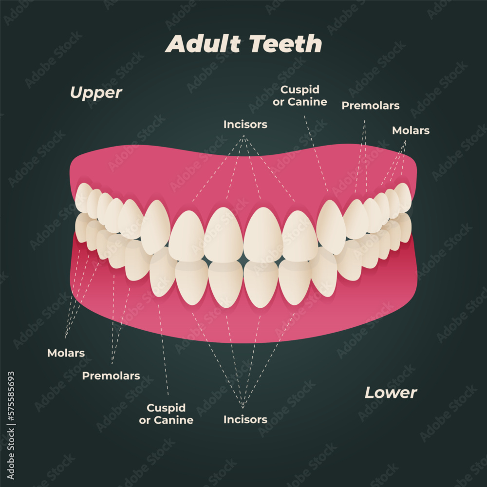
Composition of Human Teeth: Biological and Chemical Breakdown
Human teeth are highly specialized structures designed for mastication, speech, and aesthetic functions, composed of four primary layers—enamel, dentin, cementum, and pulp—each with distinct biological and chemical properties. These layers exhibit hierarchical organization from the microscopic crystallite arrangement to macroscopic anatomical features, enabling teeth to withstand mechanical stress while maintaining structural integrity. The chemical composition of each layer dictates its hardness, resilience, and susceptibility to pathological conditions such as caries or erosion. Variations in mineral density, organic matrix distribution, and water content create a gradient of mechanical properties essential for tooth functionality.The following sections dissect the biological roles and chemical constituents of each layer, supported by comparative data on mineralization, structural hierarchy, and functional adaptations.
Biological Layers of Human Teeth and Their Structural Roles
The anatomical layers of a tooth serve specialized functions critical to its longevity and performance. Enamel, the outermost layer, provides a protective barrier against physical and chemical degradation, while dentin forms the bulk of the tooth structure, offering flexibility and sensory feedback. Cementum anchors the tooth to the alveolar bone via the periodontal ligament, and the pulp contains vascular and neural tissues responsible for nourishment and sensitivity. Below is an overview of their hierarchical arrangement and functional contributions:"The enamel-dentin junction (EDJ) acts as a transitional zone where mechanical stress is distributed, preventing catastrophic failure under occlusal forces."
-
Enamel
The hardest biological tissue in the human body, enamel lacks cellular components post-development and derives its strength from tightly packed hydroxyapatite crystals aligned parallel to the tooth surface. Its primary role is to resist abrasion and acid dissolution, though it is avascular and incapable of self-repair.Key structural adaptations include:
- Prismatic arrangement: Enamel rods (prisms) are composed of elongated hydroxyapatite crystals (5–10 µm in diameter) oriented perpendicular to the EDJ, enhancing fracture resistance.
- Hunter-Schreger bands: Decussating (crossing) prism patterns in some regions deflect cracks, dissipating energy and preventing propagation.
-
Dentin
Constituting ~70% of a tooth’s volume, dentin is a semi-rigid tissue with a tubular microstructure that allows for limited fluid movement, contributing to dentinal hypersensitivity. Its organic matrix (collagen Type I) provides flexibility, while mineralized peritubular dentin surrounding tubules increases hardness.Functional attributes include:
- Tubule orientation: Radially aligned dentinal tubules (1–4 µm in diameter) transmit stimuli to the pulp, enabling sensory perception.
- Sclerotic dentin: Aging or attrition induces secondary dentin formation, reducing tubule permeability and increasing mineral density.
-
Cementum
A thin, avascular layer covering the dentin of the tooth root, cementum anchors the periodontal ligament fibers via Sharpey’s fibers. Its composition is less mineralized than enamel or dentin, allowing for limited remodeling and repair.Structural features:
- Acellular vs. cellular cementum: Acellular cementum (near the crown) is thinner and more mineralized, while cellular cementum (near the apex) contains cementocytes and supports continuous attachment.
- Resorption/reformation: Cementum undergoes physiological turnover, adapting to occlusal changes or orthodontic forces.
-
Pulp
The innermost layer houses connective tissue, odontoblasts (dentin-secreting cells), nerves, and blood vessels. Its primary functions include:
- Nutrient supply: Vascularization supports metabolic activity and dentinogenesis.
- Sensory transduction: A-delta and C-fibers mediate pain in response to thermal, mechanical, or chemical stimuli.
- Reparative dentinogenesis: Odontoblasts deposit tertiary dentin in response to injury, though this process is limited compared to developmental dentin.
Chemical Composition and Mineralization Gradients
The mechanical properties of each tooth layer correlate directly with its chemical composition, particularly the ratio of inorganic minerals, organic matrix, and water. Below is a comparative table summarizing the mineral and organic content of enamel, dentin, cementum, and pulp, alongside their functional implications:| Layer | Mineral Content (%) | Organic Content (%) | Water Content (%) | Key Functions |
|---|---|---|---|---|
| Enamel | 96–98% (hydroxyapatite: 85–90%; fluorapatite: 1–3%) | 1–2% (enamelin, amelogenin, ameloblastin) | 0.2–0.4% |
|
| Dentin | 70% (hydroxyapatite: 45–50%; carbonate-substituted apatite: 5–10%) | 18–20% (collagen Type I, phosphophoryn, dentin sialophosphoprotein) | 10–12% |
|
| Cementum | 45–50% (hydroxyapatite with higher carbonate substitution) | 30–35% (collagen Type I, proteoglycans) | 20–25% |
|
| Pulp | Trace minerals (calcium phosphate deposits in aging) | ~30% (collagen, ground substance) | ~60% |
|
"The mineral-to-matrix ratio determines a tissue’s biomechanical properties: enamel’s high mineralization sacrifices toughness for hardness, while dentin’s balanced composition enables resilience under cyclic loading."
Hierarchical Structure of Teeth: From Crystallites to Macroscopic Anatomy
The mechanical performance of teeth arises from their hierarchical organization, spanning six orders of magnitude from nanoscale crystallites to macroscopic anatomical features. This multi-scale structure enables load distribution, crack resistance, and adaptive remodeling. Below is a flowchart-style description of the hierarchical levels:-
Nanoscale (0.1–10 nm): Crystallite and Organic Matrix
- Hydroxyapatite crystallites: Plate-like or needle-shaped, aligned along the c-axis for optimal strength.
- Organic matrix: Collagen fibrils (in dentin/cementum) or enamel-specific proteins (e.g., amelogenin) template mineral deposition. "The aspect ratio (length-to-width) of hydroxyapatite crystals influences fracture toughness; elongated crystals in enamel resist shear forces."
-
Microscale (10 nm–1 µm): Fiber and Tubule Formation
- Enamel prisms: Bundles of crystallites (2–5 µm diameter) with decussating patterns (Hunter-Schreger bands) to deflect cracks.
- Dentinal tubules: Microchannels
- Herbivores (e.g., horses, cows) incorporate higher magnesium content in enamel, enhancing durability against silica-rich grasses.
- Carnivores (e.g., lions, great white sharks) exhibit larger, more tightly packed hydroxyapatite crystals, improving resistance to compressive forces during prey capture.
- Omnivores (e.g., bears, pigs) show intermediate crystallite organization, balancing abrasion resistance with flexibility.
- Horses: Enamel ridges (lophs) on molars create self-sharpening surfaces, compensating for the lack of enamel renewal. Their 6–8 mm-thick enamel resists wear from silica in grasses.
- Beavers: Orange-stained enamel (due to iron oxide deposits) forms ridges and valleys that act as "teeth within teeth," optimizing wood processing. Their incisors grow continuously to counteract gnawing forces.
- Cows: Flat, broad molars with folded enamel increase surface area for fermenting fibrous cellulose, while their low-crowned premolars adapt to softer forages.
- Lions: Canine teeth (up to 8 cm long) are conical with thin enamel, designed for piercing rather than grinding. Their carnassial pairs (P4/M1) have shearing blades for slicing meat.
- Great White Sharks: Translucent, flexible teeth (composed of fluorapatite-rich enamel) are serrated and replaceable, with dentine cores providing shock absorption during prey capture.
- Hyenas: Bone-crushing molars with enamel ridges allow them to pulverize bones, a trait absent in most carnivores.
- Pigs: Molar cusps are rounded and low, suited for crushing tubers and roots, while their canines are tusk-like for digging and dominance displays.
- Bears: Molarized premolars enable them to process both meat and vegetation, with thicker enamel on the buccal (cheek) side to resist lateral forces during chewing.
- Translucent, gelatinous teeth lack traditional enamel and are instead composed of collagen-rich dentine with microcrystalline calcium carbonate. These teeth are bioluminescent, likely used to attract prey in the deep sea’s lightless depths.
- Functional Adaptation: The lack of hardness suggests a role in chemical sensing or prey manipulation rather than mechanical processing.
- Mandibles with enamel-like chitinous ridges, hardened by calcium carbonate deposits, allowing them to chew through tough plant materials.
- Functional Adaptation: The absence of hydroxyapatite reflects an arthropod-specific mineralization strategy, optimizing for lightweight yet durable structures.
- Enamel-free, chisel-like incisors composed of bone-like dentine with keratinized tips, used for digging and sensing prey via electroreception.
- Functional Adaptation: The lack of traditional enamel aligns with their dual role in excavation and sensory perception, a rare convergence in vertebrate evolution.
- Thick (2–3 mm) enamel with high magnesium content, suggesting a diet rich in hard, fibrous plants and seeds.
- Fossil evidence: Microwear analysis on Au. africanus molars shows parallel scratches, indicative of grinding silica-rich grasses.
- Thinner (1.5–2 mm) but more uniform enamel, linked to increased meat consumption and tool-assisted food processing.
- Fossil evidence: H. habilis teeth exhibit pitting and edge damage, consistent with cutting meat with stone tools.
- Variable enamel thickness (1–2.5 mm) with reduced magnesium content, reflecting diverse diets but also higher susceptibility to enamel wear in populations consuming low-abrasive, processed foods.
- Fossil evidence: Neanderthal enamel shows thicker buccal ridges, suggesting high-meat diets, while agriculturalists exhibit thinner enamel due to reduced chewing demands.
- MSX1, PAX9: Initiation of dental lamina.
- Eda, EDAR: Tooth number and morphology (ectodermal dysplasia).
- DSPP: Dentin matrix protein critical for mineralization.
- AMELX, ENAM: Enamel matrix proteins for crystal alignment.
- Amelogenin: Forms nanospheres; regulates crystal width (25–50 nm).
- ALP Activity: Critical for phosphate availability; mutations (e.g., TNSALP gene) cause hypophosphatasia.
- Proteinase K: Degrades amelogenin to allow crystal thickening.
- Calcium/Phosphate Ratio: Maintained by ameloblast PMCA1 (plasma membrane Ca²⁺ ATPase) and NHE3 (Na⁺/H⁺ exchanger).

Comparative Anatomy of Teeth: Species-Specific Adaptations and Evolutionary Trade-offs
The composition and morphology of teeth vary dramatically across species, reflecting evolutionary adaptations to dietary niches, environmental pressures, and ecological roles. While human dentition prioritizes a balance of grinding and shearing for omnivory, other species exhibit extreme specializations—such as the crushing molars of herbivores or the needle-like canines of apex predators. These variations are not merely structural but are chemically and biomechanically optimized, with enamel thickness, mineral density, and tooth shape serving as key indicators of an organism’s ecological strategy. Understanding these adaptations provides insight into the interplay between diet, physiology, and survival, while also offering a framework for interpreting fossil records and reconstructing ancestral behaviors."Tooth morphology is a direct reflection of an organism’s evolutionary history, with enamel composition acting as a proxy for dietary specialization and environmental constraints." — Smith & Purnell, 2008 (Evolutionary Biology of Teeth)
Enamel Thickness and Mineral Density: A Spectrum of Adaptations
Enamel thickness and mineral density are primary determinants of a species’ ability to process food, with herbivores, carnivores, and omnivores exhibiting distinct trade-offs. Herbivores, which consume abrasive plant materials, typically develop thicker enamel (often 2–6 mm) to withstand grinding forces, while carnivores rely on thinner but harder enamel (0.5–2 mm) to facilitate shearing and piercing. Omnivores, such as humans, occupy an intermediate position, with enamel thickness averaging 1–2.5 mm and a mineral composition optimized for versatility.The primary mineral in enamel across most vertebrates is hydroxyapatite (Ca₁₀(PO₄)₆(OH)₂), though variations in crystallite size, orientation, and trace elements (e.g., magnesium, fluoride) influence hardness and wear resistance. For instance:
"Enamel thickness scales positively with dietary abrasiveness, while mineral density correlates with the need for rapid, forceful biting." — Ramirez-Rozzi & Bermúdez, 2012 (Journal of Mammalian Evolution)
Tooth Shape and Functional Specialization Across Taxa
Tooth morphology is closely tied to feeding mechanics, with herbivores evolving high-crowned (hypsodont) molars for grinding, carnivores developing serrated carnassials for shearing flesh, and omnivores retaining a mixed dentition for both crushing and tearing. Extreme adaptations further illustrate the relationship between form and function:- Herbivores:
- Carnivores:
- Omnivores:
Extreme Adaptations: Unconventional Teeth in Unique Ecological Niches
Some species exhibit teeth that defy typical mammalian patterns, reflecting highly specialized diets or environmental pressures:- Glass Squid (Vampyroteuthis infernalis):
- Beetles (e.g., Dynastes hercules):
- Platypus (Ornithorhynchus anatinus):
Dietary Evolution and Enamel Composition in Hominins
Fossil evidence demonstrates that enamel thickness and mineral density in early hominins shifted in response to dietary changes, particularly the transition from hard, abrasive foods (e.g., seeds, uncooked plants) to softer, processed foods (e.g., cooked tubers, meat). Key observations include:- Australopithecines (e.g., Australopithecus africanus):
- Early Homo (e.g., Homo habilis):
- Modern Humans (Homo sapiens):
"The thinning of hominin enamel over the past 2 million years correlates with the adoption of cooking and tool use, reducing the need for extreme abrasion resistance." — Teaford & Walker, 1984 (American Journal of Physical Anthropology)
Side-by-Side Comparison: Human, Lion, and Cow Dental Adaptations
The following table contrasts key dental traits across a herbivore (cow), carnivore (lion), and omnivore (human), illustrating how structural and chemical differences align with ecological roles.| Feature | Human (Homo sapiens) | Lion (Panthera leo) | Cow (Bos taurus) |
|---|