What Is Dysentery Understanding Its Forms Symptoms And Management
Table of Contents
- Definition and Basic Characteristics of Dysentery
- Etiological Classification and Comparative Analysis
- Pathophysiological Progression: Exposure to Symptom Onset
- Symptoms and Clinical Manifestations of Dysentery
- Gastrointestinal Manifestations
- Systemic Manifestations
- Severe Complications
- Diagnostic Methods and Laboratory Techniques for Dysentery
- Stool Sample Collection Protocols
- Microscopic Examination Techniques
- Molecular Diagnostic Techniques
- Treatment Protocols and Management Strategies for Dysentery
- Tiered Treatment Algorithm by Severity
- Antimicrobial Selection and Resistance Patterns
- Risks of Antidiarrheal Use in Dysentery
- Supportive Care and Complications Management
- FAQ
- What is dysentery?
- What is the dysentery disease?
- What does dysentery mean?
- What is dysentery called today?
- What is dysentery caused by?
- What is dysentery and how do you get it?
Dysentery remains a critical global health challenge, characterized by severe gastrointestinal distress that disrupts lives and strains healthcare systems. This acute inflammatory condition of the intestines—triggered by bacterial pathogens like Shigella or parasitic agents such as Entamoeba histolytica—exemplifies the intersection of infectious disease, sanitation disparities, and antimicrobial resistance. Beyond its clinical urgency, dysentery serves as a case study in how environmental factors, travel risks, and diagnostic precision converge to dictate patient outcomes, demanding both preventive vigilance and evidence-based intervention.
The disease manifests through a spectrum of symptoms, from mild discomfort to life-threatening complications like toxic megacolon, underscoring the necessity for rapid identification and stratified care. Advances in molecular diagnostics and antimicrobial stewardship now offer refined tools to combat its spread, yet regional variations in resistance patterns and resource limitations persist as formidable barriers. Understanding dysentery’s pathophysiology, transmission dynamics, and evolving treatment paradigms is essential for clinicians, public health practitioners, and travelers alike in mitigating its impact.
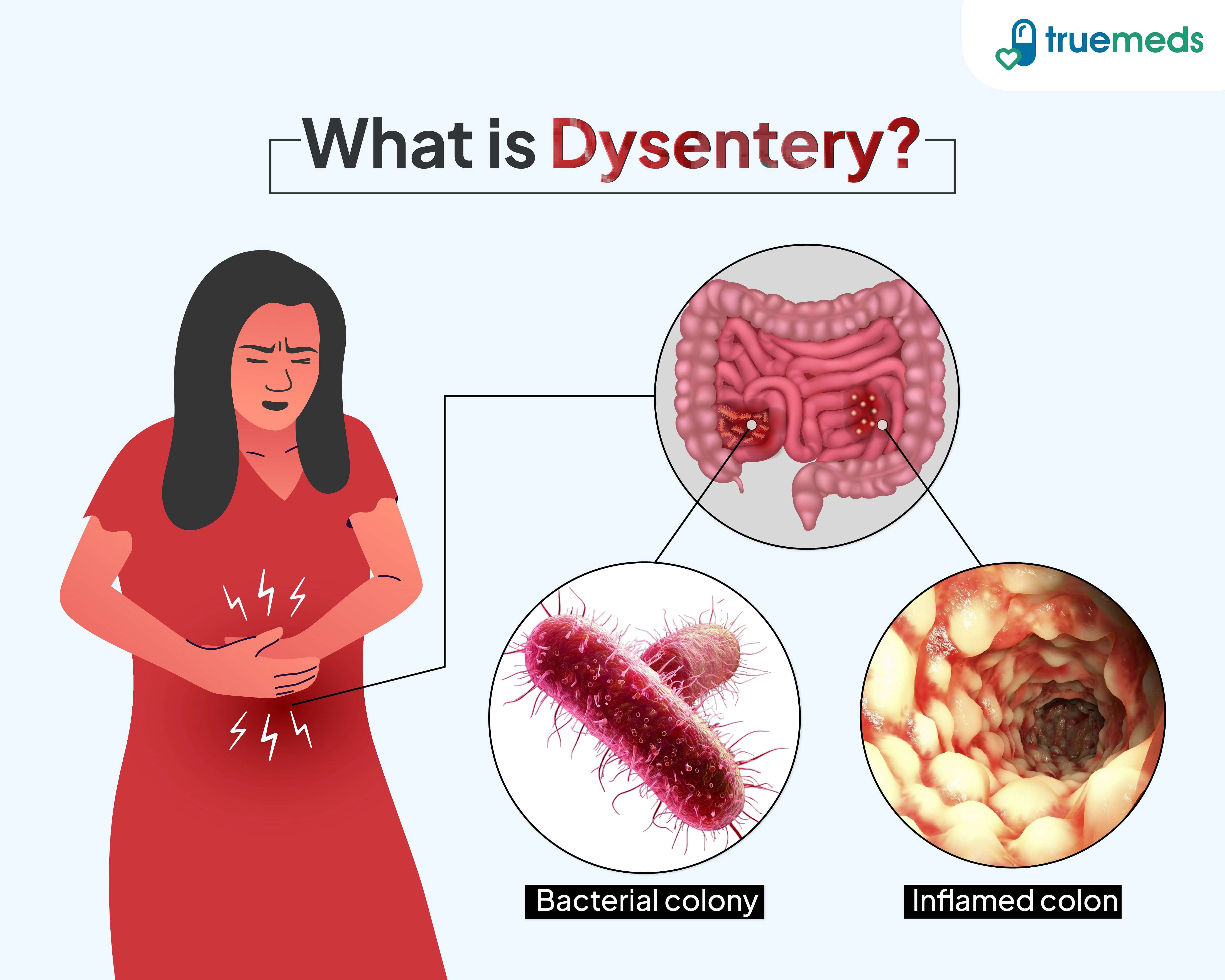
Definition and Basic Characteristics of Dysentery
Dysentery represents a group of infectious diseases primarily characterized by inflammation of the intestines, leading to severe diarrhea often accompanied by blood or mucus in stool. The condition is classified into two distinct forms based on etiology: bacterial dysentery, caused by pathogenic bacteria, and amoebic dysentery, attributed to protozoan parasites. Both forms share common clinical manifestations but differ in transmission, pathogenesis, and treatment approaches. Understanding these distinctions is critical for accurate diagnosis, public health intervention, and targeted therapeutic strategies.
The differentiation between bacterial and amoebic dysentery hinges on the causative agent, mode of transmission, and symptomatic presentation. While bacterial dysentery typically manifests as an acute, self-limiting illness, amoebic dysentery may progress to chronic infection with systemic complications if untreated. Below, a structured comparison outlines the key features of each form, followed by a visual representation of the disease progression from exposure to clinical onset.
Etiological Classification and Comparative Analysis
Dysentery is categorized based on the infectious agent responsible for intestinal inflammation. The following table provides a systematic comparison of bacterial and amoebic dysentery, highlighting their defining characteristics:| Type | Primary Causative Agent | Transmission Route | Key Symptoms | Incubation Period Range |
|---|---|---|---|---|
| Bacterial Dysentery |
|
|
|
12 hours to 7 days (typically 1–3 days for Shigella) |
| Amoebic Dysentery | Entamoeba histolytica (trophozoite stage) |
|
|
2–4 weeks (range: 1 day to months; asymptomatic carriage possible) |
Bacterial dysentery is typically acute, contagious, and associated with high fecal-oral transmission rates, whereas amoebic dysentery may present as chronic or asymptomatic and is linked to poor sanitation and environmental persistence of cysts. The presence of fever and systemic toxicity is more indicative of bacterial etiology, while bloody diarrhea with systemic abscess formation suggests amoebic infection.
Pathophysiological Progression: Exposure to Symptom Onset
The progression from initial exposure to clinical manifestation of dysentery follows a predictable sequence influenced by host immunity, infectious dose, and environmental factors. Below is a flowchart description outlining the stages, risk factors, and critical decision points:1. Exposure to Pathogen
2. Infectious Dose and Colonization
3. Incubation Period
4. Clinical Manifestations
5. Diagnostic and Therapeutic Interventions
Visual Flowchart Representation (Descriptive):
```
[Exposure → Risk Factors (Sanitation/Immunity) → Infectious Dose]
↓
[Incubation Period] → [Mucosal Invasion/Colonization]
↓
[Symptom Onset: Diarrhea → Bloody Stool/Fever → Systemic Spread]
↓
[Diagnosis → Treatment → Recovery/Complications]
```
Critical Nodes:
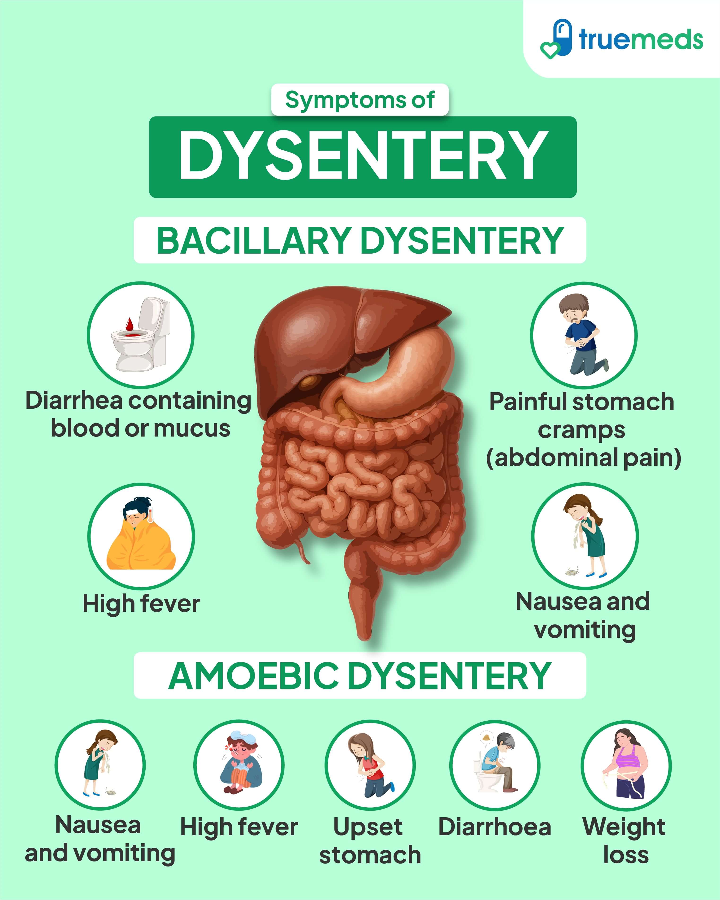
Symptoms and Clinical Manifestations of Dysentery
Dysentery presents with a heterogeneous spectrum of clinical features, ranging from mild, self-resolving gastrointestinal disturbances to life-threatening systemic complications. The severity and symptom profile are influenced by the causative pathogen (Shigella, Entamoeba histolytica, Campylobacter, or Salmonella), host immunity, and environmental factors. Early recognition of symptom clusters—particularly those indicative of systemic involvement or severe complications—is critical for timely intervention and prevention of adverse outcomes. Below, the manifestations are categorized into gastrointestinal, systemic, and severe complications, with illustrative case examples to highlight diagnostic nuances.Gastrointestinal Manifestations
The hallmark of dysentery lies in its distinct gastrointestinal symptoms, which differentiate it from other diarrheal illnesses. These features arise from colonic inflammation, mucosal ulceration, and altered motility. Key presentations include:-
Abrupt onset of diarrhea
Stool frequency typically exceeds 3–5 times daily, with a sudden onset (within hours) rather than the gradual progression seen in viral gastroenteritis. The stool may initially be watery but rapidly progresses to small volumes with high urgency. -
Tenesmus
A persistent, painful sensation of incomplete bowel evacuation, often accompanied by straining. This symptom reflects rectal inflammation and is pathognomonic for invasive dysentery (e.g., shigellosis or amoebiasis). -
Mucus and/or blood in stool
- Shigellosis: Small-volume stools with streaks of bright red blood or mucus ("currant jelly" appearance) due to colonic ulceration.
- Amoebic dysentery: Stools may contain "anchovy paste"-like mucus with flecks of blood, often alternating with constipation.
- Bacillary dysentery (e.g., Salmonella): Blood may be less prominent, with a higher likelihood of systemic symptoms.
-
Lower abdominal cramping
Colicky pain localized to the left iliac fossa or periumbilical region, often exacerbated by defecation. Pain may mimic diverticulitis or appendicitis, necessitating differential diagnosis. -
Systemic toxicity without fever
In Shigella infections, systemic symptoms (e.g., malaise, myalgia) may precede fever, reflecting bacterial toxin-mediated inflammation rather than a classical infectious response.
The presence of fecal leukocytes (WBCs >15/hpf on microscopy) strongly suggests bacterial or amoebic dysentery, distinguishing it from viral or osmotic diarrhea. Stool cultures or antigen tests (e.g., Shigella latex agglutination) confirm the etiology.
Systemic Manifestations
Systemic involvement in dysentery reflects the body’s inflammatory response to infection, with severity correlating to pathogen virulence and host susceptibility. Key features include:-
Fever
Typically spiking (38–40°C) and biphasic in Shigella or Salmonella infections, with chills and night sweats. Amoebic dysentery may present with a low-grade or intermittent fever unless liver abscess complicates the course. -
Dehydration signs
- Mild: Dry mucous membranes, slight tachycardia (HR >100 bpm), orthostatic hypotension.
- Moderate: Sunken eyes, reduced skin turgor, oliguria (<0.5 mL/kg/h in adults), and postural hypotension (SBP drop ≥20 mmHg).
- Severe: Hypotension (SBP <90 mmHg), altered mental status, and anuria—requiring IV fluid resuscitation.
-
Extraintestinal symptoms
- Arthralgia/arthritis: Seen in Salmonella or Campylobacter infections due to immune complex deposition.
- Hepatomegaly: More common in amoebic dysentery (30–40% of cases) due to hepatic abscess formation.
- Neurological: Shigella toxin may cause Guillain-Barré syndrome or Reactive Arthritis (HLA-B27-associated).
-
Toxic appearance
Lethargy, confusion, or coma in severe cases, particularly in children or immunocompromised patients, signaling sepsis or toxic megacolon.
A leukocytosis with left shift (WBC >15,000/μL, >80% neutrophils) suggests bacterial dysentery, while eosinophilia may indicate Strongyloides coinfection. Blood cultures are positive in <5% of cases unless bacteremia complicates Salmonella or Campylobacter infection.
Severe Complications
Unchecked dysentery can progress to life-threatening complications, particularly in high-risk populations (e.g., children <5 years, HIV/AIDS, or malnourished individuals). Early recognition of red flags is essential for intervention.| Complication | Pathophysiology | Clinical Presentation | Diagnostic Confirmation | ||||||||||||||
|---|---|---|---|---|---|---|---|---|---|---|---|---|---|---|---|---|---|
| Toxic Megacolon | Colonic dilation (>6 cm) secondary to severe inflammation, leading to impaired motility and risk of perforation. Most common in Shigella or Campylobacter infections; less frequent with amoebiasis. |
|
Abdominal X-ray/CT showing transverse colon >6 cm; emergency surgical consultation required. |
||||||||||||||
| Sepsis | Bacteremia (e.g., Salmonella, Shigella) or systemic inflammatory response syndrome (SIRS) due to toxin-mediated shock. |
|
Lactate >2 mmol/L, SIRS criteria + suspected infection; IV broad-spectrum antibiotics (e.g., ceftriaxone + metronidazole). |
||||||||||||||
| Amoebic Liver Abscess | Extension of colonic inflammation via portal circulation; Entamoeba histolytica trophozoites invade hepatic parenchyma. |
|
Ultrasound/CT showing hypoechoic lesion; serology (amoebic ELISA) or aspiration of pus with trophozoites on microscopy. |
||||||||||||||
| Hemolytic-Uremic Syndrome (HUS) | Shiga toxin (Stx1/St Diagnostic procedures must adhere to strict protocols to ensure specimen integrity and reliable results. Contamination, improper handling, or delayed processing can lead to false negatives or misidentification of pathogens. Below are structured methodologies for stool sample collection, microscopic examination, and molecular diagnostics, followed by a comparative analysis of diagnostic tools to guide clinical decision-making. Stool Sample Collection ProtocolsProper collection and transport of stool specimens are foundational to accurate dysentery diagnosis. Improper handling—such as delayed transport, exposure to extreme temperatures, or use of non-sterile containers—can degrade pathogen viability or introduce contaminants. For bacterial dysentery (e.g., Shigella or Salmonella), specimens should be collected within 48 hours of symptom onset, as bacterial counts decline rapidly after antibiotic use or dehydration.Key Collection Guidelines: - Sample Quantity: - Transport Conditions: - Patient Preparation: Critical Note: Specimens from patients with bloody diarrhea or fever should be prioritized for immediate culture, as these symptoms strongly suggest invasive pathogens like Shigella or Entamoeba histolytica. Microscopic Examination TechniquesMicroscopy remains a cornerstone of dysentery diagnosis, particularly in resource-limited settings, due to its low cost and rapid turnaround. However, its accuracy depends on proper staining techniques and examiner experience. Common microscopic methods include wet mounts, iodine staining, and trichrome stains, each targeting specific pathogens.1. Wet Mount Preparation 2. Iodine Staining (Lugol’s Iodine) 3. Trichrome Stain 4. Modified Acid-Fast Stain (for Cryptosporidium and Cyclospora) Challenges in Microscopy: False negatives are common due to intermittent parasite shedding (e.g., only 1 in 3 stools may test positive for E. histolytica). Repeat testing (3 samples) may be required for accurate diagnosis. Molecular Diagnostic TechniquesMolecular methods offer high sensitivity and specificity, particularly for bacterial dysentery (Shigella, Salmonella, Campylobacter) and parasitic infections (Entamoeba, Cryptosporidium). These techniques are increasingly used in high-resource settings but require specialized equipment and trained personnel.1. Polymerase Chain Reaction (PCR) 2. Loop-Mediated Isothermal Amplification (LAMP) 3. Antigen Detection Kits (Immunoassays)
Treatment Protocols and Management Strategies for DysenteryThe management of dysentery requires a structured, severity-based approach to ensure effective resolution while minimizing complications. Treatment protocols must balance antimicrobial therapy, supportive care, and monitoring for systemic deterioration, particularly in resource-limited settings where resistance patterns and access to advanced interventions vary. Below is a tiered algorithm outlining evidence-based interventions categorized by clinical severity, alongside critical considerations for antibiotic stewardship and regional resistance trends.Tiered Treatment Algorithm by SeverityThe following algorithm stratifies dysentery management into mild, moderate, and severe categories, incorporating oral and intravenous therapies, dietary adjustments, and surgical interventions where necessary. The approach prioritizes rehydration, pathogen-specific antimicrobials, and symptom control while mitigating risks such as antibiotic resistance and toxic megacolon.Visual Representation of the Algorithm: ┌───────────────────────────────────────────────────────────────┐ Key Considerations for Each Tier: Antimicrobial Selection and Resistance PatternsThe choice of antimicrobials depends on regional resistance profiles, local guidelines, and pathogen identification. Below are evidence-based recommendations and global trends in resistance:First-Line Antimicrobials by Pathogen:
Regional Adaptations: Risks of Antidiarrheal Use in DysenteryAntidiarrheal agents such as loperamide are contraindicated in dysentery unless diarrhea is non-inflammatory (e.g., watery, no blood/mucus). Their use in invasive dysentery carries significant risks:"Antidiarrheals prolong the intestinal transit time of pathogens, increasing mucosal invasion, toxin absorption, and systemic complications such as hemolytic-uremic syndrome (HUS) in Shigella infections. They are absolutely contraindicated in:Alternatives for Symptom Control: Supportive Care and Complications ManagementBeyond antimicrobials, dysentery management requires vigilance forDysentery stands as a sentinel of public health vulnerabilities, where the interplay of microbial pathogenesis, environmental exposure, and healthcare access determines its trajectory. From the microscopic identification of causative agents to the nuanced management of resistant strains, the disease exemplifies the critical balance between clinical acumen and systemic prevention. As global travel and antimicrobial resistance reshape infectious disease landscapes, the lessons from dysentery—ranging from rigorous diagnostic protocols to judicious antimicrobial use—serve as a blueprint for addressing emerging gastrointestinal threats. Vigilance in sanitation, early recognition of red flags, and adaptive treatment strategies remain the cornerstones of combating this persistent yet preventable condition. FAQWhat is dysentery?Dysentery is an infectious intestinal disease characterized by severe diarrhea, often with blood or mucus in the stool, abdominal pain, and fever. It’s caused by bacteria (like Shigella or E. coli) or parasites (such as Entamoeba histolytica) and can lead to dehydration if untreated. What is the dysentery disease?Dysentery is a group of diseases causing inflammation of the intestines, leading to bloody or watery diarrhea, stomach cramps, and fever. It spreads through contaminated food, water, or poor hygiene, and can range from mild to life-threatening without proper treatment. What does dysentery mean?"Dysentery" comes from the Greek dys- (bad) and enteron (intestine), meaning "bad intestines." Medically, it refers to diarrhea with blood or mucus, typically caused by infections that damage the intestinal lining. What is dysentery called today?Today, dysentery is often specified by its cause: bacillary dysentery (bacterial, e.g., Shigella), amoebic dysentery (parasitic, e.g., Entamoeba histolytica), or shigellosis for Shigella-specific cases. The term "dysentery" is still used broadly but less commonly than before. What is dysentery caused by?Dysentery is primarily caused by bacterial infections (e.g., Shigella, E. coli, Salmonella) or parasitic infections (e.g., Entamoeba histolytica). Poor sanitation, contaminated food/water, and close contact with infected individuals are common transmission routes. What is dysentery and how do you get it?Dysentery is an infection causing severe diarrhea with blood or mucus, often accompanied by fever and abdominal pain. You can get it by ingesting contaminated food or water, touching surfaces with fecal matter, or through person-to-person contact, especially in areas with poor hygiene. |

Leave a Comment
Comments are moderated before appearing. The data you submit is processed according to the Privacy Policy of Voltefac.