What Is A Cervix And Its Critical Role In Female Reproductive Health
Table of Contents
- Anatomical Overview of the Cervix
- Location and Position Relative to the Uterus and Vagina
- Structural Composition: Ectocervix, Endocervix, and Cervical Canal
- Histological Comparison of Cervical Layers
- Cervical Adaptations During the Menstrual Cycle
- Physiological Functions and Hormonal Influence on the Cervix
- Dual Role of the Cervix in Pregnancy and Menstruation
- Hormonal Influence on Cervical Mucus, Elasticity, and pH Across the Menstrual Cycle
- Dilation of the Cervical Os During Labor: Stages and Physiological Changes
- Three Key Markers of Cervical Health Linked to Hormonal Balance
- Clinical Significance and Common Conditions of the Cervix
- Five Common Cervical Conditions
- Pap Smear Procedure: Step-by-Step Overview
- Cervical Health Across Life Stages
- Pregnancy-Related Cervical Changes: Diagnostic Checklist
- Comparative Analysis of Cervical Changes Across Life Stages
- Postpartum Cervical Recovery: Timeline and Clinical Considerations
- FAQ
- What is a cervix check and why is it done?
- What causes a cervix infection, and what are the symptoms?
- What does a cervix look like in a healthy woman?
- What is the cervix, and where is it located on a woman’s body?
- What is a cervix polyp, and how is it treated?
- What happens during a cervix biopsy, and is it painful?
The cervix, a small yet pivotal structure within the female reproductive system, serves as the gateway between the uterus and vagina, playing an indispensable role in fertility, pregnancy, and menstrual regulation. Positioned at the lower end of the uterus, this dynamic organ undergoes cyclical transformations in response to hormonal fluctuations, adapting its function to facilitate conception, protect fetal development, or facilitate menstruation. Beyond its reproductive functions, the cervix acts as a sentinel of health, with its mucus production, structural integrity, and cellular composition offering critical insights into hormonal balance and potential pathological changes. Understanding its anatomy, physiology, and clinical significance empowers individuals to recognize early warning signs of dysfunction and prioritize preventive care.
From its layered epithelial tissues to its adaptive response during labor, the cervix exemplifies the body’s remarkable capacity for self-regulation. However, its vulnerability to infections, structural abnormalities, and age-related degeneration underscores the necessity of proactive health monitoring. This exploration delves into the cervix’s anatomical intricacies, hormonal interplay, and clinical relevance, equipping readers with a comprehensive framework to appreciate its multifaceted contributions to reproductive and overall well-being.

Anatomical Overview of the Cervix
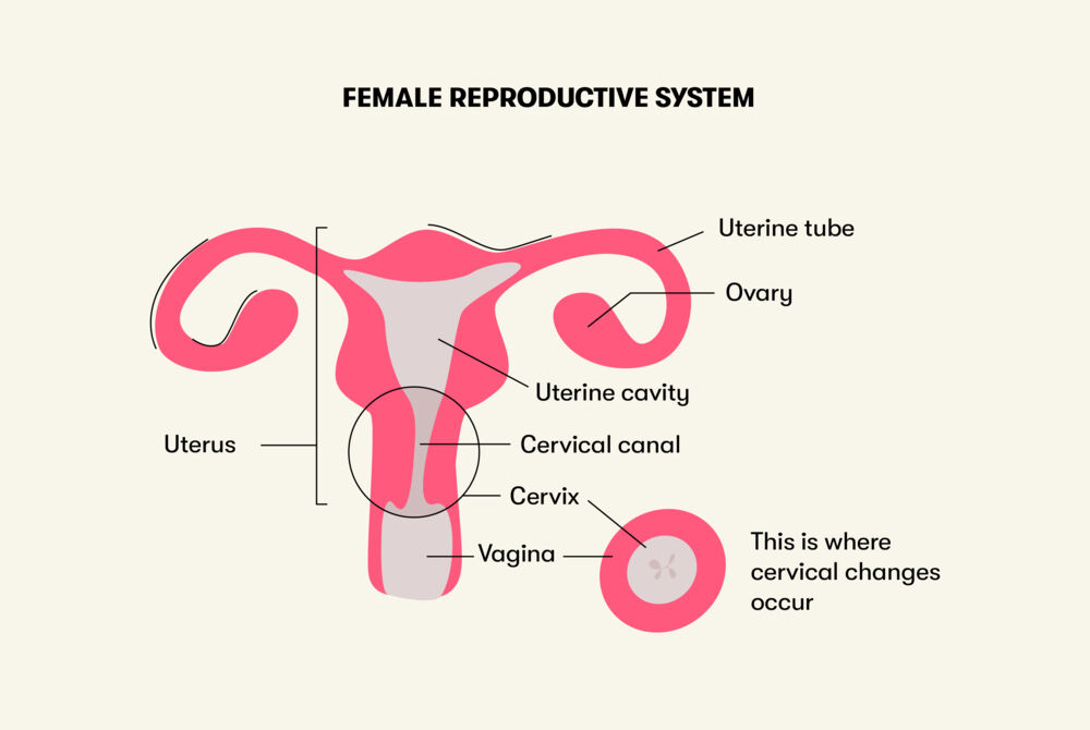
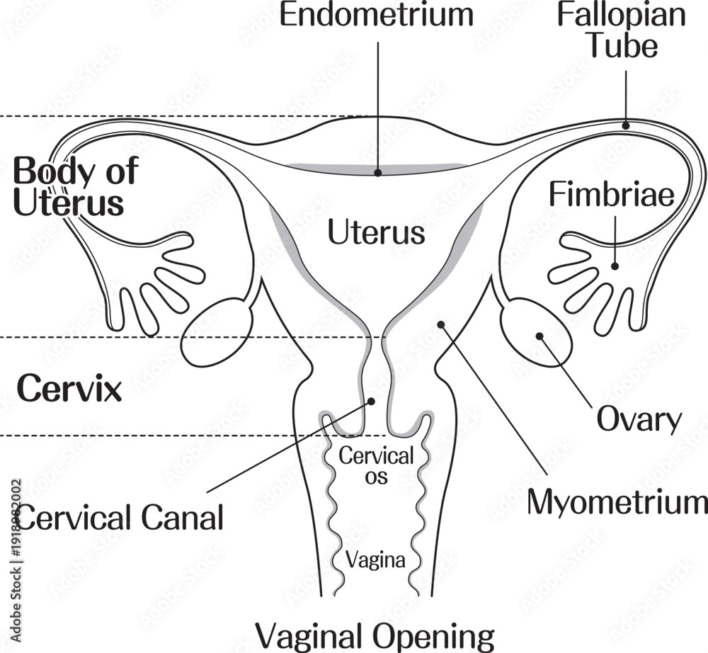
The cervix is a critical component of the female reproductive system, serving as the gateway between the uterus and the vagina while playing essential roles in fertility, pregnancy, and menstrual regulation. Positioned at the lower end of the uterus, it extends into the vaginal canal, forming a narrow passage that connects the uterine cavity to the external genitalia. Structurally, the cervix is divided into distinct regions—each with specialized functions—enabling its dual role in reproductive physiology and barrier protection. Understanding its anatomical intricacies, including histological composition and cyclical adaptations, is fundamental to comprehending its contributions to reproductive health and disease prevention.Location and Position Relative to the Uterus and Vagina
The cervix is a cylindrical, fibrous structure situated at the inferior end of the uterus, protruding into the superior aspect of the vagina. In anatomical terms, it forms the cervical os (plural: ostia), the openings that regulate passage between the uterine cavity and vaginal lumen. The external os (vaginal opening) is typically slit-like in nulliparous women and circular in parous women due to childbirth-induced remodeling. The internal os (uterine opening) connects the cervix to the uterine corpus, where the endometrial lining resides.The cervix’s position is stabilized by pelvic ligaments, including the cardinal (transverse cervical) ligaments and uterosacral ligaments, which anchor it to the pelvic sidewall and sacrum, respectively. Its anterior lip (facing the bladder) and posterior lip (facing the rectum) are palpable during a bimanual pelvic examination, aiding in clinical assessments of size, consistency, and positional abnormalities.
Structural Composition: Ectocervix, Endocervix, and Cervical Canal
The cervix is histologically and functionally divided into three primary regions, each adapted to its role in reproduction and protection:1. Ectocervix (Exocervix)
2. Endocervix
3. Cervical Canal
Histological Comparison of Cervical Layers
The cervix’s functional diversity is underpinned by its layered histology, summarized below in comparative form:| Layer | Cell Types Present | Primary Functions | Key Anatomical Landmarks |
|---|---|---|---|
| Ectocervical Epithelium |
|
|
|
| Endocervical Epithelium |
|
|
|
| Stroma |
|
|
|
Cervical Adaptations During the Menstrual Cycle
The cervix undergoes cyclical morphological and functional changes driven by hormonal fluctuations (estrogen and progesterone), optimizing conditions for either sperm survival (fertile window) or barrier protection (non-fertile phases). These adaptations are primarily reflected in cervical mucus characteristics, which follow a predictable pattern:1. Menstrual Phase (Days 1–5)
2. Follicular Phase (Days 6–14)
Physiological Functions and Hormonal Influence on the Cervix
The cervix serves as a dynamic organ with critical roles in reproductive health, adapting its structure and function in response to hormonal fluctuations throughout the menstrual cycle, pregnancy, and menopause. Its dual capacity to protect the uterine cavity during gestation and facilitate menstrual flow underscores its physiological complexity. Hormonal regulation—primarily by estrogen and progesterone—dictates cervical mucus production, cervical os elasticity, and pH balance, all of which influence fertility, pregnancy maintenance, and labor progression. Understanding these mechanisms provides insight into cervical health markers and age-related functional decline.Dual Role of the Cervix in Pregnancy and Menstruation
The cervix exhibits distinct physiological adaptations to support pregnancy and menstruation, reflecting its adaptive role in reproductive biology.Pregnancy Protection via Mucus Plug Formation
During pregnancy, the cervix undergoes structural and biochemical changes to safeguard the uterine environment. The mucus plug, a dense, gelatinous barrier composed of cervical mucus, forms in the cervical canal. This plug is rich in immunoglobulins (IgA and IgG) and antimicrobial peptides, creating a physical and biochemical barrier against ascending infections (e.g., bacterial vaginosis, group B Streptococcus). The plug also maintains a low-pH environment (3.5–4.5), inhibitory to sperm and pathogens. Its stability relies on elevated progesterone levels, which thicken mucus consistency and reduce cervical os permeability. Disruption of the plug—often signaled by bloody show—typically precedes labor, as hormonal shifts (e.g., prostaglandin release) soften cervical tissue (cervical ripening).
Menstrual Shedding and Endometrial Lining Removal
During menstruation, the cervix facilitates the expulsion of the shed endometrial lining through coordinated contractions of the uterine myometrium and cervical dilation. The cervical os dilates slightly (1–2 mm) to allow menstrual blood and tissue passage, while cervical mucus becomes thinner and more watery due to declining progesterone and rising estrogen. This fluidity aids in lubrication and reduces friction during uterine contractions. Additionally, the cervix’s vascularization increases, enhancing blood flow to the endometrial interface for efficient sloughing. Post-menstruation, the cervix returns to a firm, closed state as estrogen peaks in the follicular phase, preparing for potential fertilization.
Hormonal Influence on Cervical Mucus, Elasticity, and pH Across the Menstrual Cycle
Estrogen and progesterone exert opposing effects on cervical mucus production, elasticity, and pH, creating a cyclical pattern that optimizes fertility or maintains pregnancy. Below is a timeline of hormonal shifts and their corresponding cervical changes:-
Menstrual Phase (Days 1–5)
- Hormonal Context: Low estrogen and progesterone; follicular phase begins with rising FSH.
- Cervical Mucus: Scant, sticky, and opaque ("dry" phase); pH ~4.0–4.5 (acidic).
- Os Firmness: Closed and rigid due to low estrogen.
- Physiological Role: Inhibits sperm survival; prepares uterus for endometrial regeneration.
-
Follicular Phase (Days 6–14)
- Hormonal Context: Estrogen surges (peaking pre-ovulation); progesterone remains low.
- Cervical Mucus:
- Early Follicular: Thick, cloudy, and elastic ("egg-white" consistency appears as estrogen rises).
- Pre-Ovulatory Peak: Clear, stretchy, and alkaline (pH 6.0–8.0), facilitating sperm motility.
- Spinnbarkeit: Mucus can stretch >8 cm due to high hyaluronic acid content.
- Os Firmness: Softens and begins to open slightly (1–2 mm) as estrogen peaks.
- Physiological Role: Optimal sperm transport and survival; cervical secretions provide a hospitable environment for conception.
-
Luteal Phase (Days 15–28)
- Hormonal Context: Progesterone dominates (post-ovulation); estrogen declines slightly.
- Cervical Mucus:
- Early Luteal: Thickens, becomes opaque, and reduces in volume ("dry" phase resumes).
- Late Luteal: pH drops to ~4.0–5.0; mucus remains inhibitory to sperm.
- Os Firmness: Returns to firm and closed state; progesterone-induced cervical rigidity prepares for potential implantation.
- Physiological Role: Protects against polyspermy; maintains uterine quiescence unless pregnancy occurs.
-
Pregnancy (Post-Implantation)
- Hormonal Context: Sustained high progesterone; human chorionic gonadotropin (hCG) later supports corpus luteum.
- Cervical Mucus: Thickens into the mucus plug; pH remains acidic (3.5–4.5).
- Os Firmness: Firmly closed until labor onset; progesterone receptors in cervical tissue inhibit contractions.
- Physiological Role: Isolates uterine cavity; prevents preterm labor via inhibitory effects on prostaglandins.
Dilation of the Cervical Os During Labor: Stages and Physiological Changes
Labor progression is marked by cervical dilation, a process driven by uterine contractions, prostaglandins, and hormonal shifts. The cervical os transitions from a closed (0 cm) to fully dilated (10 cm) state through distinct stages, each associated with specific physiological adaptations:The cervical os dilates in response to oxytocin-induced myometrial contractions and prostaglandin E2, which soften cervical collagen (cervical ripening). Dilation occurs in three phases:
- Latent Phase (0–3 cm): Slow, irregular contractions; cervix effaces (thins) and dilates gradually. Cervical mucus may contain bloody show due to vascular engagement.
- Active Phase (4–7 cm): Contractions intensify (frequency: 3–5 minutes); cervical os opens rapidly. Prostaglandins peak, enhancing collagen breakdown.
- Transition Phase (8–10 cm): Strong, frequent contractions; full effacement occurs. The internal os dilates last, aligning with fetal descent.
Key Physiological Changes:
- Collagen Remodeling: Prostaglandins and matrix metalloproteinases degrade cervical stroma, reducing rigidity.
- Vascular Engagement: Increased blood flow to the cervix may cause edema or ecchymosis ("chadwick’s sign").
- Neural Feedback: Stretch receptors in the cervix trigger Ferguson reflex, amplifying oxytocin release.
Three Key Markers of Cervical Health Linked to Hormonal Balance
Non-invasive assessment of cervical health relies on observable markers influenced by hormonal status. Three critical indicators—mucus clarity, os firmness, and vascularity—provide insights into reproductive function and potential dysfunctions (e.g., hormonal imbalances, infections, or structural abnormalities).-
Cervical Mucus Clarity and Consistency
- Normal Findings:
- Follicular Phase: Clear, stretchy, and alkaline (pH 6.0–8.0); absence of blood or pus.
- Luteal Phase: Thick, opaque, and scant; pH <5.0.
- Postmenopausal: Minimal or absent mucus; dryness due to estrogen deficiency.
- Abnormal Findings:
- Persistent Watery Mucus: May indicate estrogen dominance or cervical insufficiency.
- Blood-Streaked Mucus: Could signal polyps, infection (e.g., trichomoniasis), or implantation bleeding.
-
Clinical Significance and Common Conditions of the Cervix
The cervix plays a critical role in reproductive and gynecological health, serving as a gateway between the uterus and vagina while also acting as a barrier against infections. Dysfunction or pathological changes in the cervix can manifest through structural abnormalities, infections, or neoplastic transformations, often requiring early detection for effective management. This section explores five prevalent cervical conditions, diagnostic methodologies, preventive self-assessment techniques, and the progression from HPV infection to cervical cancer, alongside a clinical case study to illustrate recurrent cervical infections.
Five Common Cervical Conditions
Cervical conditions vary widely in etiology, ranging from inflammatory processes to neoplastic growths. Below is a structured overview of five frequently encountered conditions, organized for clarity in clinical and patient education contexts.
Condition Name Primary Symptom(s) Underlying Cause Diagnostic Approach Cervical Stenosis - Dysmenorrhea (painful menstruation) due to obstructed menstrual flow
- Recurrent pelvic pain or cramping
- Secondary dyspareunia (pain during intercourse)
- Possible infertility or recurrent miscarriages (in severe cases)
- Chronic inflammation or infection (e.g., pelvic inflammatory disease)
- Post-surgical scarring (e.g., following LEEP or cone biopsy)
- Radiation therapy for cervical cancer
- Congenital abnormalities
- Pelvic examination to assess cervical opening (os) patency
- Hysterosalpingography (HSG) to evaluate uterine cavity and fallopian tubes
- Transvaginal ultrasound to measure cervical canal length
- MRI for complex cases or suspected congenital stenosis
Cervical Ectropion (Eversion) - Postcoital or intermenstrual spotting (due to exposed columnar epithelium)
- Increased vaginal discharge, often watery or mucous-like
- Mild pelvic discomfort (rarely painful)
- Pruritus (itching) secondary to irritation
- Hormonal fluctuations (e.g., pregnancy, oral contraceptives, or menopause)
- Trauma during childbirth or surgical procedures
- Chronic cervical inflammation
- Visual inspection during speculum examination (red, velvety appearance of ectocervix)
- Colposcopy with acetic acid application to confirm abnormal vascular patterns
- Exclusion of other causes (e.g., cervical polyps, infection)
Cervical Polyps - Intermenstrual or postmenopausal bleeding
- Increased vaginal discharge (clear, white, or blood-tinged)
- Pelvic pressure or discomfort (if polyp is large)
- Asymptomatic in ~30% of cases (incidental finding)
- Chronic cervical inflammation or infection (e.g., Chlamydia trachomatis, HPV)
- Hormonal imbalances (e.g., estrogen dominance)
- Endometrial hyperplasia extending to the cervix
- Unknown etiology in ~50% of cases
- Pelvic examination with speculum to visualize polyp(s)
- Colposcopy with biopsy if polyp appears atypical
- Histopathological analysis to rule out dysplasia or malignancy
- Endometrial biopsy if associated with abnormal uterine bleeding
Cervicitis - Purulent or malodorous vaginal discharge
- Pelvic pain or lower abdominal discomfort
- Postcoital bleeding or spotting
- Dysuria (painful urination) if urethra is involved
- Bacterial infections (Neisseria gonorrhoeae, Chlamydia trachomatis, Mycoplasma genitalium)
- Viral infections (HPV, HSV)
- Fungal infections (Candida species)
- Allergic reactions (e.g., to spermicides, lubricants)
- Iatrogenic (post-surgical or post-procedural)
- Pelvic examination with speculum to assess cervical erythema, edema, or discharge
- Nucleic acid amplification tests (NAATs) for Chlamydia and Gonorrhea
- HPV testing if high-risk strains are suspected
- Vaginal pH testing and wet mount microscopy for fungal/bacterial identification
- Cervical cultures for atypical pathogens
Cervical Intraepithelial Neoplasia (CIN) - Asymptomatic in early stages (detected via screening)
- Abnormal Pap smear results (e.g., LSIL/HSIL)
- Progressive symptoms if untreated (e.g., postcoital bleeding, watery discharge)
- Persistent high-risk HPV infection (types 16, 18, 31, 33)
- Chronic immune suppression (e.g., HIV)
- Smoking (increases carcinogenic risk)
- Early-onset sexual activity or multiple partners
- Pap smear with reflex HPV testing (co-testing)
- Colposcopy with directed biopsy of abnormal areas
- Endocervical curettage (ECC) for sampling endocervical canal
- HPV genotyping to identify high-risk strains
Pap Smear Procedure: Step-by-Step Overview
The Papanicolaou (Pap) smear is a cornerstone of cervical cancer screening, designed to detect precancerous changes or malignant cells through microscopic examination of cervical samples. The procedure involves systematic collection, preparation, and analysis of cells from the transformation zone, where most cervical dysplasia originates.Sample Collection Techniques:
1. Patient Positioning and Preparation
- The patient is placed in the lithotomy position on an examination table.
- A speculum is inserted to visualize the cervix, which is then cleaned with a saline-moistened swab to remove mucus or blood.
2. Cell Sampling Methods
- Traditional Method (Conventional Pap):
A wooden or plastic spatula is used to scrape cells from the ectocervix (outer portion), followed by an end
Cervical Health Across Life Stages
The cervix undergoes dynamic physiological and anatomical transformations throughout a woman’s lifespan, influenced by hormonal fluctuations, reproductive events, and aging. These changes affect cervical function, susceptibility to conditions, and clinical management strategies. Understanding these variations is critical for early detection of abnormalities, personalized care, and patient education across puberty, reproductive years, pregnancy, postpartum recovery, and menopause.
Pregnancy-Related Cervical Changes: Diagnostic Checklist
During pregnancy, the cervix exhibits predictable softening, vascular changes, and structural adaptations to support fetal development and labor. These physical signs are clinically significant for assessing gestational progression, identifying complications, and guiding prenatal care.
-
Goodell’s Sign: Softening of the cervix, detectable as early as 6 weeks gestation due to increased vascularity and cervical gland hypertrophy. This change is attributed to progesterone and estrogen-induced edema and glandular secretion.
Clinical relevance: Confirms early pregnancy and may indicate cervical insufficiency if excessive softening is noted prematurely.
-
Chadwick’s Sign: Bluish-purple discoloration of the cervix and vagina (cervical congestion) caused by increased blood flow and vascular engorgement. Typically appears by 6–8 weeks.
Clinical relevance: Supports pregnancy diagnosis and differentiates from non-pregnancy-related cervical vascular changes (e.g., varicosities).
-
Hegar’s Sign: Softening and compressibility of the lower uterine segment (between the cervix and uterus), palpable during bimanual examination. Occurs around 6–12 weeks due to cervical and uterine isthmus relaxation.
Clinical relevance: Indicates uterine expansion; absence may suggest ectopic pregnancy or fibroids.
-
Mucus Plug Formation: Thick, tenacious cervical mucus seals the cervical os to protect the uterine cavity from ascending infections. Estrogen-induced cervical gland activity increases mucus production, forming a plug that may be expelled (bloody show) before labor.
Clinical relevance: Loss of the plug signals impending labor but does not guarantee immediate delivery.
-
Cervical Effacement and Dilation: Progressive thinning (effacement) and opening (dilation) of the cervix during labor, measured in centimeters (0–10 cm). Effacement is complete at 100%, dilation at 10 cm.
Clinical relevance: Critical for staging labor; abnormal progression may indicate dystocia or fetal malposition.
-
Operculum Formation: A thin membrane of cervical mucus and tissue that partially covers the os in late pregnancy. Rupture may precede labor or occur during delivery.
Clinical relevance: Inspection of the operculum can reveal signs of infection or premature rupture of membranes (PROM).
Comparative Analysis of Cervical Changes Across Life Stages
The cervix adapts structurally and functionally in response to hormonal shifts, mechanical stress, and aging. Below is a comparative overview of key life stages, highlighting anatomical alterations, functional consequences, and associated health risks.
Life Stage Anatomical Changes Functional Impact Health Risks Puberty - Cervix elongates and narrows (nulliparous shape).
- Increased vascularity and glandular activity due to estrogen surge.
- Transformation zone (squamocolumnar junction) extends outward, increasing exposure to HPV.
- Establishment of menstrual flow regulation via cervical mucus changes.
- Enhanced susceptibility to sexually transmitted infections (STIs) due to columnar epithelium vulnerability.
- HPV infection and cervical dysplasia (peak incidence: late teens to early 20s).
- Pelvic inflammatory disease (PID) from untreated STIs (e.g., Chlamydia, gonorrhea).
Reproductive Years (Non-Pregnant) - Cervical os remains small and slit-like (nulliparous) or slightly dilated (parous).
- Cyclic changes in cervical mucus (thin and elastic during ovulation, thick and viscous post-ovulation).
- Ectocervix dominated by stratified squamous epithelium; endocervix lined with columnar epithelium.
- Facilitates sperm transport during ovulation via alkaline, watery mucus.
- Acts as a barrier to pathogens via acidic vaginal environment and mucus plug.
- Cervicitis (infectious or chemical).
- Cervical intraepithelial neoplasia (CIN) from persistent HPV infection.
- Polyps or leiomyomas (fibroids) affecting menstrual flow.
Childbirth - Permanent dilation of the cervical os (from ~2–3 cm to 10 cm during labor).
- Thinning and shortening of the cervix (effacement).
- Microtears or lacerations (e.g., cervical trauma during delivery).
- Reduced muscle tone and structural laxity post-delivery.
- Impaired barrier function against ascending infections.
- Increased risk of pelvic organ prolapse (POP) due to weakened pelvic floor support.
- Cervical insufficiency (incompetent cervix) in subsequent pregnancies.
- Chronic pelvic pain or dyspareunia from scar tissue.
- Higher susceptibility to HPV-related dysplasia due to altered epithelial integrity.
Menopause - Atrophy of cervical epithelium (thinning of squamous and columnar layers).
- Reduced vascularity and glandular activity (dryness, loss of rugae).
- Shortening and narrowing of the cervical canal.
- Calcification of cervical stroma (visible on imaging).
- Loss of estrogen-dependent protective effects (e.g., reduced mucus production).
- Increased pH of vaginal environment, predisposing to infections.
- Atrophic vaginitis and cervicitis.
- Persistent HPV infection or recurrent dysplasia.
- Cervical stenosis or synechiae (scarring) from chronic inflammation.
Postpartum Cervical Recovery: Timeline and Clinical Considerations
The cervix undergoes gradual structural and functional restoration following childbirth, influenced by hormonal recovery, tissue healing, and pelvic floor rehabilitation. Below is a standardized timeline for key recovery milestones, supported by clinical evidence.
-
Immediate Postpartum (0–6 weeks):
-
Os Closure: The cervical os begins to close but remains patent (visible on speculum exam). Complete closure may take 6–12 weeks, though the external os often retains a slit-like appearance in parous women.
Evidence: Ultrasound studies show median os
The cervix emerges not merely as a passive conduit within the female reproductive system but as a dynamic and resilient organ central to fertility, pregnancy, and menstrual health. Its ability to modulate cervical mucus consistency in sync with hormonal cycles, dilate progressively during labor, and signal underlying imbalances through subtle symptomatic cues underscores its indispensable role. By recognizing the interplay between anatomy, physiology, and clinical markers—from the cyclical shifts of the menstrual cycle to the early warning signs of conditions like HPV infection—individuals can adopt informed approaches to cervical care. Whether navigating pregnancy, menopause, or routine health assessments, awareness of the cervix’s adaptive mechanisms fosters proactive engagement with reproductive wellness. Ultimately, this exploration highlights the cervix as a mirror of systemic health, where vigilance and education serve as the cornerstones of long-term vitality.
FAQ
What is a cervix check and why is it done?
A cervix check, often called a Pap smear or pelvic exam, is a routine test where a healthcare provider examines the cervix for abnormalities like cell changes or infections. It helps detect early signs of cervical cancer, sexually transmitted infections (STIs), or other issues. Women typically start getting screened around age 21 or earlier if at risk, and frequency depends on age and health history.
What causes a cervix infection, and what are the symptoms?
A cervix infection is usually caused by bacteria, viruses (like HPV or herpes), or yeast overgrowth, often spread through sexual contact. Symptoms may include unusual vaginal discharge (with odor, color, or texture changes), pain during sex or urination, bleeding between periods, or itching. Many infections have no symptoms, so regular screenings are important.
What does a cervix look like in a healthy woman?
A healthy cervix is typically pink or reddish-pink, smooth, and slightly moist, with a small opening (os) that may appear as a tiny dot or slit. Its shape resembles a doughnut or barrel, and it can change slightly in size and firmness during the menstrual cycle or pregnancy. During a pelvic exam, a speculum is used to view it clearly.
What is the cervix, and where is it located on a woman’s body?
The cervix is the lower, narrow part of the uterus that connects to the vagina, acting as a passage for menstrual blood, sperm, and childbirth. Located at the top of the vaginal canal, it’s about 2–3 inches long and sits between the uterus (above) and the vaginal opening (below). It’s not visible externally but can be examined during a pelvic exam.
What is a cervix polyp, and how is it treated?
A cervix polyp is a small, benign growth of tissue that can develop on the cervix, often due to hormonal changes or chronic inflammation. They may cause spotting, bleeding after sex, or discharge, but many cause no symptoms. Treatment usually involves a simple in-office procedure (like a polyp removal with forceps or a loop electrosurgical excision) to prevent complications.
What happens during a cervix biopsy, and is it painful?
A cervix biopsy involves taking small tissue samples from the cervix, often after abnormal Pap test results, to check for cancer or precancerous cells. The procedure uses a local anesthetic to numb the area, so discomfort is usually mild (like cramping). You may feel pressure or a pinch, and slight bleeding or spotting afterward is normal. Results take days to weeks.
-
Os Closure: The cervical os begins to close but remains patent (visible on speculum exam). Complete closure may take 6–12 weeks, though the external os often retains a slit-like appearance in parous women.
- Normal Findings:
Leave a Comment
Comments are moderated before appearing. The data you submit is processed according to the Privacy Policy of Voltefac.