What Is Gallbladder Anatomy Function And Health Impact
Table of Contents
- Anatomy and Location of the Gallbladder
- Anatomical Position and Relations
- Structural Breakdown and Functional Roles
- Comparative Analysis: Gallbladder vs. Other Digestive Organs
- Physical Examination: Locating the Gallbladder
- Physiological Functions of the Gallbladder
- Bile Storage, Concentration, and Release Mechanisms
- Compositional Changes in Bile Before and After Gallbladder Storage
- Comparative Physiology: Gallbladder Function Across Species
- Flowchart: Bile Pathway from Liver to Intestinal Excretion
- Common Conditions and Symptoms of Gallbladder Disorders
- Five Prevalent Gallbladder Disorders and Their Underlying Causes
- Warning Signs: Acute Versus Chronic Gallbladder Issues
- Diagnostic Process for Gallbladder Diseases
- Dietary and Lifestyle Influences on Gallbladder Health
- Foods Associated with Gallstone Formation and Their Mechanisms
- Impact of Rapid Weight Loss, Fasting, and Crash Diets on Gallbladder Function
- Hydration, Fiber Intake, and Exercise in Gallbladder Health
- Medical and Surgical Interventions for Gallbladder Disorders
- Cholecystectomy: Surgical Techniques and Post-Operative Management
- Non-Surgical Treatments for Gallbladder Disorders
- Long-Term Outcomes After Gallbladder Removal
- Cultural and Historical Perspectives on the Gallbladder
- Historical Medical Practices and Gallbladder Knowledge Across Civilizations
- Cultural Diets and Gallbladder Disease Prevalence
- FAQ
- What is the gallbladder called in Tagalog?
- What are gallbladder polyps and what do they mean?
- What is gallbladder disease and what causes it?
- What is gallbladder sludge and is it dangerous?
- What is a gallbladder stone and how is it treated?
- What is gallbladder surgery called and how is it done?
The gallbladder, a small yet critical pear-shaped organ nestled beneath the liver, serves as the body’s primary bile reservoir, playing an indispensable role in digestion and metabolic regulation. Positioned strategically within the biliary system, this organ stores, concentrates, and releases bile—a complex fluid essential for fat emulsification and nutrient absorption. Beyond its physiological functions, the gallbladder’s health directly influences dietary habits, disease susceptibility, and even cultural dietary traditions across civilizations. Understanding its structure, role in digestion, and vulnerability to disorders provides foundational insights into both preventive healthcare and modern medical interventions.
From its anatomical relationship with surrounding organs to its biochemical interactions with bile components, the gallbladder exemplifies the delicate balance between form and function in human physiology. Its disorders, ranging from gallstones to inflammatory conditions, underscore the importance of lifestyle modifications, early diagnostics, and advanced surgical techniques. Exploring these dimensions reveals not only the organ’s biological significance but also its broader implications for public health and medical innovation.
Anatomy and Location of the Gallbladder
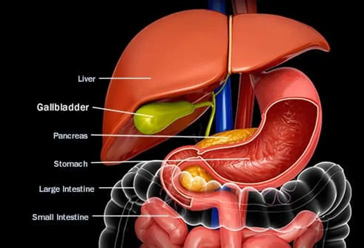
The gallbladder is a small, pear-shaped sac located beneath the liver, playing a critical role in bile storage and concentration. Its anatomical positioning, structural divisions, and functional relationships with adjacent organs—such as the liver, bile ducts, and duodenum—determine its physiological efficiency. Understanding its precise location and morphology is essential for clinical assessments, diagnostic imaging, and surgical planning.Anatomical Position and Relations
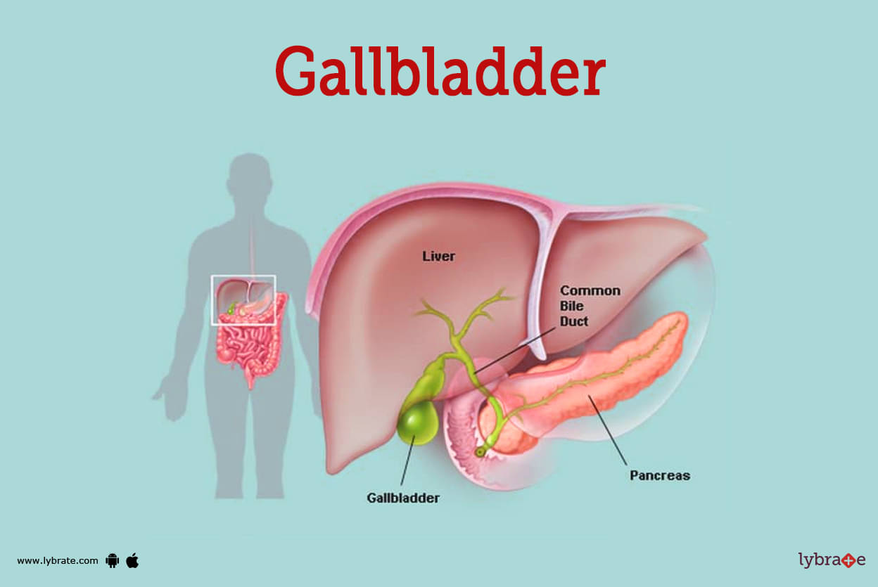
The gallbladder resides in the gallbladder fossa, a depression on the posterior-inferior surface of the liver, specifically on the visceral surface of the right lobe (segment IV). It lies in close proximity to several key structures:- Liver: The gallbladder is embedded in the liver’s bed, sharing a fascial connection via the peritoneum, which separates it from the liver parenchyma.
Labeled Diagram Description:
A cross-sectional anatomical illustration should depict:
1. The liver with its right and left lobes, highlighting the gallbladder fossa.
2. The gallbladder with its three distinct regions (fundus, body, neck) and the cystic duct.
3. The common hepatic duct, common bile duct, and their junction with the pancreatic duct.
4. Adjacent structures: duodenum, right kidney, and colon, with directional labels (e.g., "anterior," "posterior").
Structural Breakdown and Functional Roles
The gallbladder is divided into three primary regions, each with specialized functions:1. Fundus (Blind End)
2. Body (Middle Segment)
3. Neck (Narrow Transition Zone)
Bile Concentration Mechanism:
The gallbladder epithelium absorbs ~90% of water and electrolytes from hepatic bile, increasing its bile salt concentration up to 10-fold. This process is mediated by:
Comparative Analysis: Gallbladder vs. Other Digestive Organs
The following table compares the gallbladder’s physical and histological characteristics with the liver, pancreas, and stomach, emphasizing its unique role in digestion:| Feature | Gallbladder | Liver | Pancreas | Stomach |
|---|---|---|---|---|
| Size (Adult) | 7–10 cm (length), 3–5 cm (width) | ~15 cm (max diameter) | 12–15 cm (length) | 25–30 cm (length) |
| Shape | Pear-shaped, sac-like | Lobulated, wedge-shaped | Elongated, glandular | J-shaped, muscular sac |
| Primary Tissue | Simple columnar epithelium (mucosa) | Hepatocytes (parenchymal cells) | Acinar cells (exocrine) + Islets (endocrine) | Simple columnar epithelium (mucosa) + muscularis externa |
| Function | Bile storage/concentration | Bile production, metabolism | Enzyme secretion (digestive/exocrine), insulin/glucagon (endocrine) | Mechanical/chemical digestion (acid, pepsin) |
| Blood Supply | Cystic artery (branch of right hepatic artery) | Hepatic artery + portal vein | Splenic artery + superior mesenteric artery | Left/right gastric arteries + short gastrics |
| Innervation | Vagus nerve (parasympathetic) + sympathetic fibers | Vagus + hepatic plexus | Vagus + celiac plexus | Vagus + enteric nervous system |
| Histological Layers | Mucosa, muscularis (smooth muscle), serosa | Hepatic lobules, connective tissue | Acini, islets, ducts | Mucosa, submucosa, muscularis externa, serosa |
| Key Pathologies | Cholecystitis, cholelithiasis, polyps | Cirrhosis, hepatitis, fatty liver | Pancreatitis, diabetes, tumors | Gastritis, ulcers, cancer |
| Clinical Examination Landmark | Right subcostal region (Murphy’s sign) | Right upper quadrant (liver edge) | Epigastric region (Cullen’s sign) | Epigastric/left upper quadrant (succussion splash) |
Physical Examination: Locating the Gallbladder
Palpation of the gallbladder is primarily performed to assess for cholecystitis, gallstones, or tumor infiltration. The procedure requires systematic examination of the right upper quadrant (RUQ) using specific techniques:Step-by-Step Palpation Procedure:
1. Patient Positioning:
2. Landmark Identification:
3. Initial Palpation:
4. Murphy’s Sign (Diagnostic Maneuver):
5. Advanced Techniques:
Key Landmarks for Reference:
Note on Sensitivity:
Physiological Functions of the Gallbladder
The gallbladder serves as a critical accessory organ in the digestive system, primarily responsible for storing, concentrating, and releasing bile—a complex fluid essential for fat emulsification and nutrient absorption. Its physiological functions are tightly regulated by hormonal and neural mechanisms, ensuring efficient digestion while adapting to variations in dietary intake. The composition of bile undergoes significant modifications during storage, reflecting the gallbladder’s role in maintaining digestive homeostasis. Comparative analysis across species reveals evolutionary adaptations that optimize bile handling in diverse physiological contexts, from omnivorous diets in humans to herbivorous or carnivorous regimens in other mammals.Bile Storage, Concentration, and Release Mechanisms
The gallbladder’s primary function is to store and concentrate bile produced by the liver, which is continuously secreted into the biliary tree at a rate of 500–1,000 mL/day in humans. This process involves active absorption of water and electrolytes (primarily sodium and chloride) via apical Na+/H+ exchangers (NHE3) and Cl-/HCO3− exchangers (AE2), reducing bile volume by 80–90% while increasing its osmolarity and bile salt concentration up to 10-fold. The concentrated bile is stored in the gallbladder’s fundus and body, where mucin-rich secretions prevent precipitation of cholesterol and bilirubin crystals.Release of bile from the gallbladder is triggered by cholecystokinin (CCK), a peptide hormone secreted by I-cells in the duodenum in response to dietary fats and proteins. CCK binds to CCK-A receptors on gallbladder smooth muscle, inducing contraction via calcium-dependent pathways while simultaneously relaxing the sphincter of Oddi, allowing bile to flow into the duodenum. The timing and magnitude of bile release are synchronized with meal ingestion, ensuring optimal digestion of dietary lipids. Secretin, another gastrointestinal hormone, may also play a modulatory role by enhancing bicarbonate secretion from the pancreas, indirectly influencing bile flow dynamics.
Key Regulatory Pathways:
CCK secretion → Triggered by long-chain fatty acids (LCFAs) and amino acids in the duodenum. Gallbladder contraction → Mediated by IP3/Ca2+ signaling in smooth muscle cells. Sphincter of Oddi relaxation → Facilitated by nitric oxide (NO) and VIP (vasoactive intestinal peptide).
Compositional Changes in Bile Before and After Gallbladder Storage
Bile composition undergoes significant alterations during storage in the gallbladder, primarily due to water and electrolyte reabsorption. Freshly secreted hepatic bile (liver-derived) contains:After concentration in the gallbladder, vesicular bile exhibits the following changes:
Critical Thresholds for Gallstone Formation:
Cholesterol saturation index >1.0 (indicates supersaturation). Bile salt:phospholipid:cholesterol molar ratio (ideal: 10:4:1; pathological: <8:4:1). Mucin hypersecretion → Provides a scaffold for crystal nucleation.
Comparative Physiology: Gallbladder Function Across Species
The gallbladder’s structure and function exhibit marked variations across mammals, reflecting evolutionary adaptations to dietary habits and metabolic demands. Below is a comparative analysis of key differences:-
Anatomical Presence and Size:
- Humans and primates: Single-chambered gallbladder with a fundus, body, and neck; capacity ~30–50 mL.
- Herbivores (cows, horses): Often absent or vestigial (e.g., horses lack a gallbladder; cows have a small, elongated sac).
- Carnivores (dogs, cats): Elongated, sac-like gallbladder with a larger capacity relative to body size (~1–2 mL/kg), reflecting high-fat diets.
- Omnivores (pigs, rats): Intermediate structure, with multiple diverticula in some species (e.g., pigs).
-
Bile Composition and Storage Efficiency:
- Humans: High cholesterol content in bile, increasing gallstone risk; CCK-dependent release.
- Ruminants (cows, sheep): Low cholesterol, high bile salt concentration; gallbladder absent or reduced due to continuous bile flow for microbial fermentation.
- Carnivores (dogs): Higher bile salt reabsorption efficiency; gallbladder stores more concentrated bile to digest animal fats.
- Rodents (rats, mice): Rapid bile turnover; gallbladder contracts frequently due to coprophagy (re-ingestion of feces for nutrient recovery).
-
Hormonal Regulation:
- Humans/Dogs: CCK is the primary stimulant for gallbladder contraction.
- Ruminants: Minimal hormonal control due to lack of a gallbladder; bile flows continuously into the duodenum.
- Birds (e.g., chickens): No gallbladder; bile is stored in the liver’s bile ducts and released in pulses.
- Reptiles (e.g., snakes): Multichambered gallbladder with segmented storage for intermittent feeding.
-
Disease Susceptibility:
- Humans: High risk of cholesterol gallstones (75% of cases) due to supersaturated bile and Western diet.
- Dogs: Predisposition to pigmented gallstones (bilirubin-based) linked to hemolytic anemia.
- Cows: Biliary sludge common in high-production dairy cows due to rapid bile acid turnover.
- Cats: Xanthine gallstones (rare in humans) due to purine metabolism differences.
Flowchart: Bile Pathway from Liver to Intestinal Excretion
The following flowchart outlines the anatomical and physiological pathway of bile, highlighting the gallbladder’s role in storage and regulated release:-
Bile Production (Liver):
- Hepatocytes synthesize and secrete bile acids (from cholesterol), bilirubin, and phospholipids into canaliculi.
- Bile acids are conjugated with glycine or taurine for solubility.
-
Bile Transport to Gallbladder:
- Bile flows through right and left hepatic ducts → common hepatic duct → cystic duct (connects to gallbladder).
- Sphincter of Luschka (hepatic duct valves) may regulate flow.
-
Gallbladder Storage and Concentration:
- Active absorption of water and electrolytes via apical transporters (NHE3, AE2).
- Mucin secretion by gallbladder epithelium prevents precipitation.
- Bile remains stagnant until hormonal stimulation.
-
Release into Duodenum:
- CCK secretion (triggered by duodenal fats/proteins) → gallbladder contraction + sphincter of Oddi relaxation.
- Bile enters duodenum via common bile duct, aiding lipid emulsification by bile salt micelles.
-
Enterohepatic Circulation:
- 95% of bile salts are reabsorbed
- Pain: Sudden, severe right upper quadrant (RUQ) or epigastric pain radiating to the right scapula or shoulder (referred pain via phrenic nerve).
- Nausea/Vomiting: Persistent, often preceded by fatty meals; vomiting may relieve pain temporarily.
- Fever/Chills: Indicates infection or inflammation (temperature >38°C).
- Murphy’s Sign: Inspiratory arrest during deep palpation of the RUQ (highly specific for cholecystitis).
- Jaundice: Rare unless common bile duct obstruction (CBD) is present (e.g., Mirizzi syndrome).
- Systemic Toxicity: Hypotension or tachycardia suggests gangrenous or perforated gallbladder (emergency).
- Pain: Intermittent, dull RUQ or epigastric discomfort (often postprandial, especially after high-fat meals).
- Nausea: Mild, not typically accompanied by vomiting.
- Indigestion/Dyspepsia: Bloating, belching, or early satiety due to impaired bile release.
- Jaundice: Uncommon unless CBD stones cause obstruction (painless jaundice warrants urgent evaluation).
- Asymptomatic Gallstones: Detected incidentally on imaging (20% of adults in Western populations).
- Polyps: Often asymptomatic; symptoms arise if malignant or obstructive.
- Dietary: High-fat meals (stimulate CCK release, increasing gallbladder contraction).
- Pregnancy: Progesterone-induced gallbladder stasis (3rd trimester risk).
- Rapid Weight Loss: Alters bile composition, promoting stone formation.
- Critical Illness: Prolonged fasting or parenteral nutrition disrupts bile flow.
-
Ultrasound (US)
The first-line imaging modality for suspected gallbladder disease due to its accessibility, lack of radiation, and high sensitivity (95% for gallstones). Key findings include:
- Gallstones (hyperechoic with posterior acoustic shadowing).
- Gallbladder wall thickening (>3 mm) or pericholecystic fluid (cholecystitis).
- Sludge (low-level echoes in dependent areas, often post-fasting).
- Limitations: Operator-dependent; may miss CBD stones if not dilated.
-
Magnetic Resonance Cholangiopancreatography (MRCP)
A non-invasive MRI-based technique for evaluating bile duct anatomy and obstructions. Indications include:
- Suspected CBD stones or strictures (e.g., in patients with jaundice).
- Anomalies of the biliary tree (e.g., choledochal cysts).
- Preoperative planning for complex cases (e.g., Mirizzi syndrome).
- Advantage: No contrast required; superior to ERCP for ductal visualization.
-
Hepatobiliary Iminodiacetic Acid (HIDA) Scan
A nuclear medicine test using technetium-99m to assess gallbladder function. Used when:
- Ultrasound is inconclusive (e.g., acalculous cholecystitis in critically ill patients).
- Chronic acalculous cholecystitis is suspected (ejection fraction <35%).
- Suspected sphincter of Oddi dysfunction.
- Mechanism: Delayed or absent gallbladder visualization suggests obstruction or dysfunction.
-
Computed Tomography (CT) Scan
Reserved for complex cases or when ultrasound is non-diagnostic. Provides detailed cross-sectional images for:
- Gallbladder wall calcification (porcelain gallbladder, high cancer risk).
- Complications (e.g., perforation, abscess).
- Alternative diagnoses (e.g., hepatic abscess, pancreatitis).
- Disadvantage: Lower sensitivity for small stones; exposes patients to radiation.
-
Endoscopic Ultrasound (EUS)
An invasive but highly sensitive modality for:
- Characterizing gallbladder polyps (differentiating benign vs. malignant).
- Guiding fine-needle aspiration (FNA) for suspected cancer.
- Evaluating CBD stones or strictures not visualized on MRCP.
- Indication: High clinical suspicion of malignancy or indeterminate polyps.
-
Liver Function Tests (LFTs)
Elevated enzymes indicate biliary obstruction or inflammation:
- Alkaline Phosphatase (ALP): Rises with CBD obstruction (sensitivity >90%).
- Bilirubin (Direct): Conjugated hyperbilirubinemia suggests CBD stones or strictures.
- Alanine Aminotransferase (ALT) / Aspartate Aminotransferase (AST): Mild-moderate elevation in cholecystitis; marked elevation suggests cholangitis or hepatitis.
-
Inflammatory Markers
- White Blood Cell Count (WBC): Leukocytosis (>10,000/mm³) in acute cholecystitis or cholangitis.
- C-reactive Protein (CRP): Elevated in inflammation (non-specific but useful for monitoring).
-
Lipase
Elevated in concurrent pancreatitis (common with CBD stones). -
Tumor Markers (if malignancy suspected)
- CA 19-9: Elevated in gallbladder cancer (though non-specific).
- Stimulate hepatic cholesterol synthesis via insulin resistance and de novo lipogenesis.
- Reduce bile acid secretion, increasing bile cholesterol saturation.
- Promote visceral adiposity, linked to elevated serum cholesterol and gallbladder sludge.
- Elevate LDL cholesterol and reduce HDL, altering bile lipid ratios.
- Induce low-grade inflammation, impairing gallbladder emptying.
- Inhibit lecithin-cholesterol acyltransferase (LCAT), reducing cholesterol solubilization in bile.
- Reduce bile acid reabsorption in the ileum, leading to hepatic cholesterol overproduction.
- Decrease gallbladder emptying frequency, promoting bile stasis.
- Alter gut microbiota composition, increasing secondary bile acid deconjugation (e.g., deoxycholic acid), which is lithogenic.
- Directly increase hepatic cholesterol secretion into bile, exceeding bile acid capacity for solubilization.
- Promote cholesterol crystal nucleation via supersaturation.
- Associated with elevated serum triglycerides, further impairing gallbladder motility.
- Enhance hepatic de novo lipogenesis, increasing VLDL-triglyceride production.
- Reduce bile acid synthesis, shifting bile toward cholesterol saturation.
- Promote metabolic syndrome, a known risk factor for gallstones.
- Bile Acid Malabsorption: Prolonged fasting reduces enterohepatic circulation of bile acids, forcing the liver to secrete cholesterol-rich bile.
- Gallbladder Stasis: Skipping meals or extreme calorie restriction leads to infrequent CCK (cholecystokinin) release, reducing gallbladder emptying.
- Inflammatory Response: Rapid fat mobilization releases free fatty acids, triggering low-grade inflammation that may precipitate gallstone formation.
- A study in Obesity Surgery (2015) reported that patients undergoing bariatric surgery (e.g., Roux-en-Y gastric bypass) experienced a 30% incidence of new gallstones within 6 months post-operation due to rapid weight loss (RR: 1.30, 95% CI: 1.10–1.52).
- Research in The American Journal of Clinical Nutrition (2010) demonstrated that individuals following very-low-calorie diets (<800 kcal/day) had a 2.5-fold increase in gallstone risk compared to gradual weight loss (OR: 2.50, 95% CI: 1.30–4.80).
- Mechanism: Water intake increases bile volume and flow, diluting cholesterol concentration. Dehydration thickens bile, increasing lithogenic potential.
- Recommendations:
- Daily Int
- Patient assessment for comorbidities (e.g., diabetes, cardiovascular disease) and adjustment of anticoagulant medications.
- Administration of prophylactic antibiotics to reduce infection risk.
- Marking the gallbladder fundus and cystic duct for surgical orientation.
- Insertion of a Veress needle or Hasson trocar to establish pneumoperitoneum (CO₂ insufflation to 12–15 mmHg for abdominal cavity distension).
- Placement of four trocars (10–12 mm ports) under direct visualization, typically in the umbilicus, epigastrium, and bilateral subcostal regions.
- Mobilization of the gallbladder fundus using electrocautery or harmonic shears.
- Identification and division of the cystic artery and duct using clips, staples, or energy devices (e.g., LigaSure).
- Achievement of the "critical view of safety"—clear visualization of the cystic duct and artery junction with no residual gallbladder tissue—before resection.
- Removal of the gallbladder through the umbilical port or a small extension incision.
- Irrigation of the abdominal cavity and verification of hemostasis.
- Closure of trocar sites, with larger ports requiring subcutaneous sutures.
- A 7–10 cm right subcostal incision (Kocher’s incision).
- Direct visualization and manual dissection of the gallbladder, cystic duct, and artery.
- Higher risk of wound complications (infection, hernia) and longer recovery (5–7 days vs. 1–2 days for laparoscopic).
- Intraoperative:
- Bile duct injury (0.3–0.5% in laparoscopic cases, higher in open or emergency surgeries).
- Hemorrhage from hepatic artery or portal vein injury.
- Conversion to open surgery (5–10% due to adhesions or anatomical challenges).
- Postoperative:
- Infectious: Wound infection (1–5%), abscess formation, or cholangitis.
- Biliary: Bile leak (1–3%), retained stones, or stricture (0.5–1%).
- General: Pneumonia, deep vein thrombosis (DVT), or pulmonary embolism (PE), particularly in high-risk patients.
- Port-Site Hernia: Occurs in <1% of laparoscopic cases, typically at 10–12 mm trocar sites.
- Pain Management:
- Oral analgesics (e.g., acetaminophen, NSAIDs) for port-site discomfort; opioids reserved for severe pain.
- Avoidance of NSAIDs in patients with renal impairment or anticoagulation.
- Dietary Progression:
- Clear liquids on the day of surgery, advancing to a low-fat diet within 24–48 hours.
- Gradual reintroduction of normal diet as tolerated, with emphasis on small, frequent meals to prevent biliary stasis.
- Activity Restrictions:
- Avoid heavy lifting (>10 lbs or 4.5 kg) and strenuous activity for 2–4 weeks.
- Gradual resumption of light exercise (e.g., walking) to promote bowel motility and reduce DVT risk.
- Follow-Up:
- Routine clinical assessment at 2–4 weeks post-surgery to monitor for complications.
- Imaging (e.g., ultrasound) if symptoms of bile leak or retained stones persist.
- Mechanism: Oral administration of bile acids (e.g., ursodeoxycholic acid [UDCA] or chenodeoxycholic acid) to dissolve cholesterol stones by altering bile composition.
- Efficacy and Limitations:
- Effective for small (<10 mm), radiolucent cholesterol stones in a functional gallbladder.
- Complete dissolution achieved in 50–70% of cases over 6–24 months, with relapse rates of 30–50% after cessation.
- Contraindications: Pigment stones, gallbladder polyps, or biliary obstruction.
- Patient Selection:
- Ideal for elderly or high-risk patients with symptomatic but non-emergent stones.
- Requires regular ultrasound monitoring (every 3–6 months) to assess stone size and response.
- Indications: CBD stones, cholangitis, or pancreatitis secondary to biliary obstruction.
- Procedure:
- Endoscopic cannulation of the ampulla of Vater and injection of contrast to visualize the biliary tree.
- Sphincterotomy (incision of the Oddi sphincter) to facilitate stone removal using baskets or balloons.
- Optional placement of a nasobiliary drain or stent for persistent leaks or strictures.
- Complications: Pancreatitis (5–10%), perforation (0.5–1%), or post-ERCP bleeding (1–3%).
- Indications: High-risk surgical candidates with acute cholecystitis or gallbladder perforation.
- Procedure:
- Transgastric or transduodenal puncture of the gallbladder under EUS guidance.
- Placement of a drainage catheter (internal or external) to decompress the organ.
- Outcomes: Temporary relief for 4–6 weeks, with eventual need for cholecystectomy in stable patients.
- Indications: Severe acute cholecystitis in patients unfit for surgery or ERCP.
- Procedure:
- Ultrasound-guided aspiration of gallbladder contents followed by catheter placement.
- Used as a bridge to definitive surgery or in palliative care.
- Complications: Bleeding, bile leak, or catheter displacement.
- Bile Composition Changes:
- Loss of bile acid reabsorption in the ileum, leading to increased fecal bile acid excretion and potential malabsorption of fat-soluble vitamins (A, D, E, K).
- Compensation: The liver increases bile acid synthesis to maintain enterohepatic circulation, typically stabilizing within 6–12 months.
- Gastrointestinal Symptoms:
- Post-Cholecystectomy Syndrome (PCS): Persistent symptoms (e.g., bloating, diarrhea, or upper abdominal pain) in 5–40% of patients, often due to:
- Sphincter of Oddi dysfunction (SOD).
- Bile reflux into the stomach (leading to
- Bittersweet nightshade (Solanum dulcamara) for bile flow regulation.
- Myrrh and frankincense in poultices to "cool" inflammatory processes.
- Therapeutic fasting to reduce liver burden, a practice later adopted in Greek medicine.
- Cheese, pork, and legumes (considered "phlegmatic" or "melancholic" in humor theory).
- Wine, which he warned could "thicken" bile.
- Bitter herbs: Kasani (Cichorium intybus), Tulsi (Ocimum sanctum), and Bhumyamalaki (Phyllanthus niruri) to stimulate bile flow.
- Panchakarma detox: A five-step purification process including therapeutic vomiting (vamana) to remove excess bile.
- Dietary restrictions: Avoidance of sour, salty, and fried foods, which were believed to aggravate pitta.
- Liver-gallbladder disharmony: Symptoms like abdominal distension or bitter taste in the mouth were treated with acupuncture at points like GB-34 (Yanglingquan) to "unblock" bile flow.
- Herbal formulas:
- Yin Chen Hao Tang (Artemisia capillaris decoction) for jaundice.
- Long Dan Xie Gan Tang (a bitter, cooling formula) for "heat" in the gallbladder.
- Dietary therapy: Emphasis on light, easily digestible foods (e.g., congee, steamed vegetables) and avoidance of raw, cold, or greasy foods, which were deemed "stagnant."
- Stone dissolution: A mixture of vinegar, honey, and myrrh.
- Surgical innovation: Al-Zahrawi (Abulcasis, 10th century) documented early laparotomy techniques, though gallbladder surgery remained experimental.
- Maya and Aztec medicine: Used chicle (sap from Manilkara zapota) as a bile stimulant and copal resin in poultices for abdominal pain.
- Native American tribes: Employed yarrow (Achillea millefolium) and goldenseal (Hydrastis canadensis) for liver-gallbladder support, often in smudging rituals to "cleanse" the body.
- High intake of refined carbohydrates, trans fats, and red meat (linked to bile supersaturation).
- Low fiber and polyunsaturated fats (e.g., olive oil) may reduce stone formation.
- Rapid weight cycling (e.g., crash diets) increases cholesterol stone risk.
- High fiber (whole grains, legumes) and monounsaturated fats (olive oil, nuts) promote bile flow.
- Moderate fish consumption (omega-3s may reduce inflammation).
- Limited processed meats and dairy.
- High rice and noodle consumption may dilute bile concentration.
- Low-fat diets reduce cholesterol stone risk but increase pigment stone prevalence (linked to hepatitis B/C in regions like China).
- Fermented foods (e.g., kimchi, miso) may support gut-liver axis health.
- High
The gallbladder’s multifaceted role—spanning anatomy, physiology, pathology, and cultural influences—highlights its significance as a cornerstone of digestive health. From ancient medical practices to cutting-edge surgical advancements, the study of this organ bridges historical perspectives with contemporary medical challenges. Recognizing its functional intricacies, susceptibility to dietary and lifestyle factors, and the evolving landscape of treatment options empowers individuals to make informed health decisions. As research continues to unravel its complexities, the gallbladder remains a pivotal subject in both clinical practice and preventive medicine, underscoring the interplay between biology, behavior, and healthcare outcomes.
FAQ
What is the gallbladder called in Tagalog?
The gallbladder in Tagalog is called pangatahan or pangatahang bato (when referring to gallstones).
What are gallbladder polyps and what do they mean?
Gallbladder polyps are small growths on the inner lining of the gallbladder, often benign but sometimes precancerous. They can be cholesterol-based, inflammatory, or adenomatous (a higher-risk type). Most are harmless, but large or growing polyps may require removal or monitoring.
What is gallbladder disease and what causes it?
Gallbladder disease refers to conditions like cholecystitis (inflammation), gallstones, or polyps that affect the gallbladder. Causes include gallstones blocking bile flow, infections, or chronic irritation. Symptoms may include pain in the upper right abdomen, nausea, or jaundice.
What is gallbladder sludge and is it dangerous?
Gallbladder sludge is a thick, bile-like fluid containing cholesterol crystals, calcium, and mucus. It’s often seen in pregnant women or after rapid weight loss but usually isn’t dangerous unless it progresses to gallstones or causes blockages.
What is a gallbladder stone and how is it treated?
A gallbladder stone (or gallstone) is a hard deposit of cholesterol, bilirubin, or calcium that forms in the gallbladder. Treatment depends on size and symptoms: small stones may pass on their own, while larger ones often require surgery (cholecystectomy) or medication to dissolve them.
What is gallbladder surgery called and how is it done?
Gallbladder surgery is called a cholecystectomy, usually performed laparoscopically (minimally invasive) with small abdominal incisions. The entire gallbladder is removed since it’s not essential for survival; bile then flows directly from the liver to the intestines. Recovery typically takes 1–2 weeks.
Common Conditions and Symptoms of Gallbladder Disorders
The gallbladder, though small, plays a critical role in digestion by storing and concentrating bile, aiding in fat emulsification and nutrient absorption. Dysfunction in this organ often manifests as acute or chronic conditions, ranging from benign gallstones to life-threatening complications. Understanding these disorders—including their etiologies, clinical presentations, and diagnostic pathways—enables early intervention and improved patient outcomes. This section examines five prevalent gallbladder pathologies, their distinguishing symptoms, and the systematic approach required for accurate diagnosis.Five Prevalent Gallbladder Disorders and Their Underlying Causes
Gallbladder disorders typically arise from structural abnormalities, inflammatory processes, or metabolic imbalances. The following conditions account for the majority of clinical presentations and vary in severity, from asymptomatic to emergent:Cholelithiasis (Gallstones)
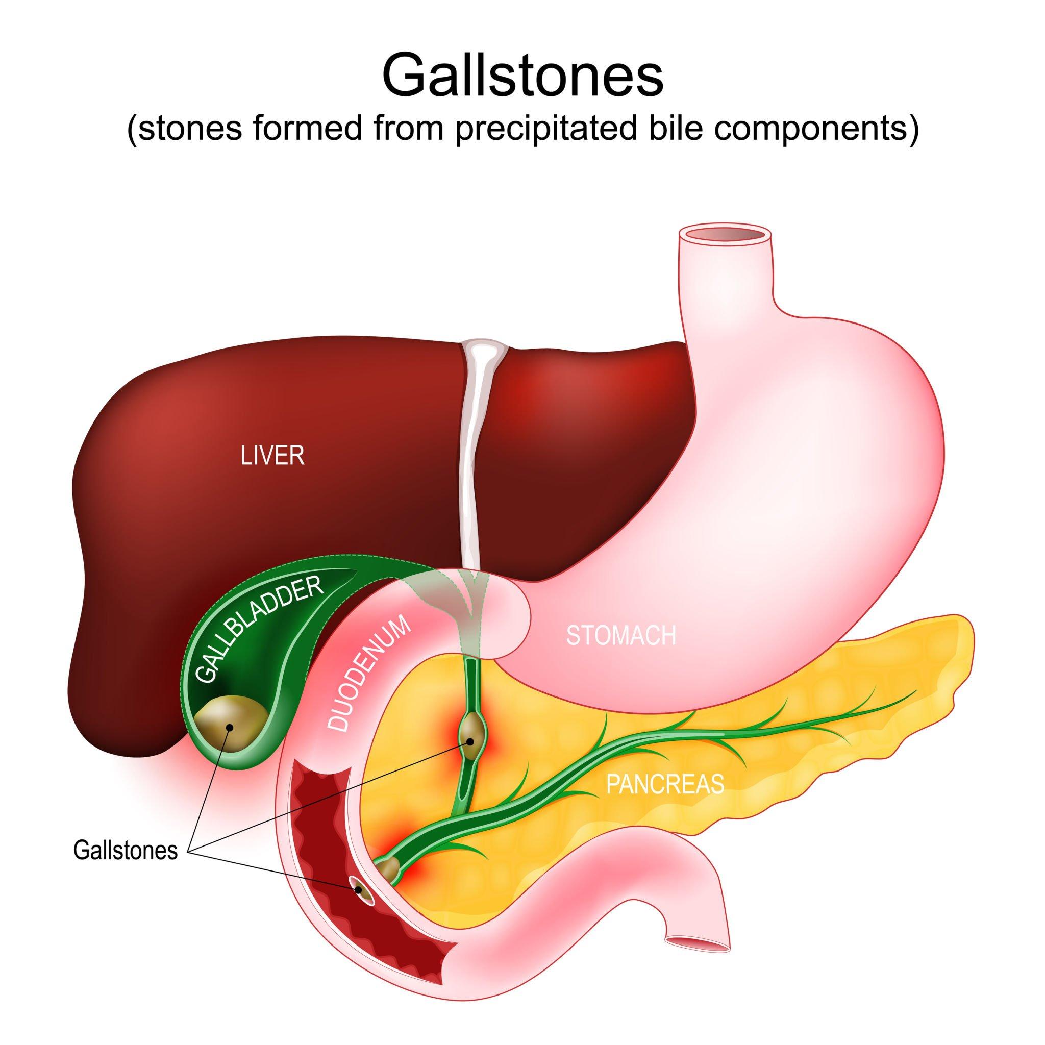
Gallstones form due to an imbalance in bile composition, primarily involving cholesterol supersaturation, pigment stone formation (from bilirubin excess), or gallbladder stasis. Risk factors include obesity, rapid weight loss, metabolic syndrome, cirrhosis, and genetic predisposition. Cholesterol stones (80% of cases) result from excess cholesterol in bile, while pigment stones (20%) stem from hemolytic anemia or biliary infections.
Cholecystitis (Gallbladder Inflammation)
Acute cholecystitis occurs when gallstones obstruct the cystic duct, leading to bile stasis, bacterial overgrowth (E. coli, Klebsiella), and subsequent inflammation. Chronic cholecystitis develops from repeated episodes of obstruction or chemical irritation, resulting in fibrosis and gallbladder wall thickening. Risk factors mirror those of cholelithiasis, with additional contributions from prolonged fasting or critical illness.
Gallbladder Polyps
Polyps are mucosal projections that may be inflammatory (pseudopolypoid), cholesterol (adenomyomatosis), or neoplastic (adenomatous or malignant). Adenomatous polyps (>10 mm) carry a higher risk of malignant transformation, necessitating surveillance. Underlying causes include chronic inflammation, bile stasis, or genetic syndromes (e.g., familial adenomatous polyposis).
Acalculous Cholecystitis
This condition involves gallbladder inflammation without gallstones, often seen in critically ill patients (e.g., post-surgery, sepsis, or trauma). Pathophysiology includes bile stasis, ischemia, or direct mucosal injury. Risk factors encompass severe illness, prolonged parenteral nutrition, or immunosuppression.
Gallbladder Cancer (Cholangiocarcinoma or Adenocarcinoma)
Primary gallbladder malignancies are rare (<1% of cancers) but aggressive, with risk factors including chronic cholecystitis, gallstones, porcelain gallbladder, and Salmonella typhi infection. Adenocarcinomas arise from the epithelial lining, while cholangiocarcinomas originate in bile ducts. Symptoms often mimic benign conditions, delaying diagnosis until advanced stages.
Warning Signs: Acute Versus Chronic Gallbladder Issues
Clinical presentation varies significantly between acute and chronic gallbladder disorders, with pain patterns, systemic symptoms, and jaundice serving as key discriminators. The following summary highlights critical warning signs:Acute Gallbladder Disorders (e.g., Acute Cholecystitis, Emphysematous Cholecystitis)
Chronic Gallbladder Disorders (e.g., Chronic Cholecystitis, Gallstones)Triggers for Symptom Exacerbation
Diagnostic Process for Gallbladder Diseases
Accurate diagnosis relies on a combination of clinical evaluation, laboratory tests, and advanced imaging. The following modalities are standardized in gallbladder pathology assessment:Imaging Techniques
The initial diagnostic step involves non-invasive imaging to visualize gallbladder anatomy and pathology. Selection depends on clinical suspicion and resource availability:
Blood tests support clinical findings and guide further evaluation:
Dietary and Lifestyle Influences on Gallbladder Health
The gallbladder’s function is intricately linked to dietary patterns and lifestyle choices, with specific nutritional factors either promoting or mitigating gallstone formation and biliary dysfunction. Research indicates that dietary composition—particularly the balance of macronutrients, fiber, and cholesterol—directly influences bile saturation and gallbladder motility. Concurrently, lifestyle behaviors such as rapid weight loss, hydration levels, and physical activity modulate bile composition and gallbladder contractility. Traditional dietary frameworks, such as the Mediterranean diet, demonstrate protective effects against gallbladder disorders, whereas modern processed food consumption correlates with increased risk. This section examines the mechanistic relationships between diet, lifestyle, and gallbladder pathology, supported by epidemiological and clinical evidence.Foods Associated with Gallstone Formation and Their Mechanisms
Dietary components contribute to gallstone formation primarily through alterations in bile composition, gallbladder hypomotility, or inflammatory responses. Cholesterol supersaturation in bile, a key driver of lithogenesis, is exacerbated by diets high in refined carbohydrates, trans fats, and cholesterol, while low fiber intake impairs bile acid reabsorption and promotes stasis. Below is a comparative table of high-risk foods, their mechanistic pathways, and supporting evidence.| Food Category | Key Components | Mechanism of Gallstone Promotion | Epidemiological/Clinical Evidence |
|---|---|---|---|
| Refined Carbohydrates | High glycemic index (e.g., white bread, pastries, sugary beverages) | A meta-analysis in Gut (2016) demonstrated a 20% increased risk of gallstones per 10% increase in refined carbohydrate intake, independent of obesity (OR: 1.20, 95% CI: 1.05–1.37). |
|
| Trans Fats and Saturated Fats | Partially hydrogenated oils, red meat, full-fat dairy | The Nurses’ Health Study (2008) found that women consuming ≥4g/day of trans fats had a 39% higher risk of symptomatic gallstones (RR: 1.39, 95% CI: 1.10–1.76). |
|
| Low-Fiber Diets | Processed meats, fast foods, white rice, pasta without whole grains | A prospective cohort study in Journal of Clinical Gastroenterology (2019) showed that each 10g/day increment in fiber intake reduced gallstone risk by 12% (HR: 0.88, 95% CI: 0.82–0.95). |
|
| High-Cholesterol Foods | Egg yolks, organ meats, shellfish, butter | The Physicians’ Health Study (2002) reported a 1.5-fold increase in gallstone risk for individuals consuming ≥7 egg yolks/week (RR: 1.50, 95% CI: 1.10–2.05). |
|
| Fructose-Rich Sweeteners | High-fructose corn syrup, agave nectar, sodas | A study in Diabetologia (2017) found that fructose intake ≥25% of total calories was associated with a 40% higher prevalence of gallstones in adults with prediabetes (OR: 1.40, 95% CI: 1.02–1.92). |
Impact of Rapid Weight Loss, Fasting, and Crash Diets on Gallbladder Function
Rapid weight loss disrupts gallbladder physiology through metabolic and hormonal adaptations that prioritize energy conservation over biliary function. When caloric intake drops precipitously, hepatic cholesterol synthesis decreases, but bile acid production also declines, leading to relative cholesterol supersaturation. Concurrently, gallbladder hypomotility results from reduced dietary stimulation, while visceral fat loss alters adipokine profiles (e.g., leptin reduction), further impairing gallbladder contractility. Fasting and crash diets exacerbate these effects by inducing ketosis, which shifts bile composition toward lithogenic profiles and increases the risk of acute cholecystitis.Key Mechanisms:Clinical Evidence:
Hydration, Fiber Intake, and Exercise in Gallbladder Health
Hydration, fiber consumption, and physical activity collectively modulate gallbladder function through mechanical, hormonal, and metabolic pathways. Adequate hydration dilutes bile, reducing cholesterol saturation, while dietary fiber enhances bile acid excretion and promotes regular gallbladder emptying. Exercise, particularly aerobic activity, improves insulin sensitivity and reduces visceral adiposity, both of which are protective against gallstone formation. Below are evidence-based recommendations for each factor.Hydration:
Medical and Surgical Interventions for Gallbladder Disorders
Gallbladder disorders, particularly cholelithiasis (gallstones) and cholecystitis (inflammation), often require targeted medical or surgical interventions to alleviate symptoms, prevent complications, and restore biliary function. While non-surgical approaches may address mild or asymptomatic cases, surgical removal of the gallbladder (cholecystectomy) remains the gold standard for definitive treatment. Advances in minimally invasive techniques and endoscopic therapies have expanded treatment options, enabling tailored approaches based on patient anatomy, comorbidities, and disease severity. This section examines the procedural steps, risks, and post-operative considerations for cholecystectomy, alongside non-surgical alternatives, and evaluates long-term outcomes, including dietary adaptations and potential complications.Cholecystectomy: Surgical Techniques and Post-Operative Management
Cholecystectomy is the most common surgical intervention for gallbladder disorders, with laparoscopic cholecystectomy accounting for over 90% of procedures due to its reduced recovery time and lower complication rates compared to open surgery. The choice between laparoscopic and open approaches depends on factors such as the presence of acute inflammation, anatomical variations, or prior abdominal surgeries. Robotic-assisted techniques are emerging as a hybrid option, combining precision with minimally invasive benefits.Steps in Laparoscopic Cholecystectomy:
The procedure involves the following sequential stages:
1. Preoperative Preparation:
2. Port Placement and Pneumoperitoneum:
3. Dissection and Critical View of Safety:
4. Specimen Extraction and Closure:
Open Cholecystectomy:
Reserved for cases with complex anatomy, severe inflammation (e.g., gangrenous cholecystitis), or failed laparoscopic attempts, this technique involves:
Risks and Complications:
Post-Operative Care Instructions:
Patients undergoing laparoscopic cholecystectomy are typically discharged within 24 hours, while open surgery may require 3–5 days of hospitalization. Key recovery guidelines include:
Non-Surgical Treatments for Gallbladder Disorders
Non-surgical interventions are considered for patients with asymptomatic gallstones, high surgical risk, or those who refuse surgery. These approaches aim to dissolve stones, relieve obstruction, or manage symptoms without removing the gallbladder.Bile Acid Dissolution Therapy:
Endoscopic Procedures:
Endoscopic retrograde cholangiopancreatography (ERCP) with sphincterotomy is primarily used for common bile duct (CBD) stones or obstruction, but may also address gallbladder pathology in select cases.
1. ERCP with Stone Extraction:
2. Endoscopic Ultrasound (EUS)-Guided Gallbladder Drainage:
Percutaneous Cholecystostomy:
Long-Term Outcomes After Gallbladder Removal
Removal of the gallbladder (cholecystectomy) eliminates the reservoir for bile storage, leading to continuous bile flow into the duodenum. While this adaptation is generally well-tolerated, patients may experience physiological and dietary adjustments over time.Physiological Adaptations:
Cultural and Historical Perspectives on the Gallbladder
The gallbladder, though often overlooked in modern medicine, has held profound significance across civilizations—both as a physiological organ and a symbolic entity. Ancient medical traditions interpreted its functions through empirical observation, spiritual beliefs, and dietary practices, while its cultural representations in art and literature reflected deeper metaphors of emotion, fate, and health. Historical accounts reveal how civilizations from Egypt to China and beyond developed unique approaches to gallbladder-related ailments, often intertwined with broader societal norms. This section explores the intersection of gallbladder pathology, cultural diets, and symbolic interpretations, alongside a chronological timeline of key advancements in its study and treatment.Historical Medical Practices and Gallbladder Knowledge Across Civilizations
Ancient civilizations attributed diverse roles to the gallbladder, often linking its dysfunction to broader theories of bodily humors, energy flows, or spiritual imbalances. Below are key examples of how different cultures approached its diagnosis and treatment:"The gallbladder is the seat of choleric bile, which governs anger and passion—a principle recognized in both Ayurveda and Hippocratic medicine."Ancient Egypt (c. 1600 BCE – 30 BCE)
The Ebers Papyrus and Edwin Smith Papyrus, two of the oldest known medical texts, describe gallbladder-related conditions under the framework of imyut (disease) caused by imbalances in the body’s four humors. Priests-physicians treated symptoms like jaundice or abdominal pain with herbal remedies, including:
Surgical interventions were rare but documented; the Edwin Smith Papyrus includes a case of a "lacerated gallbladder" treated with wound cleaning and binding, though survival rates were low.
Ancient Greece and Rome (5th century BCE – 5th century CE)
Hippocrates (c. 460–370 BCE) classified the gallbladder as a reservoir for chole (bile), linking its excess to the "choleric" temperament—associating it with irritability and aggression. Galen (2nd century CE) expanded this by correlating gallbladder stones (cholelithiasis) with dietary habits, particularly overconsumption of:
Roman physicians like Celsus (1st century CE) documented the first known surgical attempt to remove gallstones via lithotomy (stone crushing), though mortality remained high due to infection.
Ayurveda (1500 BCE – Present)
In Ayurvedic medicine, the gallbladder (pitta dosha storage site) regulates digestion and emotional balance. The Charaka Samhita (c. 300 BCE) describes kamala (jaundice) as a pitta disorder treated with:
Traditional Chinese Medicine (TCM) (2000 BCE – Present)
TCM views the gallbladder (dan) as a "decision-making" organ linked to courage and judgment, while its bile (dan ye) supports liver function. The Huangdi Neijing (Yellow Emperor’s Inner Canon, c. 3rd century BCE) associates gallbladder disorders with:
Islamic Golden Age (8th–14th centuries CE)
Physicians like Avicenna (Canon of Medicine, 1025 CE) synthesized Greek, Ayurvedic, and Persian knowledge, describing gallstones as al-jamra (pebbles) and recommending:
Pre-Columbian and Indigenous American Traditions
Cultural Diets and Gallbladder Disease Prevalence
Dietary patterns significantly influence gallbladder disease risk, with epidemiological studies revealing stark contrasts between populations. The following table summarizes key correlations:| Population/Diet Type | Gallbladder Disease Prevalence | Key Dietary Factors | Cultural Context |
|---|---|---|---|
| Western (High-Fat, Processed) | High (cholelithiasis: ~10–20% in adults; cholecystitis: ~200,000+ surgeries/year in the U.S.) | Industrialization and urbanization led to sedentary lifestyles and reliance on convenience foods. | |
| Mediterranean (Plant-Based, Olive Oil) | Low to moderate (~5–10% cholelithiasis) | Traditional diets emphasize seasonal, locally sourced foods with minimal processing. | |
| East Asian (High-Carb, Low-Fat) | Moderate (~8–12% in Japan; higher in urbanized regions) | Historical reliance on steamed or boiled foods reduced fat intake; modern urbanization has increased fast-food consumption. | |
| Indigenous Populations (Traditional Diets) | Low (pre-colonial); rising with Westernization |
Leave a Comment
Comments are moderated before appearing. The data you submit is processed according to the Privacy Policy of Voltefac.