What Is The Duodenum Anatomy Function And Clinical Insights
Table of Contents
- Anatomy and Location of the Duodenum
- Segmentation and Structural Features of the Duodenum
- Duodenal Dimensions Across Age Groups
- Visualization of Duodenal Anatomy: Key Features for Illustration
- Physiological Functions and Role in Digestion of the Duodenum
- Chemical and Mechanical Processing of Chyme
- Hormonal Regulation of Digestive Secretions
- Comparative Absorptive Capabilities of the Duodenum, Jejunum, and Ileum
- Integration of Bile and Pancreatic Juices with Chyme
- Clinical Significance and Common Pathologies of the Duodenum
- Prevalent Duodenal Disorders and Their Etiologies
- Symptomatic Presentation and Diagnostic Correlation
- Diagnostic Procedures for Duodenal Pathologies
- Duodenal Histology and Microscopic Features
- Histological Layers of the Duodenal Wall
- Comparison of Duodenal, Stomach, and Jejunal Mucosa
- Role of Brunner’s Glands in Alkaline Mucus Production
- Microscopic Structure of Duodenal Villi and Cellular Composition
- Duodenal Surgery and Interventions
- Surgical Approaches for Duodenal Obstructions, Perforations, and Malignancies
- Comparative Analysis: Minimally Invasive vs. Open Surgical Techniques for Duodenal Resections
- Step-by-Step Guide for Upper Gastrointestinal Endoscopy with Duodenal Biopsy
- FAQ
- what is the duodenum in the body?
- what is the duodenum function?
- what is the duodenum and what does it do?
- what is the duodenum part of?
- what is the duodenum jejunum and ileum?
- what is the duodenum bulb?
The duodenum serves as the critical gateway of the digestive system, linking the stomach to the small intestine while orchestrating essential processes that transform ingested food into absorbable nutrients. Positioned as the first segment of the small intestine, this C-shaped structure plays a pivotal role in neutralizing acidic chyme, activating digestive enzymes, and regulating hormone secretion—all while maintaining a delicate balance between mechanical processing and chemical digestion. Its intricate anatomy, from the duodenal bulb to the ascending segment, houses specialized structures like Brunner’s glands and villi, which collectively enable efficient nutrient absorption and protection against digestive acids. Understanding its functions not only illuminates the complexities of human physiology but also underscores its vulnerability to pathologies such as ulcers and Crohn’s disease, which can disrupt digestive harmony.
Beyond its physiological significance, the duodenum’s clinical relevance extends to diagnostic challenges and surgical interventions, from endoscopy-guided biopsies to complex procedures like the Whipple operation. Whether examining its histological layers or exploring the hormonal feedback mechanisms that govern digestion, the duodenum emerges as a cornerstone of gastrointestinal health—one whose proper function is indispensable to overall well-being. This exploration delves into its anatomical precision, digestive contributions, pathological risks, and therapeutic approaches, offering a comprehensive perspective on a structure often overshadowed yet indispensable.
Anatomy and Location of the Duodenum
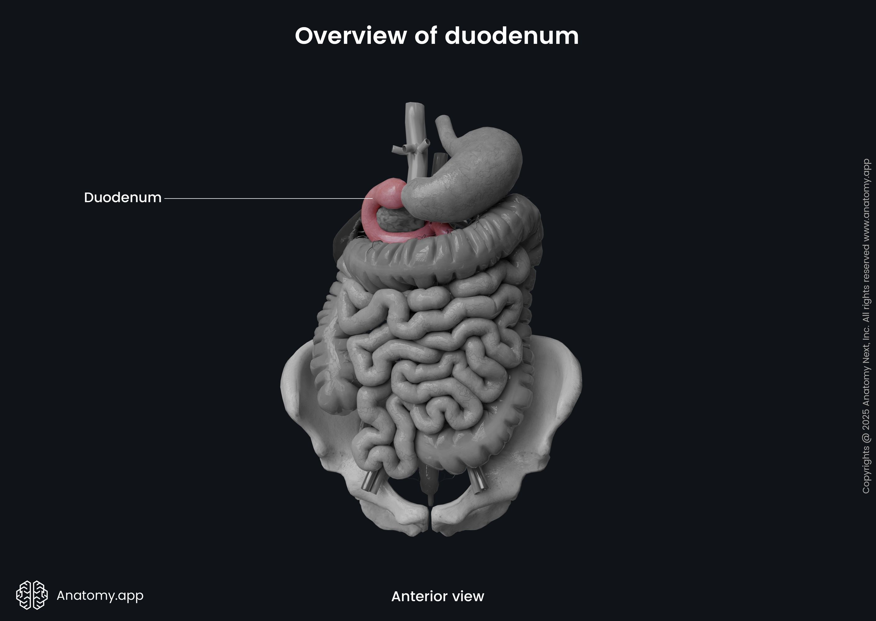
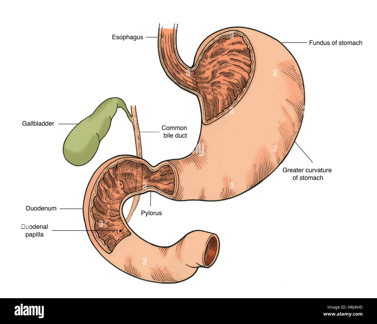
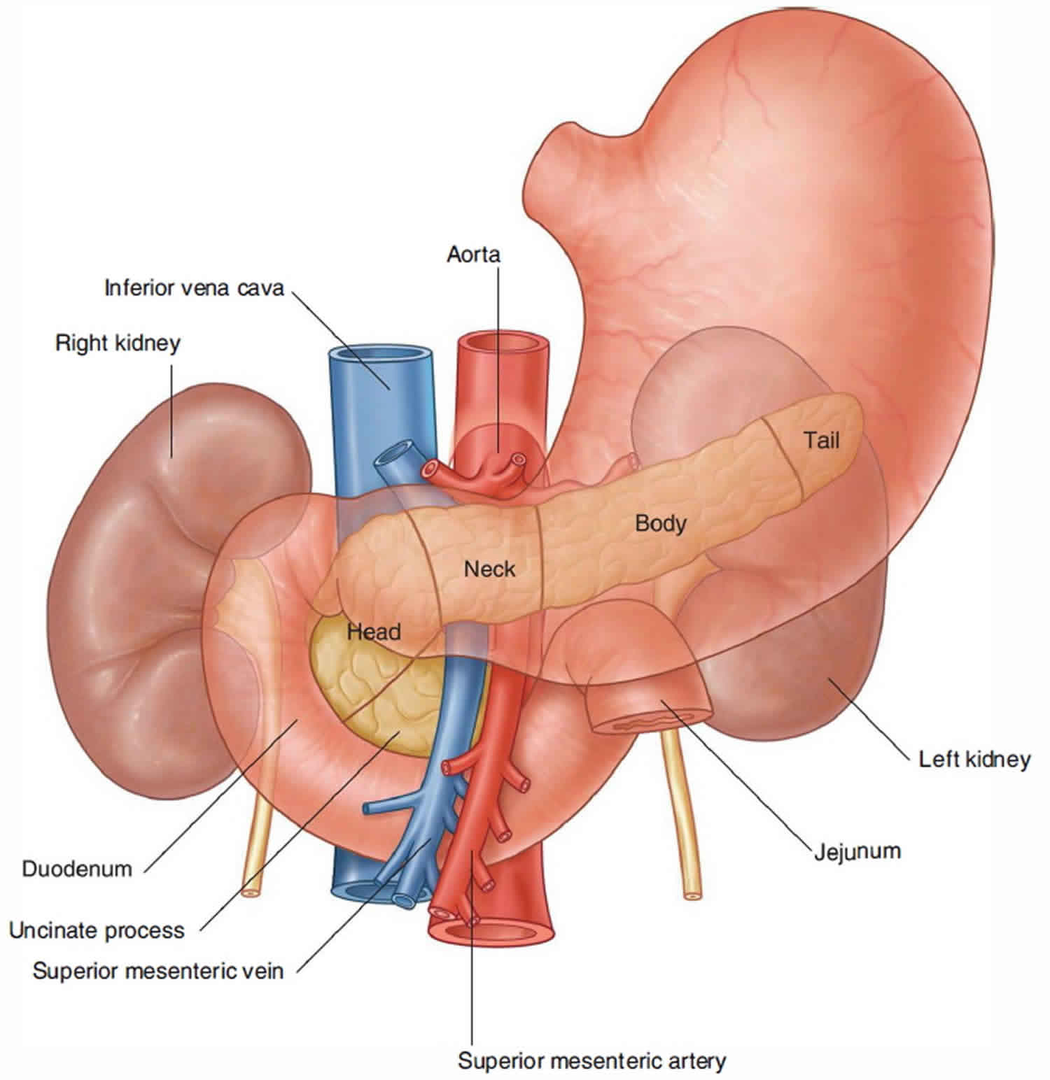
The duodenum is the first and shortest segment of the small intestine, forming a C-shaped curve that encircles the head of the pancreas. Its strategic position between the stomach and jejunum makes it a critical junction for digestive and absorptive processes. Structurally, the duodenum is divided into four distinct segments, each with specialized functions and anatomical landmarks. Understanding its precise location and segmentation is essential for clinical assessments, surgical planning, and diagnosing gastrointestinal disorders.The duodenum begins at the pyloric sphincter, where it receives chyme from the stomach, and ends at the duodenojejunal flexure, marking the transition to the jejunum. Its retroperitoneal position (except for the duodenal bulb) allows it to maintain close anatomical relationships with the liver, gallbladder, pancreas, and major blood vessels such as the gastroduodenal artery and superior mesenteric vessels.
Segmentation and Structural Features of the Duodenum
The duodenum is anatomically divided into four segments, each characterized by unique structural adaptations and functional roles. These segments—duodenal bulb (first part), descending (second part), horizontal (third part), and ascending (fourth part)—reflect its dual role in digestion and nutrient absorption.The duodenum’s curvature and sphincters (e.g., pyloric, hepatopancreatic) regulate the flow of bile, pancreatic juices, and chyme, ensuring efficient enzymatic digestion.Step-by-step breakdown of the four segments:
1. Duodenal Bulb (First Part)
2. Descending Duodenum (Second Part)
3. Horizontal Duodenum (Third Part)
4. Ascending Duodenum (Fourth Part)
Duodenal Dimensions Across Age Groups
The duodenum undergoes significant morphological changes from infancy to old age, influencing its functional capacity and susceptibility to pathological conditions. Below is a comparative table of its length, diameter, and key anatomical landmarks across three age groups, based on anatomical studies and radiographic data.| Parameter | Infant (0–2 years) | Adult (18–65 years) | Elderly (≥65 years) |
|---|---|---|---|
| Total Length | 10–12 cm (proportional to body size) | 25–30 cm (average 25 cm) | 20–28 cm (slight atrophy with age) |
| Diameter | 1.5–2.5 cm (wider relative to body) | 3–5 cm (varies by segment; bulb ~4 cm, descending ~3 cm) | 2.5–4 cm (possible dilation due to motility disorders) |
| Duodenal Bulb | Intraperitoneal, mobile | Retroperitoneal, fixed | May exhibit mucosal thinning |
| Major Duodenal Papilla | Located ~2–3 cm distal to pylorus | ~7–10 cm distal to pylorus (variable) | May show calcification (e.g., chronic pancreatitis) |
| Ligament of Treitz | High and mobile (easier intussusception risk) | Fixed at L2 vertebral level | May weaken, contributing to gastroparesis |
Age-related changes in the duodenum include mucosal thinning, reduced Brunner’s gland activity, and altered sphincter competence, increasing susceptibility to reflux, ulcers, and malabsorption syndromes.
Visualization of Duodenal Anatomy: Key Features for Illustration
To accurately depict the duodenum’s three-dimensional relationships, an illustration should emphasize the following elements:1. Curvature and Orientation
2. Sphincters and Orifices
3. Major Blood Vessels and Nerve Supply
4. Adjacent Organs and Landmarks
Physiological Functions and Role in Digestion of the Duodenum
The duodenum serves as the primary site for chemical digestion and initial nutrient absorption in the gastrointestinal (GI) tract. Its strategic location at the junction of the stomach and jejunum allows it to integrate gastric chyme with pancreatic, biliary, and duodenal secretions, creating an optimal environment for enzymatic breakdown. Beyond its digestive functions, the duodenum acts as an endocrine regulator through hormone secretion, coordinating pancreatic and hepatic responses to ingested nutrients. Its absorptive capabilities, though limited compared to downstream segments, play a critical role in the uptake of essential minerals and vitamins.The duodenum’s physiological role extends beyond mere transit, functioning as a highly specialized segment that ensures chyme undergoes controlled neutralization, enzymatic activation, and partial nutrient absorption before progressing to the jejunum. Its structural adaptations, including Brunner’s glands and a rich vascular network, support these processes while protecting the intestinal mucosa from acidic and enzymatic damage.
Chemical and Mechanical Processing of Chyme
The duodenum initiates the final stages of gastric chyme processing through a combination of chemical and mechanical mechanisms. Chemical digestion begins with the neutralization of acidic chyme (pH ~2–4) entering from the stomach, a process facilitated by bicarbonate-rich secretions from Brunner’s glands and pancreatic juice. This neutralization is critical to prevent mucosal damage and optimize the activity of pancreatic enzymes, which operate efficiently at a near-neutral pH (6.5–7.5). Key enzymes introduced in the duodenum include:Mechanically, the duodenum employs segmentation movements—rhythmic contractions of circular and longitudinal muscle layers—to mix chyme with digestive juices thoroughly. These contractions, regulated by the enteric nervous system, ensure uniform exposure of nutrients to enzymatic action while gradually propelling the mixture toward the jejunum. The duodenal migrating motor complex (MMC) further aids in clearing residual debris between meals, preventing bacterial overgrowth.
Hormonal Regulation of Digestive Secretions
The duodenum functions as an endocrine organ, secreting hormones that modulate pancreatic, biliary, and gastric activity in response to chyme composition. Three primary hormones—secretin, cholecystokinin (CCK), and gastrin—mediate these responses through feedback mechanisms:- Secretin is released in response to acidic chyme (pH < 4.5) and stimulates the pancreas to secrete bicarbonate-rich fluid, neutralizing duodenal pH. It also reduces gastric emptying to slow chyme delivery, allowing adequate neutralization.
These hormones create a closed-loop regulation system, ensuring digestive secretions are matched to the nutritional content of chyme. For example, a high-fat meal elicits robust CCK release, enhancing lipase and bile secretion, while acidic chyme induces secretin to balance pH.
Comparative Absorptive Capabilities of the Duodenum, Jejunum, and Ileum
The duodenum’s absorptive surface, though less extensive than the jejunum or ileum, specializes in the uptake of water-soluble vitamins, minerals, and iron, with limited absorption of carbohydrates and proteins. Key differences in absorptive function across intestinal segments are summarized below:The duodenum absorbs iron (via DMT1 and ferroportin), calcium (active transport via TRPV6 and calbindin), and folate (via proton-coupled folate transporter, PCFT), whereas the jejunum and ileum prioritize glucose/galactose (SGLT1), amino acids (PEPT1, B⁰AT1), and bile salts (ileal bile acid transporter, IBAT). The ileum uniquely reclaims vitamin B12 (intrinsic factor-mediated) and bile acids, while the jejunum handles the bulk of nutrient absorption due to its longer villi and greater surface area.Key absorptive distinctions:
| Nutrient/Mineral | Duodenum | Jejunum | Ileum |
|---|---|---|---|
| Iron | Primary site for non-heme iron uptake (ferric reduction by DcytB). | Limited absorption. | Minimal role. |
| Calcium | Active transport (1,25(OH)₂D-dependent). | Continues absorption. | Reabsorbs residual calcium. |
| Folate | PCFT-mediated uptake. | Additional absorption. | Minimal contribution. |
| Bile Acids | Negligible reabsorption. | Minimal uptake. | Primary site (IBAT-mediated). |
| Vitamin B12 | No intrinsic factor binding. | No intrinsic factor binding. | Cubilin-mediated uptake. |
| Glucose/Amino Acids | Minimal absorption. | Primary site (high SGLT1/PEPT1 activity). | Secondary absorption. |
Integration of Bile and Pancreatic Juices with Chyme
The duodenum orchestrates the synergistic action of bile and pancreatic juices to achieve efficient digestion and nutrient solubilization. This process unfolds in a stepwise, chemically regulated sequence:1. Chyme Entry and pH Neutralization
Acidic chyme (pH ~2) triggers secretin release, prompting pancreatic ductal cells to secrete ~100 mEq/L bicarbonate (via CFTR channels). Brunner’s glands contribute additional bicarbonate, raising duodenal pH to 6.5–7.5 within minutes.
2. Bile Emulsification and Micelle Formation
Bile, stored in the gallbladder and released upon CCK stimulation, contains bile salts (sodium taurocholate, glycocholate), phospholipids (lecithin), and cholesterol. These components:
3. Pancreatic Enzyme Activation and Substrate Breakdown
Pancreatic juice, rich in trypsinogen, chymotrypsinogen, and procarboxypeptidase, is activated by enterokinase (on duodenal brush border), converting trypsinogen to trypsin (a serine protease). Trypsin then autoactivates other zymogens and cleaves proteins into peptides. Concurrently:
4. Nutrient Solubilization and Transport Preparation
The resulting digestion products (fatty acids, monoglycerides, peptides, disaccharides) are incorporated into mixed micelles, which diffuse to the brush border membrane. Here, specific transporters facilitate uptake:
5. Residual Debris Propulsion
Unabsorbed chyme, now enriched with bile salts and digestive byproducts, is gradually propelled into the jejunum by peristalt
Clinical Significance and Common Pathologies of the Duodenum
The duodenum, as the first segment of the small intestine, plays a critical role in digestion and nutrient absorption, making it susceptible to a range of pathological conditions. Disorders affecting this region often arise from infectious agents, autoimmune processes, congenital abnormalities, or iatrogenic factors such as medication use. Understanding these pathologies—including their etiologies, clinical manifestations, and diagnostic approaches—is essential for accurate diagnosis and targeted management. This section examines the most prevalent duodenal disorders, their underlying mechanisms, associated symptoms, and the diagnostic modalities employed to identify them.Prevalent Duodenal Disorders and Their Etiologies
The duodenum is commonly affected by inflammatory, ulcerative, obstructive, and neoplastic conditions, each with distinct pathophysiological origins. Below are the most frequently encountered disorders, categorized by their primary mechanism of injury:Duodenal pathologies often present with overlapping symptoms, necessitating a systematic diagnostic approach to differentiate between conditions such as peptic ulcer disease, Crohn’s disease, or malignancy.
- Crohn’s Disease
- Duodenal Atresia and Stenosis
- Duodenal Neoplasms
- Duodenitis
- Duodenal Diverticula
- Duodenal Varices
Symptomatic Presentation and Diagnostic Correlation
Duodenal pathologies manifest with a spectrum of symptoms that overlap across conditions, necessitating a structured approach to correlate clinical findings with diagnostic tests. The table below summarizes common symptoms, their likely associated disorders, and recommended diagnostic methods:| Symptom | Likely Condition | Diagnostic Method |
|---|---|---|
| Epigastric pain (burning/gnawing, relieved by food) | Duodenal ulcer (PUD) | Upper endoscopy with biopsy, H. pylori testing (urease breath test, stool antigen, serology), NSAID history review. |
| Chronic diarrhea, weight loss, abdominal cramps | Crohn’s disease (duodenal involvement) | Colonoscopy with biopsy (granulomas), MRI enterography, fecal calprotectin. |
| Projectile, bilious vomiting in neonates | Duodenal atresia/stenosis | Upper GI contrast study (microcolon sign), prenatal ultrasound findings, surgical consultation. |
| Painless jaundice, dark urine, pale stools | Duodenal/periampullary adenocarcinoma | Endoscopic retrograde cholangiopancreatography (ERCP), CT/MRI with contrast, tumor marker (CA 19-9). |
| Hematemesis or melena (coffee-ground emesis) | Duodenal ulcer, varices, or neoplasm | Emergency endoscopy, cross-sectional imaging (CT angiography for varices), H. pylori testing. |
| Postprandial fullness, early satiety | Duodenal obstruction (stenosis, tumor) | Upper GI series with small-bowel follow-through, endoscopy with biopsy. |
| Iron-deficiency anemia (microcytic) without menorrhagia | Overt or occult duodenal bleeding (ulcer, angiodysplasia) | Capsule endoscopy, double-balloon enteroscopy, technetium-99m red blood cell scan. |
| Recurrent abdominal pain with food intolerance | Eosinophilic duodenitis or celiac disease | Endoscopy with biopsy (eosinophil count, villous atrophy), serum IgA tTG antibodies. |
Symptom duration and response to therapy (e.g., proton pump inhibitors for ulcers) guide further investigation. For example, persistent symptoms despite acid suppression warrant endoscopy to rule out malignancy or Crohn’s disease.
Diagnostic Procedures for Duodenal Pathologies
Accurate diagnosis of duodenal disorders relies on a combination of endoscopic, histological, and radiologic techniques, each with specific indications and limitations. Below are the primary diagnostic modalities, their procedural steps, and key considerations:Diagnostic accuracy depends on the timing of tests (e.g., H. pylori eradication therapy may falsely negate serological tests) and the expertise of the operator (e.g., deep duodenal biopsies require advanced endoscopic techniques).
- Biopsy and Histopathology
Duodenal Histology and Microscopic Features
The duodenum exhibits a complex histological architecture tailored to its dual roles in digestion and protection against acidic chyme from the stomach. Its wall comprises four primary layers—mucosa, submucosa, muscularis externa, and serosa—each with specialized structures that facilitate nutrient absorption, secretion, and mechanical processing. The duodenal mucosa, in particular, distinguishes itself through unique adaptations, including Brunner’s glands, villi, and crypts of Lieberkühn, which collectively optimize digestive efficiency while safeguarding the intestinal epithelium.Histological Layers of the Duodenal Wall
The duodenal wall follows the standard gastrointestinal tract (GIT) layering but incorporates modifications that reflect its transitional role between the stomach and small intestine. The mucosa is the innermost layer, characterized by a simple columnar epithelium with villi and crypts, underlain by the lamina propria and muscularis mucosae. Below this lies the submucosa, a dense connective tissue layer housing Brunner’s glands, blood vessels, and nerves. The muscularis externa consists of an inner circular and outer longitudinal muscle layer, responsible for segmentation and peristalsis, while the serosa (or adventitia in retroperitoneal segments) provides structural support and lubrication.The mucosa is the most functionally critical layer, featuring:
Comparison of Duodenal, Stomach, and Jejunal Mucosa
The duodenal mucosa differs markedly from the stomach and jejunum in epithelial composition, glandular secretions, and vascular patterns. Below is a comparative table highlighting these distinctions:| Feature | Duodenum | Stomach | Jejunum |
|---|---|---|---|
| Epithelial Cell Types |
|
|
|
| Glandular Secretions |
|
|
|
| Vascular Patterns |
|
|
|
| Protective Mechanisms | Brunner’s glands secrete alkaline mucus to neutralize gastric acid, while villi and crypts maintain rapid epithelial turnover (~3–5 days). |
Mucous bicarbonate barrier and tight junctions prevent autodigestion; parietal cells secrete HCl (pH 1.5–3.5). |
Goblet cells produce mucus; rapid cell turnover and Peyer’s patches enhance immune defense. |
Role of Brunner’s Glands in Alkaline Mucus Production
Brunner’s glands, exclusive to the duodenum, are compound tubuloacinar glands located in the submucosa, primarily concentrated in the duodenal bulb and proximal duodenum. Their secretions consist of:Mechanism of Action:
1. Stimulation: Acidic chyme entering the duodenum triggers secretin release from S-cells in the mucosa, which binds to Brunner’s gland cells via G-protein-coupled receptors, activating adenylate cyclase and increasing cAMP.
2. Secretion: Chloride ions are exchanged for bicarbonate via CFTR channels, while mucus glycoproteins are exocytosed into the duodenal lumen.
3. Protection: The alkaline mucus forms a neutralizing barrier, preventing pepsin-mediated damage and maintaining an optimal pH (6.0–7.0) for pancreatic enzymes (e.g., trypsin, amylase).
Clinical Relevance:
Microscopic Structure of Duodenal Villi and Cellular Composition
Duodenal villi are leaf-shaped or finger-like projections (1–1.6 mm tall) that maximize surface area for digestion and absorption. Their structure includes:-
Duodenal Surgery and Interventions
The duodenum, as a critical segment of the upper gastrointestinal tract, frequently requires surgical intervention due to obstructive pathologies, traumatic perforations, or neoplastic growths. Advances in endoscopic and laparoscopic techniques have expanded treatment options, allowing for less invasive procedures with improved patient outcomes. Surgical approaches range from minimally invasive endoluminal stenting to complex resections like the Whipple procedure, each tailored to the underlying pathology and patient-specific factors. This section explores the surgical strategies for duodenal obstructions, perforations, and malignancies, compares minimally invasive versus open techniques, and outlines procedural protocols for upper gastrointestinal endoscopy with biopsy. Additionally, it addresses post-operative complications and their management, emphasizing evidence-based protocols to optimize recovery and minimize morbidity.Surgical Approaches for Duodenal Obstructions, Perforations, and Malignancies
Duodenal obstructions often result from neoplastic processes (e.g., pancreatic head adenocarcinoma, duodenal adenocarcinoma), strictures (post-inflammatory or post-surgical), or external compression (e.g., chronic pancreatitis). Perforations may arise from peptic ulcer disease, trauma, or iatrogenic injuries, while malignancies necessitate resection with negative margins. The choice of surgical intervention depends on the etiology, anatomical involvement, and patient’s overall health.Endoluminal Stenting
For malignant obstructions or high-risk surgical candidates, self-expandable metal stents (SEMS) are deployed via endoscopy to restore luminal patency. Stent selection depends on lesion length and location:
1. Pre-operative assessment: CT enterography or MRI to evaluate tumor extent and vascular involvement.
2. Endoscopic placement: Under fluoroscopic guidance, the stent is advanced across the obstruction using a guidewire.
3. Post-procedure monitoring: Contrast studies to confirm patency; stent patency typically lasts 3–6 months for malignant cases.
Whipple Procedure (Pancreaticoduodenectomy)
Indicated for duodenal adenocarcinoma, periampullary tumors, or chronic pancreatitis with duodenal involvement, this procedure involves resection of the distal stomach, duodenum, pancreatic head, gallbladder, and common bile duct, followed by reconstructive anastomoses. Key variations include:
1. Kocher maneuver: Mobilization of the duodenum and head of the pancreas.
2. Resection margins: En bloc removal of the specimen with negative margins (≥1 cm for adenocarcinoma).
3. Reconstruction: End-to-side pancreaticojejunostomy, hepaticojejunostomy, and gastrojejunostomy (or duodenojejunostomy in pylorus-preserving variants).
Duodenal Resection for Benign Pathologies
For traumatic perforations or benign strictures, segmental duodenal resection with primary anastomosis is performed. In cases of extensive damage (e.g., gunshot wounds), diversion procedures (e.g., gastrojejunostomy) may be necessary to bypass the injured segment.
Comparative Analysis: Minimally Invasive vs. Open Surgical Techniques for Duodenal Resections
The advent of laparoscopic and robotic-assisted surgery has reduced the invasiveness of duodenal resections, though open techniques remain standard for complex cases. Below is a comparative analysis of laparoscopic, robotic, and open approaches based on clinical outcomes and patient recovery profiles.| Parameter | Laparoscopic Duodenal Resection | Robotic-Assisted Duodenal Resection | Open Duodenal Resection |
|---|---|---|---|
| Indications | Early-stage duodenal adenocarcinoma, benign strictures, select pancreatic head tumors. | Complex pancreaticoduodenectomies, locally advanced tumors, or cases requiring precise vascular dissection. | Advanced malignancies, extensive vascular involvement, or prior abdominal surgeries with adhesions. |
| Surgical Risks |
|
|
|
| Recovery Time | Hospital stay: 5–7 days; return to normal activity: 3–4 weeks. | Hospital stay: 7–10 days; return to normal activity: 4–6 weeks (due to longer operative time). | Hospital stay: 10–14 days; return to normal activity: 6–8 weeks. |
| Success Rates (R0 Resection) | 80–85% for early-stage duodenal cancer. | 85–90% for pancreaticoduodenectomy (comparable to open). | 85–90% for experienced surgeons; higher in high-volume centers. |
| Cost Considerations | $15,000–$25,000 (excluding robotic equipment). | $30,000–$50,000 (robotic platform adds ~$10,000–$20,000). | $20,000–$40,000 (varies by complexity and complications). |
Step-by-Step Guide for Upper Gastrointestinal Endoscopy with Duodenal Biopsy
Endoscopic biopsy of the duodenum is essential for diagnosing malignancies, inflammatory conditions (e.g., celiac disease), or infectious processes (e.g., Helicobacter pylori). The procedure requires sterile technique, precise instrument handling, and patient sedation to ensure safety and diagnostic accuracy.Equipment Required:
Patient Positioning and Preparation:
Procedure Steps:
1. Endoscope Insertion:
The duodenum exemplifies the precision of human anatomy and physiology, where structural intricacy and functional specialization converge to sustain digestive efficiency. From its strategic location as the stomach’s exit point to its role in activating pancreatic enzymes and absorbing vital minerals, this segment of the small intestine embodies a harmonious interplay of mechanical and chemical processes. Clinical awareness of its vulnerabilities—whether due to bacterial infections, autoimmune disorders, or iatrogenic injuries—highlights the necessity of early diagnosis and targeted interventions, from proton pump inhibitors to advanced surgical techniques. As research continues to unravel its microscopic features and hormonal interactions, the duodenum remains a focal point for both medical education and therapeutic innovation, reinforcing its status as a linchpin of gastrointestinal function.
FAQ
what is the duodenum in the body?
Q: What exactly is the duodenum in the human body?
what is the duodenum function?
Q: What is the main function of the duodenum?
what is the duodenum and what does it do?
Q: What is the duodenum, and what does it do in digestion?
what is the duodenum part of?
Q: What part of the digestive system is the duodenum?
what is the duodenum jejunum and ileum?
Q: What are the differences between the duodenum, jejunum, and ileum?
what is the duodenum bulb?
Q: What is the duodenum bulb, and where is it located?
Leave a Comment
Comments are moderated before appearing. The data you submit is processed according to the Privacy Policy of Voltefac.