What Heart Rate Is Too Low Understanding Medical Thresholds And Risks
Table of Contents
- Physiological Ranges and Clinical Thresholds for Low Heart Rates
- Normal Resting Heart Rate Ranges by Age and Fitness Level
- Medical Definition and Classification of Bradycardia
- Clinical Guidelines on Intervention for Low Heart Rates
- Causes of Low Heart Rate: Medical and Lifestyle Factors
- Medical Causes of Low Heart Rate by System
- Lifestyle and Environmental Factors Contributing to Low Heart Rate
- Symptoms and When to Seek Medical Attention in Low Heart Rate Conditions
- Symptom Checklist for Low Heart Rate: Severity, Urgency, and Associated Heart Rate Ranges
- Asymptomatic Bradycardia: Harmless vs. Silent Pathology
- FAQ
- What heart rate is considered too low while you're sleeping?
- What heart rate is too low when you're at rest?
- What heart rate is too low while taking metoprolol?
- What heart rate is too low at night?
- What heart rate is too low for an athlete?
- What heart rate is too low for a baby?
A low heart rate can signal optimal cardiovascular health in trained athletes or pose serious risks in vulnerable populations, blurring the line between physiological adaptation and pathological concern. Understanding the spectrum of normal resting heart rates—ranging from 60 to 100 beats per minute (bpm) in adults—requires contextual awareness of age, fitness level, and underlying medical conditions. While bradycardia, defined as a resting heart rate below 60 bpm, may be benign in endurance athletes, it can also indicate life-threatening conditions such as heart block or electrolyte imbalances when accompanied by symptoms like dizziness, fatigue, or fainting. This exploration dissects the medical thresholds, causal mechanisms, and diagnostic nuances separating harmless bradycardia from emergencies requiring immediate intervention.
The interplay between medical and lifestyle factors further complicates the assessment, as chronic conditions like hypothyroidism or medications such as beta-blockers may suppress heart rate, while dehydration or sleep apnea can trigger transient episodes. Athletes often exhibit physiological bradycardia due to enhanced stroke volume, whereas pathological bradycardia in elderly patients may stem from degenerative conduction system disorders. This guide provides structured frameworks—including comparative tables, symptom checklists, and self-monitoring protocols—to empower individuals in recognizing when a low heart rate warrants medical evaluation versus when it reflects a natural adaptation to training or aging.
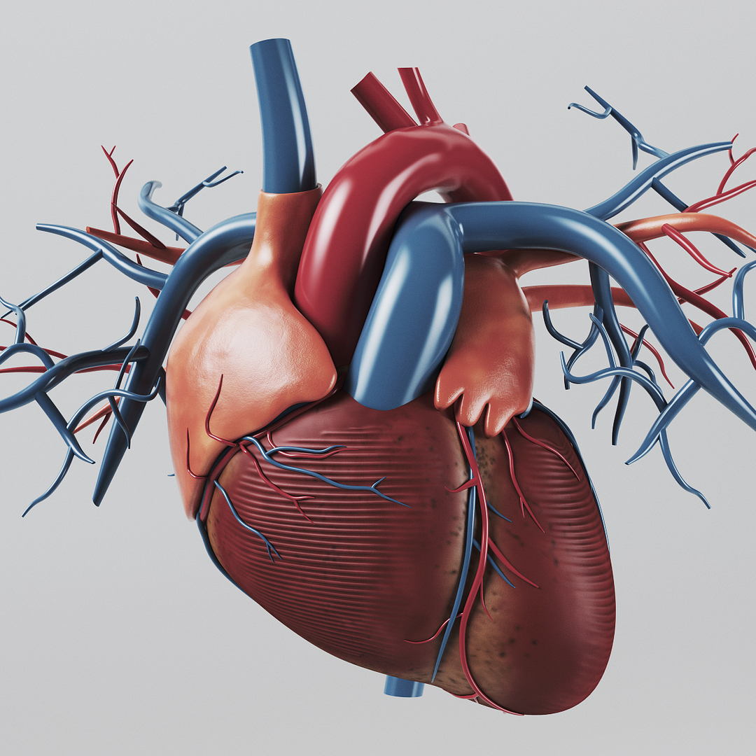
Physiological Ranges and Clinical Thresholds for Low Heart Rates
Heart rate (HR) reflects the rhythmic contractions of the heart, serving as a critical indicator of cardiovascular health. While individual variations exist, deviations from expected ranges—particularly abnormally low rates—may signal underlying physiological adaptations or pathological conditions. Understanding these thresholds across demographics is essential for accurate diagnosis, risk stratification, and timely intervention. This section delineates the normal and bradycardic ranges for adults, children, and athletes, alongside the medical definitions and clinical implications of chronic low heart rates.
Normal Resting Heart Rate Ranges by Age and Fitness Level
Resting heart rate (RHR) varies significantly with age, physical conditioning, and health status. Athletes, for instance, often exhibit lower RHRs due to enhanced cardiac efficiency, whereas infants and young children have higher baseline rates to meet metabolic demands. Below is a comparative table summarizing average resting heart rates across key demographic groups, including thresholds for potential bradycardia.
| Age Group | Normal Resting Heart Rate (bpm) | Low Threshold (bpm) | Potential Causes of Low Rates |
|---|---|---|---|
| Newborns (0–1 month) | 70–190 | <70 (persistent) | Congenital heart defects, hypoxia, maternal medications, or transient sinus bradycardia. |
| Infants (1–12 months) | 80–160 | <80 (with symptoms) | Infections (e.g., sepsis), metabolic disorders, or increased intracranial pressure. |
| Toddlers (1–2 years) | 80–130 | <70 | Hypothyroidism, sleep apnea, or vagal tone elevation. |
| Children (3–10 years) | 70–110 | <60 (with symptoms) | Athletic training, congenital conduction delays, or medication side effects (e.g., beta-blockers). |
| Adolescents (11–18 years) | 60–100 | <50 (with symptoms) | Genetic predisposition, sleep disorders, or autonomic dysfunction. |
| Adults (18–65 years) | 60–100 | <60 (symptomatic) or <50 (asymptomatic in athletes) | Sinus bradycardia, sick sinus syndrome, or electrolyte imbalances (e.g., hyperkalemia). |
| Seniors (>65 years) | 60–100 (may trend higher due to reduced cardiac reserve) | <50 (with symptoms) or <40 (pathological) | Cardiac fibrosis, medications (e.g., digoxin), or degenerative conduction system diseases. |
| Endurance Athletes (trained) | 40–60 (RHR) | <40 (rare, may indicate excessive training or pathology) | Physiological adaptation (e.g., "athlete’s heart"), but risk of arrhythmias or cardiac remodeling. |
Key Observations:
Medical Definition and Classification of Bradycardia
Bradycardia is defined as a resting heart rate below the lower limit of normal for age, typically accompanied by symptoms or signs of hemodynamic compromise. The distinction between sinus bradycardia and pathological bradycardia hinges on underlying mechanisms, patient context, and clinical presentation.- Sinus Bradycardia:
A slow but regular heart rhythm originating from the sinoatrial (SA) node, often benign in athletes or during sleep. Causes include:
- Pathological Bradycardia:
Reflects abnormal conduction or primary cardiac disease, such as:
Bradycardic Thresholds by Population:
Clinical Guidelines on Intervention for Low Heart Rates
Medical societies provide structured criteria for identifying bradycardia requiring intervention. The American Heart Association (AHA) and European Society of Cardiology (ESC) emphasize symptom correlation and underlying etiology in decision-making. Below are key guidelines:"Bradycardia necessitating intervention is defined by the presence of symptoms (e.g., syncope, dyspnea, chest pain, or hypotension) or evidence of hemodynamic instability, rather than heart rate alone. Asymptomatic bradycardia in athletes or trained individuals may not require treatment unless associated with structural heart disease or exercise intolerance. For pathological bradycardia:Additional Considerations:
Adults: Persistent HR <50 bpm with symptoms or <40 bpm (regardless of symptoms) warrants pacemaker evaluation. Pediatrics: HR <60 bpm in infants or <50 bpm in children with symptoms (e.g., poor feeding, lethargy) requires urgent cardiac assessment. High-Degree AV Block: Second-degree Type II or third-degree AV block always requires pacemaker therapy, even in asymptomatic patients. Drug-Induced Bradycardia: Medication adjustments (e.g., reducing beta-blocker doses) or temporary pacing may be necessary." —Adapted from AHA/ACC/HRS 2022 Guidelines for Cardiovascular Disease in the Young and ESC 2021 Guidelines on Cardiac Pacing and Resynchronization Therapy.
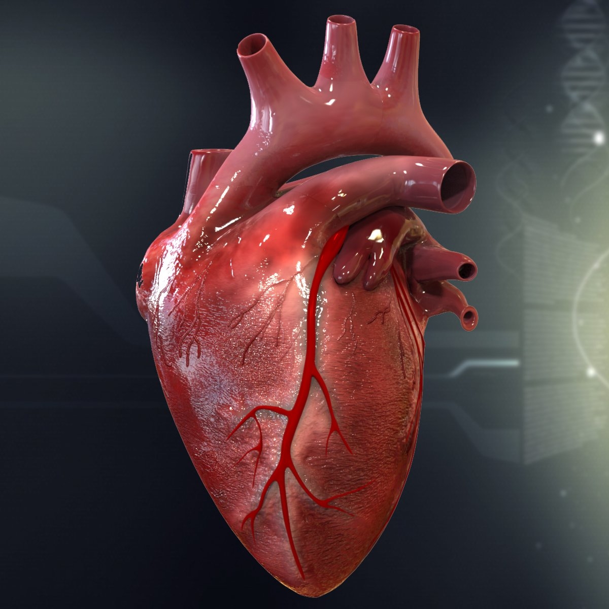
Causes of Low Heart Rate: Medical and Lifestyle Factors
A low heart rate, or bradycardia, arises from a complex interplay of medical conditions, physiological adaptations, and external influences. While some causes are benign or even adaptive (e.g., athletic training), others reflect underlying pathologies requiring immediate intervention. Understanding the systemic origins—ranging from cardiovascular disorders to metabolic imbalances—and the reversible versus chronic nature of lifestyle-related factors is critical for accurate diagnosis and management. This section categorizes medical etiologies by anatomical or functional systems, examines the impact of lifestyle and environmental triggers, and distinguishes between physiological and pathological bradycardia through structured comparisons.Medical Causes of Low Heart Rate by System
Low heart rates often stem from disruptions in cardiac conduction, autonomic nervous system dysregulation, or systemic diseases affecting cardiac function. Below is a categorized breakdown of medical causes, emphasizing their mechanistic pathways and clinical relevance.Cardiovascular System:
- Structural Heart Disease
Neurological System:
- Central Nervous System Pathologies
Endocrine and Metabolic Disorders:
Infectious and Inflammatory Causes:
Genetic and Congenital Disorders:
Neoplastic and Toxic Causes:
Lifestyle and Environmental Factors Contributing to Low Heart Rate
Lifestyle and environmental exposures can modulate heart rate through autonomic adaptations, metabolic shifts, or direct toxic effects. These factors are further classified as reversible (temporary or treatable) or chronic (persistent or progressive), with implications for management strategies.Reversible Causes:
- Dehydration and Hypovolemia:
- Sleep Apnea:
- Medications:
Chronic Causes:
- Chronic Alcohol Use:
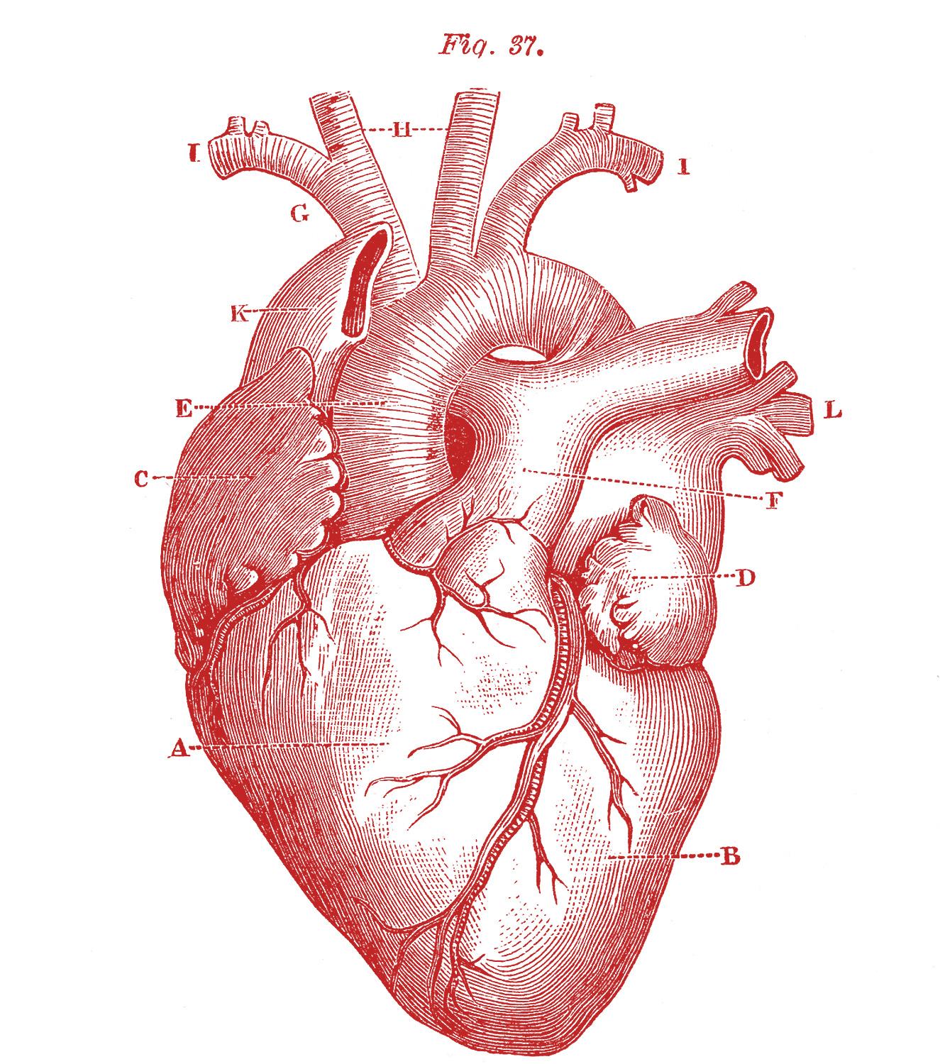
Symptoms and When to Seek Medical Attention in Low Heart Rate Conditions
A low heart rate (bradycardia) may present with a spectrum of symptoms ranging from mild discomfort to life-threatening complications, depending on underlying causes and individual physiology. While some individuals—particularly trained athletes—experience asymptomatic bradycardia without adverse effects, others may exhibit subtle or pronounced signs that necessitate urgent medical evaluation. Recognizing symptom patterns, severity, and associated heart rate ranges is critical for differentiating benign conditions from those requiring intervention. This section provides a structured symptom checklist, guidelines for self-monitoring, and distinctions between harmless and clinically significant bradycardia, including scenarios where silent bradycardia may indicate serious underlying pathology.Symptom Checklist for Low Heart Rate: Severity, Urgency, and Associated Heart Rate Ranges
The following table categorizes common symptoms of bradycardia by severity, urgency for medical attention, and typical heart rate ranges where they may manifest. Symptoms are not exclusive to bradycardia and may overlap with other conditions, but their presence—especially in conjunction with a slow pulse—warrants further assessment.| Symptom | Possible Cause | Red Flag (Requires Immediate Care?) | Associated Heart Rate Range (bpm) |
|---|---|---|---|
| Dizziness or lightheadedness (especially upon standing) | Reduced cardiac output, hypotension, or autonomic dysfunction (e.g., carotid sinus hypersensitivity). | Yes, if accompanied by syncope or heart rate <50 bpm with symptoms. | 40–60 (may drop further with exertion or position change). |
| Fatigue or weakness (persistent, worsening over days/weeks) | Chronic hypoxia due to inadequate perfusion (e.g., heart block, sick sinus syndrome). | Yes, if fatigue is debilitating or associated with chest discomfort. | Typically <50 bpm at rest, may fluctuate. |
| Syncope (fainting or near-fainting) | Transient cerebral hypoperfusion (e.g., complete heart block, severe bradycardia-tachycardia syndrome). | Yes; syncope with bradycardia is a medical emergency. | Often <40 bpm during episodes (may be intermittent). |
| Shortness of breath (dyspnea) at rest or with minimal exertion | Pulmonary congestion due to reduced cardiac output (e.g., heart failure with bradycardia). | Yes, if accompanied by chest pain or orthopnea. | Varies; often <50 bpm with exertional symptoms. |
| Chest discomfort or pressure (may mimic angina) | Ischemic heart disease or reduced coronary perfusion (e.g., bradycardia-induced myocardial ischemia). | Yes; chest pain with bradycardia requires immediate evaluation. | Typically <50 bpm, but may occur at higher rates if symptomatic. |
| Confusion or cognitive impairment (e.g., memory lapses) | Chronic cerebral hypoperfusion (e.g., advanced conduction disease). | Yes, if progressive or associated with other red flags. | Often <40 bpm with persistent symptoms. |
| Palpitations (paradoxical sensation of "skipped" or "fluttering" beats) | Bradycardia-tachycardia syndrome (e.g., atrial fibrillation with slow ventricular response). | Yes, if palpitations alternate with severe bradycardia. | Ventricular rate <50 bpm during bradycardic phases. |
| Nausea or vomiting | Vasovagal response or severe hypotension (e.g., high-degree block). | Yes, if recurrent or associated with syncope. | Often <40 bpm during episodes. |
| Asymptomatic (no symptoms) | Physiologic (e.g., trained athletes) or pathologic (e.g., early sick sinus syndrome). | No, unless heart rate is <40 bpm at rest or drops significantly with exertion. | Varies; athletes may have <40 bpm at rest, while others may have <50 bpm without symptoms. |
Asymptomatic Bradycardia: Harmless vs. Silent Pathology
Asymptomatic bradycardia encompasses a broad spectrum, from benign adaptations in athletes to subclinical manifestations of serious cardiac disorders. Distinguishing between these scenarios requires clinical context, heart rate trends, and additional diagnostic testing.Physiologic Asymptomatic Bradycardia:
Pathologic Asymptomatic Bradycardia:
Silent bradycardia may indicate underlying conduction system disease or metabolic disorders that progress insidiously. Key scenarios include:
Diagnostic Caution:
Determining whether a low heart rate is a sign of exceptional cardiovascular efficiency or an early warning of systemic dysfunction hinges on a multifaceted approach: understanding demographic norms, identifying symptomatic red flags, and distinguishing between reversible lifestyle influences and chronic medical conditions. Asymptomatic bradycardia in athletes or well-conditioned individuals often requires no intervention, while persistent symptoms—such as syncope, chest pain, or cognitive impairment—demand prompt evaluation to rule out conditions like sick sinus syndrome or third-degree heart block. By leveraging structured diagnostic tools, including manual pulse assessment, wearable technology for trend tracking, and guideline-based thresholds from organizations like the American Heart Association (AHA), individuals can navigate the complexities of bradycardia with clarity. Ultimately, the key lies in balancing vigilance with proportional response, ensuring that medical attention is sought when necessary without dismissing the potential benefits of a naturally low heart rate in healthy populations.
FAQ
What heart rate is considered too low while you're sleeping?
A resting heart rate below 40 bpm during sleep may indicate bradycardia, especially if you feel dizzy, fatigued, or have other symptoms. For most adults, a nighttime rate under 50 bpm (without medical conditions like athlete’s heart) could warrant evaluation. Always consult a doctor if you experience symptoms like fainting or shortness of breath.
What heart rate is too low when you're at rest?
A resting heart rate below 60 bpm is generally considered bradycardia, but it’s only dangerous if it causes symptoms like fatigue, dizziness, or fainting. Some people (e.g., athletes) naturally have lower rates (40–50 bpm), but persistent rates under 40 bpm without explanation should be checked by a doctor.
What heart rate is too low while taking metoprolol?
Metoprolol can lower heart rate to below 50–60 bpm, but a rate under 40–45 bpm (or lower than your baseline) may be too low, especially if you feel lightheaded, weak, or have symptoms of low blood pressure. Always report rates below your doctor’s target range or severe symptoms immediately.
What heart rate is too low at night?
A nighttime heart rate consistently below 40 bpm (without a pacemaker or athlete’s heart) may signal bradycardia and should be evaluated. Rates between 40–50 bpm might be normal for some, but if paired with symptoms like confusion or chest discomfort, seek medical attention.
What heart rate is too low for an athlete?
Athletes often have resting rates as low as 30–40 bpm due to increased cardiac efficiency, but below 30 bpm (or a sudden drop) could indicate an issue like sick sinus syndrome. If symptoms like dizziness or exercise intolerance occur, consult a doctor—even elite athletes need monitoring.
What heart rate is too low for a baby?
A newborn’s heart rate below 60 bpm is emergency-level bradycardia and requires immediate medical attention. For infants (1–12 months), rates under 70–80 bpm may signal distress, while toddlers/children under 60–70 bpm should prompt evaluation. Always call emergency services if a baby’s HR drops suddenly.
Leave a Comment
Comments are moderated before appearing. The data you submit is processed according to the Privacy Policy of Voltefac.