What Is Fibrinogen Its Role Structure And Clinical Significance
Table of Contents
- Biological Role of Fibrinogen in Blood Coagulation and Hemostatic Function
- Structural Transformation and Conversion to Fibrin
- Interaction with Thrombin, Calcium Ions, and Clotting Factors
- Comparative Analysis: Fibrinogen in Hemostasis vs. Bleeding Disorders
- Structural Composition and Synthesis of Fibrinogen
- Molecular Structure of Fibrinogen
- Synthesis and Post-Translational Modifications
- Structural Differences Between Fibrinogen and Fibrin
- Genetic Mutations and Dysfibrinogenemias
- Clinical Significance and Diagnostic Methods in Fibrinogen Evaluation
- Diagnostic Criteria for Evaluating Fibrinogen Levels
- Comparative Analysis of Fibrinogen-Related Disorders
- Interpretation of Fibrinogen Degradation Products (FDPs) and D-Dimers
- Fibrinogen in Disease Pathophysiology
- Fibrinogen Polymorphisms and Cardiovascular Risk
- Differential Role in Venous vs. Arterial Thrombosis
- Pro-Inflammatory Properties of Fibrinogen
- Feedback Loops in Sepsis-Induced Coagulopathy
- Therapeutic Applications and Emerging Research in Fibrinogen Modulation
- Mechanisms and Clinical Use of Fibrinogen Concentrates
- Engineering Fibrinogen Variants for Enhanced Functionality
- Anti-Fibrinogen Therapies in Thrombosis and Cancer Metastasis
- FAQ
- What does fibrinogen mean when it appears on a blood test?
- What is the role of fibrinogen in the blood?
- What is a fibrinogen test and why is it done?
- What does fibrinogen activity refer to in medical terms?
- What does it mean if fibrinogen levels are high?
- What are normal fibrinogen levels in the blood?
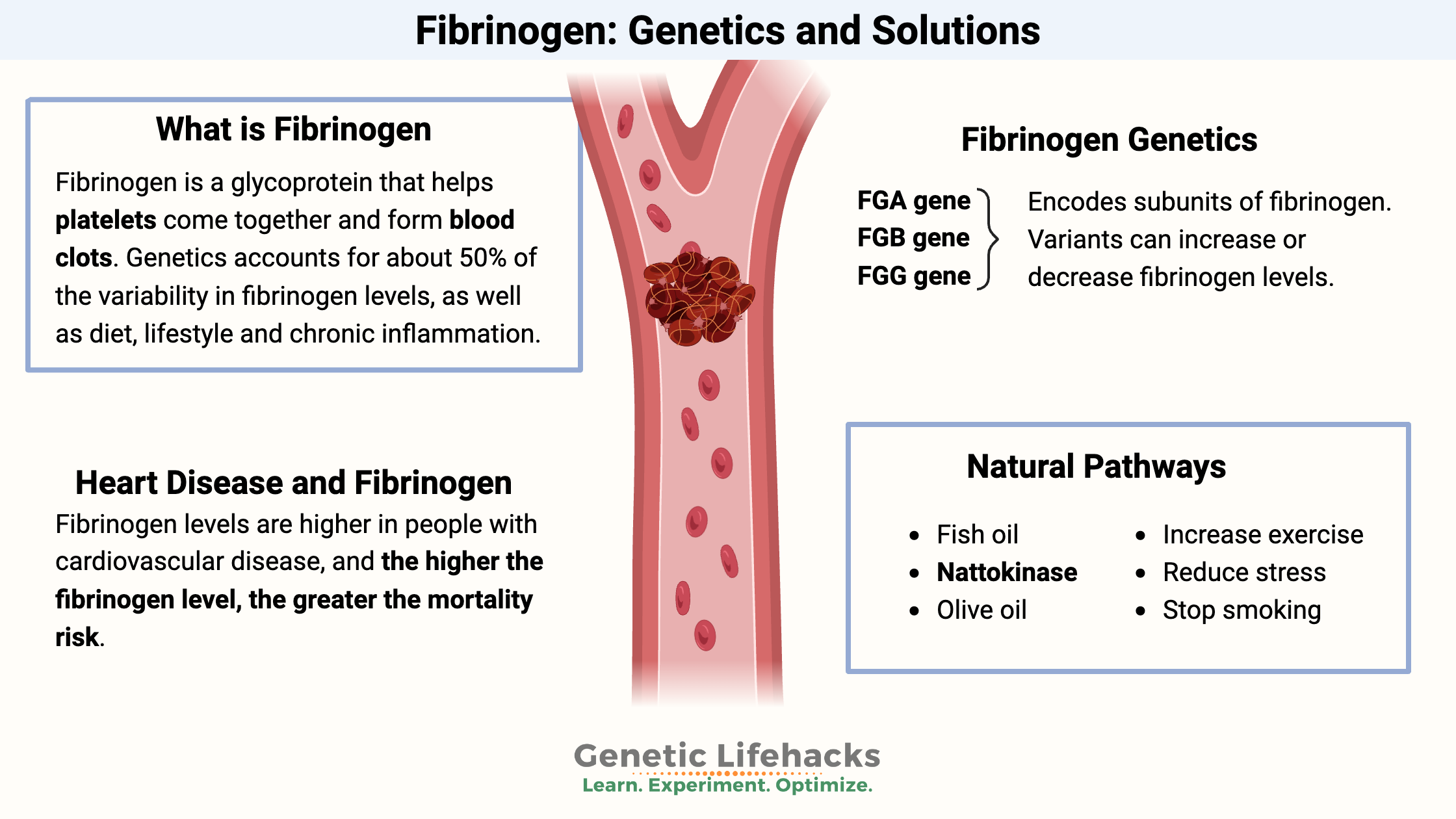
Fibrinogen, a soluble plasma glycoprotein, serves as the cornerstone of the blood coagulation cascade, orchestrating the transition from a fluid state to a stable clot essential for hemostasis. Beyond its structural role in clot formation, this multifunctional protein interacts dynamically with platelets, clotting factors, and inflammatory pathways, influencing both physiological hemostasis and pathological thrombotic events. Its synthesis in hepatocytes, regulated by precise genetic and post-translational mechanisms, underscores its systemic importance, while mutations or deficiencies in fibrinogen can precipitate bleeding disorders or heighten cardiovascular risks.
The biological conversion of fibrinogen into fibrin via thrombin-mediated cleavage exemplifies a finely tuned biochemical process critical for wound repair and vascular integrity. Comparative analyses reveal how dysregulated fibrinogen activity—whether through genetic variants, acquired deficiencies, or hypercoagulable states—contributes to diverse clinical syndromes, from congenital dysfibrinogenemias to disseminated intravascular coagulation (DIC). Diagnostic methodologies, including prothrombin time (PT), thrombin time (TT), and Clauss assays, provide critical insights into fibrinogen function, while emerging therapeutic strategies, such as fibrinogen concentrates and engineered variants, expand treatment horizons for bleeding and thrombotic disorders.
Biological Role of Fibrinogen in Blood Coagulation and Hemostatic Function
Fibrinogen is a soluble plasma glycoprotein synthesized primarily by hepatocytes and serves as the central substrate in the blood coagulation cascade, facilitating the formation of stable blood clots. Its structural transformation from a soluble precursor to an insoluble fibrin network is a critical step in hemostasis, ensuring vascular integrity and preventing excessive bleeding. Dysregulation of fibrinogen levels or function leads to severe hemorrhagic disorders, underscoring its indispensable role in maintaining physiological hemostasis. This section examines fibrinogen’s conversion mechanism, its interaction with coagulation factors, and the clinical implications of its deficiency or dysfunction.
Structural Transformation and Conversion to Fibrin
Fibrinogen is a 340-kDa hexameric protein composed of three pairs of polypeptide chains (α, β, γ), arranged in a symmetric, elongated structure stabilized by disulfide bonds. Its central E-domain (endogenous) connects to two D-domains (distal) via coiled-coil regions, forming a rod-like shape critical for polymerization. Upon activation by thrombin (Factor IIa), fibrinogen undergoes proteolytic cleavage at specific arginyl residues (Arg554–Gly555 in the α-chain and Arg14–Gly15 in the Aα-chain), generating fibrin monomers. These monomers expose binding sites for Factor XIII (transglutaminase), which cross-links glutamine and lysine residues, stabilizing the fibrin network.
The structural changes involve:
Key Structural Transition:
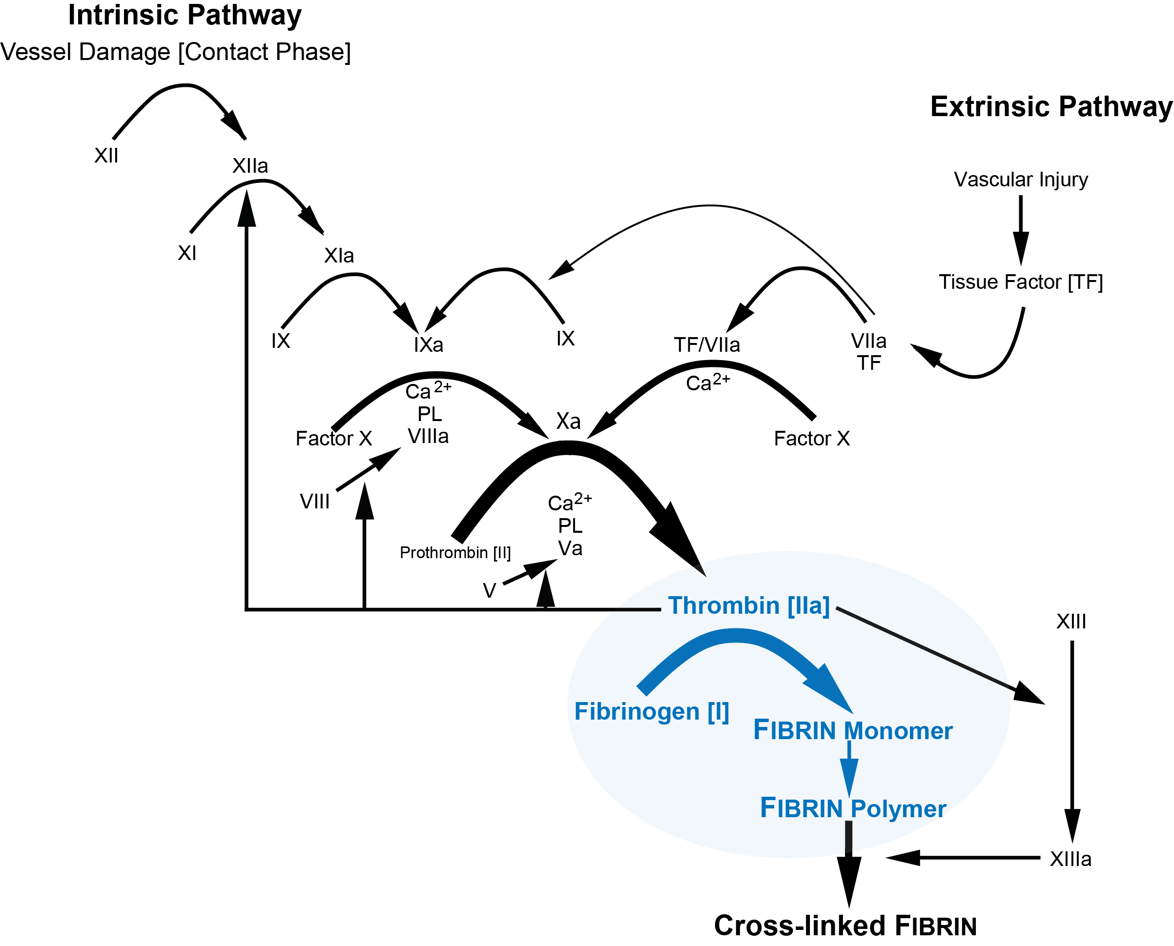
Fibrinogen → (Thrombin + Ca²⁺) → Fibrin Monomers → (Factor XIIIa) → Cross-Linked Fibrin Network.
Interaction with Thrombin, Calcium Ions, and Clotting Factors
The conversion of fibrinogen to fibrin is a tightly regulated, multi-step process dependent on enzymatic and ionic cofactors. Thrombin, generated via the intrinsic (Factor XII) or extrinsic (tissue factor, Factor III) pathways, binds to fibrinogen’s D-domains, accelerating fibrinopeptide release. Calcium ions (Ca²⁺) enhance thrombin’s catalytic efficiency and facilitate fibrin polymerization by neutralizing negative charges on fibrinogen’s surface.The sequential activation involves:
1. Thrombin binding: High-affinity interaction with fibrinogen’s γ-chains, inducing conformational changes.
2. Fibrinopeptide release: Proteolytic cleavage at Arg-Gly bonds, exposing polymerization sites.
3. Protofibril formation: Non-covalent alignment of fibrin monomers, stabilized by electrostatic and hydrophobic interactions.
4. Factor XIII activation: Thrombin cleaves Factor XIII to XIIIa, which cross-links fibrin strands in the presence of Ca²⁺.
Critical Cofactors:
Thrombin (Factor IIa): Primary protease for fibrinogen cleavage. Calcium ions (Ca²⁺): Essential for thrombin activity and Factor XIII activation. Factor XIII (transglutaminase): Mediates covalent cross-linking of fibrin.
Comparative Analysis: Fibrinogen in Hemostasis vs. Bleeding Disorders
Fibrinogen’s role in hemostasis is paramount, but its deficiency or dysfunction disrupts clot formation, leading to severe bleeding disorders. Afibrinogenemia (congenital absence of fibrinogen) and hypofibrinogenemia (reduced levels) exemplify the clinical spectrum of fibrinogen-related pathologies.| Clotting Factor | Role in Fibrinogen Activation | Deficiency Impact | Lab Test Used |
|---|---|---|---|
| Thrombin (Factor IIa) | Cleaves fibrinopeptides A/B, initiating polymerization. | Prolonged PT/APTT; risk of hemorrhage. | PT (Prothrombin Time), TT (Thrombin Time). |
| Factor XIII | Cross-links fibrin strands, stabilizing the clot. | Delayed clot retraction; recurrent bleeding. | Factor XIII activity/antigen assays. |
| Calcium (Ca²⁺) | Cofactor for thrombin and Factor XIII activation. | Impaired fibrin polymerization; bleeding diathesis. | Ionized calcium levels, PT correction. |
| Factor V/Va | Accelerates thrombin generation via prothrombinase complex. | Mild-moderate bleeding; prolonged PT/APTT. | PT, APTT, Factor V activity assays. |
Clinical Correlation:
Afibrinogenemia patients exhibit normal PT/APTT but prolonged thrombin time (TT), reflecting direct fibrinogen dysfunction.
Structural Composition and Synthesis of Fibrinogen
Fibrinogen, a soluble plasma glycoprotein, serves as the precursor to fibrin, the insoluble meshwork essential for clot formation. Its molecular architecture and biosynthesis in hepatocytes are intricately regulated to ensure hemostatic function while maintaining solubility in circulation. The structural integrity of fibrinogen is dictated by its three distinct polypeptide chains (Aα, Bβ, γ), interconnected by disulfide bonds, and specialized functional domains that mediate interactions with platelets, thrombin, and other coagulation factors. Disruptions in its synthesis or structural integrity lead to pathological conditions, including dysfibrinogenemias, which impair coagulation efficiency.Molecular Structure of Fibrinogen
Fibrinogen is a 340-kDa hexameric glycoprotein composed of two identical halves, each containing three non-identical polypeptide chains: Aα (66 kDa), Bβ (52 kDa), and γ (46 kDa). These chains are arranged in a symmetrical, elongated structure (~45 nm × 9 nm), resembling a "staple" or "dumbbell" shape. The central E (ectodomain) region is flanked by two distal D (discoid) domains, connected by coiled-coil α-helical regions formed by the Aα chains.The structural stability of fibrinogen relies on 18 disulfide bonds, including:
Functional domains critical for hemostasis include:
Synthesis and Post-Translational Modifications
Fibrinogen synthesis occurs exclusively in hepatocytes, regulated by transcriptional, translational, and post-translational mechanisms. The process involves three genes: FGA (Aα chain), FGB (Bβ chain), and FGG (γ chain), each encoding a precursor polypeptide that undergoes proteolytic processing.Transcriptional Regulation
Post-Translational Modifications
Fibrinogen undergoes critical modifications to achieve functional maturity:
Secretion Mechanism
Structural Differences Between Fibrinogen and Fibrin
The conversion of fibrinogen to fibrin involves thrombin-mediated proteolytic cleavage at specific arginyl residues, triggering polymerization. Key structural transformations include:Fibrinogen → Fibrin Transition:Key Structural Contrasts
Thrombin cleaves: Aα chain at Arg558-Val559, releasing fibrinopeptide A (FpA). Bβ chain at Arg16-Gly17, releasing fibrinopeptide B (FpB). Exposure of binding sites: Cleavage uncovers hydrophobic and ionic residues in the E domain, enabling D-E polymerization. Fibrin monomer formation: The cleaved E domain binds to D domains of adjacent monomers, forming protofibrils. Cross-linking: Factor XIIIa stabilizes the clot by forming γ-γ and α-α cross-links via transglutaminase activity.
| Feature | Fibrinogen | Fibrin |
|---|---|---|
| Solubility | Soluble in plasma (no exposed binding sites) | Insoluble polymer (exposed D and E domains) |
| Molecular Weight | 340 kDa (hexamer) | ~330 kDa (monomer); polymers up to 100+ monomers |
| Binding Sites | Masked by fibrinopeptides (FpA, FpB) | Exposed after cleavage (D-E, D-D interactions) |
| Function | Precursor; circulates in plasma | Forms clot scaffold; cross-linked by FXIIIa |
Genetic Mutations and Dysfibrinogenemias
Mutations in FGA, FGB, or FGG disrupt fibrinogen structure, leading to congenital dysfibrinogenemias, characterized by impaired coagulation, thrombosis, or bleeding. Over 500 mutations have been identified, affecting synthesis, secretion, or functional domains.Mechanisms of Pathogenicity
Examples of Dysfibrinogenemias
- Ruel dysfibrinogenemia (Bβ Arg35→Cys): Impairs thrombin cleavage, causing hypofibrinogenemia and bleeding.
- Baltimore dysfibrinogenemia (γ Arg355→Cys): Alters platelet binding, leading to thrombosis despite normal fibrinogen levels.
- Lyon dysfibrinogenemia (Aα Gly310→Val): Reduces fibrin polymerization efficiency, resulting in bleeding diathesis.
- Heterozygous FGB mutations: May present as asymptomatic carriers or with thrombotic tendencies due to altered fibrin structure.

Clinical Significance and Diagnostic Methods in Fibrinogen Evaluation
Fibrinogen plays a pivotal role in hemostasis, and its clinical assessment is essential for diagnosing coagulation disorders, monitoring therapeutic interventions, and evaluating thrombotic risks. Diagnostic evaluation of fibrinogen relies on standardized assays, each with distinct principles, reference ranges, and limitations. Understanding these methods—including prothrombin time (PT), thrombin time (TT), and the Clauss assay—enables accurate interpretation of fibrinogen-related pathologies, from congenital deficiencies to acquired dysregulations. Below, structured diagnostic criteria, comparative disorder analyses, and procedural interpretations for fibrinogen degradation products (FDPs) and D-dimers are detailed to guide clinical practice.Diagnostic Criteria for Evaluating Fibrinogen Levels
The assessment of fibrinogen levels employs multiple assays, each targeting different aspects of the coagulation cascade. Prothrombin time (PT) measures the extrinsic pathway, indirectly reflecting fibrinogen function by assessing the time required for clot formation after tissue factor activation. Thrombin time (TT) evaluates the conversion of fibrinogen to fibrin by thrombin, providing a direct measure of fibrinogen quantity and quality. The Clauss assay, considered the gold standard, quantifies fibrinogen by clotting plasma with excess thrombin, yielding a precise concentration in grams per liter (g/L). Reference ranges and limitations for each assay are summarized below:- Prothrombin Time (PT):
- Thrombin Time (TT):
- Clauss Assay:
Key Consideration: Fibrinogen levels <1.0 g/L are associated with spontaneous bleeding, while levels >6.0 g/L may predispose to thrombosis, particularly in inflammatory or malignant states.
Comparative Analysis of Fibrinogen-Related Disorders
Fibrinogen-related disorders encompass congenital deficiencies (afibrinogenemia, hypofibrinogenemia) and acquired dysfunctions (dysfibrinogenemia). The following table organizes clinical features, pathophysiological mechanisms, and therapeutic approaches for these conditions:| Condition | Pathophysiology | Symptoms | Treatment Approaches |
|---|---|---|---|
| Afibrinogenemia | Autosomal recessive mutations in FGA, FGB, or FGG genes, resulting in complete fibrinogen absence. Neonatal onset with severe bleeding diathesis. |
|
|
| Hypofibrinogenemia | Acquired (liver disease, DIC, malnutrition) or congenital (heterozygous mutations) with fibrinogen levels <1.0 g/L. May coexist with hypoprothrombinemia. |
|
|
| Dysfibrinogenemia | Qualitative defects due to fibrinogen structural abnormalities (e.g., R554C mutation), impairing polymerization or thrombin cleavage. May present as bleeding, thrombosis, or asymptomatic. |
|
|
Diagnostic Pitfall: Dysfibrinogenemia may mimic factor deficiencies due to overlapping PT/TT prolongation, necessitating functional assays (e.g., reptilase time) or genetic testing.
Interpretation of Fibrinogen Degradation Products (FDPs) and D-Dimers
FDPs and D-dimers are degradation byproducts of cross-linked fibrin, serving as biomarkers for fibrinolysis and thrombotic states. Their interpretation requires contextual analysis of clinical scenarios, particularly in acute thrombosis or disseminated intravascular coagulation (DIC). Below is a procedural outline for their evaluation:1. Pre-Analytical Considerations:
2. Assay-Specific Interpretation:
3. Clinical Correlation:
4. Differential Diagnoses:
Critical Algorithm:
In DIC, D-dimer >10,000 ng/mL FEU with PT >18 seconds and platelets <50 × 10⁹/L strongly supports consumptive coagulopathy,Fibrinogen in Disease Pathophysiology
Fibrinogen plays a central role in both hemostasis and pathological thrombosis, with its dysfunction or dysregulation contributing to cardiovascular diseases, inflammatory disorders, and sepsis-associated coagulopathy. Genetic variants, structural alterations, and pro-inflammatory properties of fibrinogen disrupt normal hemostatic balance, promoting arterial and venous thrombosis, chronic inflammation, and systemic coagulopathies. Understanding these mechanisms elucidates therapeutic targets for preventing fibrinogen-mediated complications.
Fibrinogen Polymorphisms and Cardiovascular Risk
Genetic variations in the FGB (fibrinogen β-chain) and FGG (fibrinogen γ-chain) genes alter fibrinogen structure, function, and plasma levels, increasing susceptibility to arterial thrombosis. The β148Ala polymorphism (rs1800787) and γ407Arg polymorphism (rs1800790) are among the most studied variants associated with elevated cardiovascular risk. These polymorphisms enhance fibrinogen’s prothrombotic potential through multiple mechanistic pathways:- Enhanced Platelet Aggregation
The β148Ala variant increases fibrinogen’s affinity for glycoprotein IIb/IIIa (GPIIb/IIIa) receptors on platelets, accelerating thrombus formation under shear stress. Studies demonstrate a ~20–30% higher risk of myocardial infarction (MI) and ischemic stroke in carriers of this allele, particularly in individuals with preexisting atherosclerosis.- Altered Fibrin Clot Structure
The γ407Arg polymorphism modifies fibrin polymerization, producing denser, less permeable clots that resist fibrinolysis. This structural alteration prolongs arterial occlusion, as observed in patients with unstable angina or acute coronary syndromes (ACS). Research in Circulation (2016) linked this variant to reduced clot lysis time (CLT) by ~15–25% in vitro.- Endothelial Dysfunction and Arterial Thrombosis
Fibrinogen polymorphisms exacerbate endothelial activation by amplifying the binding of von Willebrand factor (vWF) to collagen and platelets. In arterial beds, where high shear forces prevail, these interactions promote white thrombus formation (rich in platelets and fibrin), a hallmark of ACS. The β148Ala variant correlates with increased carotid intima-media thickness (IMT), a marker of subclinical atherosclerosis.
Key Insight: Fibrinogen polymorphisms do not merely elevate plasma levels but directly enhance thrombus stability and resistance to fibrinolysis, a critical distinction in arterial thrombosis management.Differential Role in Venous vs. Arterial Thrombosis
While fibrinogen is essential for both venous and arterial thrombosis, its mechanistic contributions diverge due to distinct hemodynamic and cellular environments. The following table summarizes these differences:
Feature Arterial Thrombosis Venous Thrombosis Primary Trigger Endothelial injury, plaque rupture, or turbulent flow (e.g., atherosclerosis) Stasis, hypercoagulability (e.g., Factor V Leiden), or venous endothelial damage Thrombus Composition Platelet-rich ("white thrombus") with fibrin meshwork Red blood cell-rich ("red thrombus") with fibrin strands Fibrinogen Interaction with vWF Amplified under high shear; stabilizes platelet aggregates via GPIIb/IIIa Moderate role; vWF primarily mediates platelet adhesion to exposed collagen Endothelial Cell Role Dysfunctional endothelium secretes P-selectin, enhancing fibrinogen-platelet binding Procoagulant endothelium expresses tissue factor (TF) and reduces thrombomodulin Fibrinolysis Resistance High due to dense fibrin networks (e.g., γ407Arg variant) Variable; often compensated by elevated tPA in venous systems Clinical Correlation: Arterial thrombosis (e.g., MI, stroke) is more strongly associated with fibrinogen’s platelet-binding properties, while venous thrombosis (e.g., DVT) is linked to fibrinogen’s role in TF-mediated coagulation and stasis-induced clot propagation.Pro-Inflammatory Properties of Fibrinogen
Beyond hemostasis, fibrinogen serves as a damage-associated molecular pattern (DAMP) that bridges coagulation and inflammation. Its pro-inflammatory roles include:- Leukocyte Recruitment and Activation
Fibrinogen fragments (e.g., fibrinogen degradation products [FDPs]) bind to Mac-1 (CD11b/CD18) on neutrophils, promoting adhesion to endothelial cells via ICAM-1. This interaction sustains chronic inflammation in conditions like atherosclerosis and rheumatoid arthritis. Studies in Journal of Immunology (2019) showed that fibrinogen-deficient mice exhibit reduced neutrophil extravasation in models of peritonitis.- Cytokine Release and Immune Modulation
Fibrinogen interacts with TLR4 (Toll-like receptor 4), triggering NF-κB signaling and upregulating IL-6, TNF-α, and IL-1β. This axis contributes to sepsis-induced coagulopathy and chronic wound healing dysfunction. Elevated fibrinogen levels in critically ill patients correlate with higher SOFA scores and mortality, as documented in Critical Care Medicine (2021).- Fibrinogen in Chronic Wound Healing
Persistent fibrin deposits in wounds impair granulation tissue formation by trapping growth factors (e.g., VEGF, PDGF) and inhibiting macrophage polarization to the pro-healing M2 phenotype. This mechanism underlies diabetic ulcers and pressure injuries, where fibrinogen accumulation correlates with delayed re-epithelialization.
Therapeutic Implication: Anti-fibrinolytic agents (e.g., tranexamic acid) must be used cautiously in inflammatory conditions, as they may exacerbate leukocyte trapping and tissue damage.Feedback Loops in Sepsis-Induced Coagulopathy
Sepsis triggers a vicious cycle involving fibrinogen, tissue factor (TF), and the complement system, culminating in disseminated intravascular coagulation (DIC). The following flowchart outlines the key interactions:1. Initiation Phase
Bacterial LPS activates monocytes/macrophages via TLR4, inducing TF expression on endothelial cells and leukocytes. TF initiates the extrinsic coagulation pathway, converting Factor VIIa to Factor Xa, which cleaves prothrombin to thrombin. Thrombin cleaves fibrinogen to fibrin, forming microclots that occlude microvasculature. 2. Amplification Phase
Fibrin(ogen) fragments (e.g., E-fragments) bind to complement C5, enhancing C5a generation and neutrophil extracellular trap (NET) formation. C5a further activates endothelial cells, increasing P-selectin and TF expression, perpetuating coagulation. Platelets release platelet factor 4 (PF4), which inhibits fibrinolysis by neutralizing tissue plasminogen activator (tPA). 3. Feedback Inhibition and Organ Dysfunction
Complement activation depletes Factor H, reducing regulatory control over the alternative pathway. Thrombin activates protein C (via thrombomodulin), but sepsis-induced TF overproduction overwhelms this pathway. Microthrombi in organs (e.g., lungs, kidneys) trigger ischemia-reperfusion injury, releasing additional DAMPs (e.g., HMGB1), which stimulate more TF expression. Pathophysiological Cycle:
TF ↑ → Thrombin ↑ → Fibrin(ogen) ↑ → Complement Activation ↑ → TF ↑
This loop explains why fibrinogen levels >6 g/L in sepsis are associated with ~80% mortality risk (ICU studies, 2018).
Therapeutic Applications and Emerging Research in Fibrinogen Modulation
Fibrinogen plays a central role in hemostasis, wound healing, and pathological thrombosis, making it a critical target for therapeutic intervention. Advances in recombinant production, protein engineering, and targeted biologics have expanded fibrinogen-based therapies beyond traditional coagulation management into regenerative medicine and antithrombotic strategies. This section explores clinically approved fibrinogen concentrates, experimental modifications for enhanced functionality, and emerging anti-fibrinogen approaches in preclinical and translational research.
Mechanisms and Clinical Use of Fibrinogen Concentrates
Fibrinogen concentrates represent a cornerstone in the treatment of acquired hypofibrinogenemia, congenital fibrinogen deficiencies (e.g., afibrinogenemia, hypofibrinogenemia), and massive bleeding in trauma or surgery. These products are derived from plasma or recombinant sources, with formulations such as RiaSTAP (CSL Behring) and Haemocomplettan (CSL Behring) undergoing rigorous purification to minimize pathogen transmission and immunogenicity.Mechanisms of Action
Fibrinogen concentrates restore hemostatic function by:
Direct replacement of deficient fibrinogen, achieving plasma levels of 1.0–1.5 g/L within 30–60 minutes post-infusion. Enhancing clot formation via the common pathway of coagulation, where thrombin converts fibrinogen to fibrin, stabilizing platelet aggregates. Supporting secondary hemostasis by providing a scaffold for fibrin polymerization and cross-linking by factor XIIIa. Clinical Indications and Dosing Protocols
Primary indications include:Dosing is typically weight-based, with initial boluses of 3–5 g (adults) or 50–100 mg/kg (pediatrics), followed by maintenance infusions to sustain fibrinogen >1.0 g/L. In trauma, fibrinogen concentrates are often administered in massive transfusion protocols (MTP) alongside prothrombin complex concentrates (PCCs) and platelets. For congenital deficiencies, long-term prophylaxis may require weekly infusions (e.g., 20–40 mg/kg).Trauma-induced coagulopathy (e.g., polytrauma with fibrinogen <1.0 g/L). Cardiac surgery (e.g., aortic aneurysm repair, where fibrinogen consumption exceeds 50% of baseline). Obstetric hemorrhage (e.g., placenta accreta with fibrinogen <1.5 g/L). Congenital deficiencies (e.g., afibrinogenemia with spontaneous bleeding). Efficacy and Safety
Clinical trials demonstrate that fibrinogen concentrates reduce transfusion requirements by 30–50% in trauma patients and shorten time to hemostasis in surgical bleeding. Adverse effects are rare but include:
Allergic reactions (e.g., anaphylaxis in IgA-deficient patients). Thrombotic events (controversial; linked to hypercoagulable states post-infusion). Dilutional coagulopathy if administered without volume resuscitation. Engineering Fibrinogen Variants for Enhanced Functionality
Protein engineering of fibrinogen aims to improve clot stability, thromboresistance, or targeted delivery for regenerative applications. Key strategies include site-directed mutagenesis, PEGylation, and fusion proteins to modulate fibrinogen’s structural or functional domains.1. Site-Directed Mutagenesis for Clot Stability
Fibrinogen’s D-domain (responsible for polymerization) and E-domain (thrombin cleavage site) are prime targets. Mutations such as:
R554K in the γ-chain enhance fibrin fiber thickness, increasing clot tensile strength by 40% without altering thrombin time (TT). D233A in the α-chain reduces plasminogen binding, conferring resistance to fibrinolysis while preserving hemostatic function. Double mutants (e.g., R554K + D233A) are under investigation for chronic wound healing and vascular graft applications. 2. PEGylation for Reduced Thrombogenicity
Polyethylene glycol (PEG) conjugation to fibrinogen’s N-terminal regions masks immunogenic epitopes and prolongs circulation half-life. Studies show:
20 kDa PEG-fibrinogen reduces platelet adhesion by 60% in vitro while maintaining clot formation. Site-specific PEGylation (e.g., at lysine residues) avoids steric hindrance of thrombin cleavage, preserving hemostatic efficacy. Preclinical models (e.g., arterial thrombosis in mice) demonstrate reduced thrombus burden without bleeding diathesis. 3. Fusion Proteins for Targeted Delivery
Fibrinogen is engineered as a carrier scaffold for therapeutic peptides or drugs via:
Fibrinogen-αC domain fusions with vascular endothelial growth factor (VEGF) to promote angiogenesis in ischemic tissues. Hydrogel-forming fibrinogen variants (e.g., fibrinogen-PEG hydrogels) for 3D tissue engineering, where cross-linking is triggered by thrombin or light activation. Challenges and Future Directions
Scalable production: Recombinant fibrinogen (e.g., in Pichia pastoris) must achieve >90% identity to plasma-derived forms. Immunogenicity: Non-human fibrinogen sequences (e.g., from Crotalus atrox venom-derived fibrinogen-like proteins) may elicit antibodies. Regulatory hurdles: Modified fibrinogen products require bridging studies to establish safety in healthy volunteers before disease-specific trials. Anti-Fibrinogen Therapies in Thrombosis and Cancer Metastasis
Fibrinogen is a dual-edged target: while essential for hemostasis, its overexpression or hyperactivation contributes to arterial thrombosis, venous thromboembolism (VTE), and cancer metastasis. Anti-fibrinogen strategies include neutralizing antibodies, nanobody inhibitors, and small-molecule disruptors of fibrinogen-platelet interactions.1. Antibody-Based Inhibition
Antilaminin Antibodies (e.g., Anti-LAMC1) Fibrinogen binds to laminin-5 in the extracellular matrix, promoting cancer cell adhesion and invasion. Monoclonal antibodies like mAb 19 (targeting the α2 chain of laminin-5) reduce breast cancer metastasis in mouse models by 70% without affecting hemostasis.
Anti-Fibrinogen γ-Chain Antibodies (e.g., 5G1) 5G1 binds the C-terminal region of the γ-chain, preventing fibrin polymerization and reducing thrombus formation in FeCl₃-induced arterial thrombosis models. However, off-target bleeding remains a concern.2. Nanobody Inhibitors
Single-domain antibody fragments (nanobodies) derived from llamas or alpacas offer high specificity and low immunogenicity. Examples include:
Nb-Fgn (targeting the E-domain), which inhibits fibrinogen’s interaction with GPIIb/IIIa receptors on platelets, reducing deep vein thrombosis (DVT) in rodent models by 50%. Bivalent nanobodies (e.g., Nb-Fgn-Fc) enhance avidity, enabling subcutaneous administration with prolonged half-life. 3. Small-Molecule Disruptors
Fibrinogen-Platelet Interaction Inhibitors (e.g., Tirofiban Analogs) Cilostazol metabolites and peptidomimetics (e.g., ROCK inhibitors) disrupt fibrinogen’s binding to αIIbβ3 integrin, reducing platelet aggregation without anticoagulant effects.
Fibrinogen Cleavage Mimetics (e.g., Thrombin-Like Enzymes) Batroxobin (derived from Bothrops atrox venom) selectively cleaves fibrinogen’s Aα-chain, generating non-clottable fibrin(ogen) degradation products (FDPs) that inhibit thrombosis.Preclinical Efficacy and Limitations
Key findings in animal models:ChallengesThrombosis: Anti-fibrinogen nanobodies reduce stroke volume in carotid artery thrombosis by 60% (vs. heparin). Cancer: Fibrinogen depletion via siRNA or CRISPR-Cas9 in tumor-associated fibroblasts reduces lung metastasis in murine models of melanoma. Sepsis: Fibrinogen-targeting aptamers (e.g., FD-1) improve survival in CLP-induced sepsis by 40% by preventing microthrombi.
Bleeding risk: Systemic fibrinogen inhibition increases surgical bleeding time by >2-fold. Compensatory mechanisms: Thrombosis models may underestimate fibrinogen-independent pathways (e.g., factor XII activation). Fibrinogen emerges not only as a pivotal mediator of hemostasis but also as a nexus between coagulation, inflammation, and vascular biology, bridging molecular mechanisms with clinical outcomes. Its dual role in clot stabilization and immune modulation highlights its therapeutic potential, from regenerative medicine applications to targeted anti-thrombotic interventions. As research advances—spanning genetic polymorphisms, structural engineering, and diagnostic innovations—fibrinogen remains a focal point for understanding disease pathophysiology and developing precision-based therapies. The interplay between its structural integrity, functional dynamics, and pathological implications underscores its enduring significance in both basic science and clinical practice.
FAQ
What does fibrinogen mean when it appears on a blood test?
Fibrinogen is a protein measured in blood tests that plays a key role in blood clotting. Its levels help assess clotting function, and abnormal results may indicate bleeding disorders, clotting risks, or liver disease.
What is the role of fibrinogen in the blood?
Fibrinogen is a soluble plasma protein that converts to fibrin during clotting, forming the mesh that traps platelets to create blood clots. It’s essential for wound healing but can contribute to clotting disorders if levels are too high or low.
What is a fibrinogen test and why is it done?
A fibrinogen test measures the amount of this clotting protein in your blood, often included in coagulation panels. It’s used to diagnose bleeding disorders, monitor liver function, or evaluate risks for clots (e.g., in pregnancy or surgery).
What does fibrinogen activity refer to in medical terms?
Fibrinogen activity reflects how well the protein can be converted into fibrin during clotting, often assessed via tests like the Clauss method. Reduced activity may signal liver disease, DIC, or inherited clotting disorders.
What does it mean if fibrinogen levels are high?
High fibrinogen (hyperfibrinogenemia) can indicate inflammation, infection, or chronic diseases like cancer. It may also raise clot risk, though mild elevations are often benign (e.g., during pregnancy or stress).
What are normal fibrinogen levels in the blood?
Normal fibrinogen levels typically range from 200 to 400 mg/dL, but values vary by lab. Low levels (<100 mg/dL) increase bleeding risk, while high levels (>400 mg/dL) may signal underlying health issues.
Leave a Comment
Comments are moderated before appearing. The data you submit is processed according to the Privacy Policy of Voltefac.