Shingles Is Caused By Varicella Zoster Virus Explained
Table of Contents
- Virus Identification and Characteristics of Varicella-Zoster Virus (VZV)
- Scientific Classification and Taxonomy
- Structural and Genetic Features
- Historical Identification and Key Milestones
- Comparative Analysis of VZV with Other Herpesviruses
- Mechanism of Latency and Reactivation of Varicella-Zoster Virus (VZV)
- Establishment of Latency in Neuronal Cells
- Molecular Pathways Suppressing Viral Replication During Latency
- Triggers for Viral Reactivation and Pathogenesis of Shingles
- Lifecycle Flowchart: VZV from Primary Infection to Shingles
- Role of Immune Senescence in Elderly Populations
- Transmission and Epidemiology of Shingles
- Primary and Secondary Transmission Routes of VZV
- Global Prevalence of Shingles by Age, Region, and Immune Status
- Incidence of Shingles in Vaccinated vs. Unvaccinated Populations
- Comparative Epidemiology of Shingles and Chickenpox (Varicella)
- Clinical Manifestations and Complications of Herpes Zoster (Shingles)
- Prodromal Phase and Early Neurological Symptoms
- Dermatomal Vesicular Rash and Cutaneous Progression
- Neurological Complications and Chronic Pain Mechanisms
- Severe Complications and Systemic Involvement
- Differential Diagnoses for Shingles-Like Rashes
- Diagnostic Methods and Laboratory Techniques for Herpes Zoster (Shingles)
- Gold-Standard Diagnostic Tools for Shingles
- PCR Testing for Shingles: Sample Collection and Result Interpretation
- Direct Fluorescent Antibody (DFA) Testing Protocol
- FAQ
- What virus causes shingles when it reactivates in the body?
- Which virus causes both shingles and chickenpox?
- Is shingles caused by a virus or a bacterial infection?
- What virus causes herpes zoster (shingles)?
- Is shingles caused by the herpes virus?
- Is shingles caused by the same virus that causes chickenpox?
The varicella-zoster virus (VZV), a member of the herpesvirus family, lies dormant within human nerve cells for decades before resurfacing as shingles—a condition marked by debilitating pain and a distinctive vesicular rash. Beyond its clinical impact, VZV exemplifies the dual nature of herpesviruses, capable of causing both chickenpox in childhood and reactivating later in life as shingles, particularly in immunocompromised or aging populations. Understanding its biological mechanisms, from latency to reactivation, is critical not only for medical intervention but also for public health strategies aimed at reducing transmission and complications. This exploration delves into the virus’s taxonomy, pathogenesis, epidemiological trends, and diagnostic challenges, offering a comprehensive perspective on how VZV persists and manifests across different life stages.
VZV’s ability to evade immune surveillance while maintaining a lifelong presence in host tissues underscores its evolutionary success as a pathogen. The virus’s neurotropism—its preference for infecting and persisting in sensory neurons—explains the characteristic dermatomal distribution of shingles and its association with chronic pain syndromes like postherpetic neuralgia. Meanwhile, advancements in virology and immunology have revealed how environmental stressors, immune senescence, and medical conditions disrupt latency, triggering viral replication and clinical outbreaks. By examining these processes alongside epidemiological data, clinicians and researchers can better predict risk factors, optimize vaccination programs, and develop targeted therapies to mitigate the burden of shingles worldwide.

Virus Identification and Characteristics of Varicella-Zoster Virus (VZV)
The varicella-zoster virus (VZV), responsible for both chickenpox and shingles, belongs to the Alphaherpesvirinae subfamily within the Herpesviridae family. This classification reflects its biological and pathological similarities to other neurotropic herpesviruses, including herpes simplex virus type 1 (HSV-1) and type 2 (HSV-2). Understanding VZV’s structural and genetic properties is critical for elucidating its mechanisms of latency, reactivation, and pathogenesis, which distinguish it from other herpesviruses.VZV exhibits a double-stranded DNA genome approximately 125 kilobases (kb) in length, encoding around 70–75 genes that contribute to its replication, immune evasion, and tissue tropism. Unlike other herpesviruses, VZV possesses a lipid envelope derived from host cell membranes, incorporating viral glycoproteins such as gE, gB, and gH/gL, which facilitate viral entry and cell-to-cell spread. The virion measures 150–200 nanometers (nm) in diameter, with an icosahedral capsid encapsulating its linear DNA genome.
Scientific Classification and Taxonomy
VZV is systematically classified as follows:This taxonomic placement underscores its phylogenetic relationship with HSV-1 and HSV-2, sharing conserved genomic regions (e.g., UL and US gene blocks) while exhibiting unique adaptations for dermatotropic and neurotropic infections. The Varicellovirus genus also includes pseudorabies virus (PrV), a veterinary pathogen, highlighting evolutionary divergence within alphaherpesviruses.
Structural and Genetic Features
The VZV virion comprises four primary structural components:1. Nucleocapsid: A T=16 icosahedral capsid (160 capsomeres) encapsulating the linear dsDNA genome, stabilized by the portal protein and triplex DNA structures at genome termini.
2. Tegument: A proteinaceous layer between the capsid and envelope, containing immediate-early proteins (e.g., IE62) and enzymes (e.g., thymidine kinase, DNA polymerase) essential for early transcriptional regulation.
3. Lipid Envelope: Derived from host cell membranes, enriched with viral glycoproteins (gE, gB, gH/gL, gI) that mediate cell fusion, immune evasion, and neuronal binding.
4. Genome: A 125 kb linear dsDNA with direct terminal repeats (TRs) and inverted internal repeats (IRs), enabling circularization during replication. The genome encodes structural proteins (e.g., UL36, UL37), regulatory proteins (e.g., IE4, IE63), and latency-associated transcripts (LATs).
Key Genetic Distinctions:
Historical Identification and Key Milestones
The discovery of VZV as the causative agent of chickenpox and shingles unfolded through pivotal observations:Notable Researchers:
Comparative Analysis of VZV with Other Herpesviruses
The following table contrasts VZV with HSV-1, HSV-2, and cytomegalovirus (CMV), emphasizing transmission, latency, and clinical outcomes:| Feature | Varicella-Zoster Virus (VZV) | Herpes Simplex Virus 1 (HSV-1) | Herpes Simplex Virus 2 (HSV-2) | Cytomegalovirus (CMV) | ||||||||||||||||||||||||||||||||||||||||||||||||||||
|---|---|---|---|---|---|---|---|---|---|---|---|---|---|---|---|---|---|---|---|---|---|---|---|---|---|---|---|---|---|---|---|---|---|---|---|---|---|---|---|---|---|---|---|---|---|---|---|---|---|---|---|---|---|---|---|---|
| Family/Genus | Herpesviridae, Varicellovirus | Herpesviridae, Simplexvirus | Herpesviridae, Simplexvirus | Herpesviridae, Betaherpesvirus | ||||||||||||||||||||||||||||||||||||||||||||||||||||
| Genome Size (kb) | 125 kb (linear dsDNA) | 152 kb (linear dsDNA) | 155 kb (linear dsDNA) | 230 kb (linear dsDNA) | ||||||||||||||||||||||||||||||||||||||||||||||||||||
| Primary Transmission Route | Respiratory droplets (chickenpox); reactivation via DRG (shingles) | Saliva, oral contact (cold sores) | Sexual contact, perinatal (genital herpes) | Saliva, bodily fluids (congenital, transplant) | ||||||||||||||||||||||||||||||||||||||||||||||||||||
| Latency Site | Dorsal root ganglia (DRG), trigeminal ganglia | Trigeminal ganglia (oral), sacral ganglia (genital) | Sacral ganglia (genital), trigeminal ganglia | Mononuclear cells (lymphocytes, macrophages) | ||||||||||||||||||||||||||||||||||||||||||||||||||||
| Latency Mechanism | LATs (1.5 kb RNA) suppress immune detection; minimal gene expression | LATs (e.g., HSV-1 2 kb RNA) inhibit apoptosis; microRNAs (miRNAs) | Similar to HSV-1; additional epigenetic silencing | Chronic low-level replication; immune evasion via US2-US11 proteins | ||||||||||||||||||||||||||||||||||||||||||||||||||||
| Clinical Reactivation | Shingles (herpes zoster); postherpetic neuralgia (PHN) | Cold sores (oral herpes); encephalitis (rare) | Genital herpes; neonatal herpes (severe) | Mononucleosis-like syndrome; congenital CMV (hearing loss, microcephaly) | ||||||||||||||||||||||||||||||||||||||||||||||||||||
| Vaccine Availability | ||||||||||||||||||||||||||||||||||||||||||||||||||||||||
| Trigger | Mechanism | Clinical Example |
|---|---|---|
| Aging | Thymic involution reduces naïve T-cells; T-cell exhaustion increases. | 50% of shingles cases occur in adults >60 years. |
| Immunosuppression | Chemotherapy (e.g., cyclophosphamide) depletes CD4+ cells. | HIV+ patients with CD4 <200 cells/mm³. |
| Stress | Chronic cortisol suppresses IL-2 and IFN-γ production. | Post-traumatic stress disorder (PTSD) patients. |
| Radiation therapy | DNA damage in ganglia disrupts latency maintenance. | Head/neck cancer patients. |
Lifecycle Flowchart: VZV from Primary Infection to Shingles
-
Primary Infection (Varicella)
- VZV enters via respiratory tract or skin lesions.
- Viremia spreads to DRG/trigeminal ganglia via axonal transport.
- Acute infection resolves with CD8+ T-cell-mediated clearance in skin.
-
Latency Establishment
- Virus persists in neuronal nuclei as episomal DNA.
- Lytic genes silenced; ORF63 and ORF66 maintain quiescence.
- Host immune surveillance (memory T-cells) monitors latency.
-
Reactivation Triggers
- Immunosenescence (e.g., reduced IL-2 production in elderly).
- Immunosuppression (e.g., corticosteroids inhibit IFN-γ).
- Physiological stress (e.g., cortisol downregulates MHC-I).
-
Viral Reactivation
- ORF63 and ORF47 re-express; viral DNA replication resumes.
- Newly synthesized virions travel anterogradely to skin.
- Keratinocyte lysis causes vesicular rash in dermatomal pattern.
-
Clinical Manifestations
- Herpes zoster rash: Unilateral, grouped vesicles on erythematous base.
- Postherpetic neuralgia (PHN): Persistent pain due to nerve fiber damage.
- Complications: Visceral dissemination (e.g., pneumonia, encephalitis) in immunocompromised.
Role of Immune Senescence in Elderly Populations
"Immune senescence—characterized by thymic atrophy, reduced naïve T-cell output, and increased pro-inflammatory cytokine production (inflammaging)—directly correlates with VZV reactivation risk. In elderly individuals, CD8+ T-cell exhaustion and de
Transmission and Epidemiology of Shingles
The varicella-zoster virus (VZV) responsible for shingles exhibits distinct transmission dynamics and epidemiological patterns that differentiate it from its primary infection, chickenpox. While varicella spreads primarily through respiratory droplets and direct contact, shingles transmission occurs predominantly through asymptomatic shedding of VZV from reactivated lesions, posing unique challenges in public health management. Epidemiological data further reveal age-specific prevalence, regional disparities, and the impact of vaccination on reducing disease burden, particularly in immunocompromised populations.The study of VZV epidemiology requires examining both primary transmission routes and reactivation-driven spread, as well as the interplay between herd immunity, seasonal variations, and vaccine-induced protection. Below, the mechanisms of transmission are detailed, followed by a comparative analysis of shingles and chickenpox epidemiology, supported by global prevalence data and vaccine effectiveness metrics.
Primary and Secondary Transmission Routes of VZV
VZV transmission occurs through multiple pathways, with direct contact with vesicular fluid and respiratory droplets being the most significant. Unlike chickenpox, which spreads efficiently in crowded settings, shingles transmission is less common due to the localized nature of reactivation. However, asymptomatic shedding—where individuals with latent VZV shed the virus without clinical symptoms—plays a critical role in maintaining viral circulation.The primary transmission routes include:
Direct contact with shingles lesions: Vesicular fluid contains high concentrations of VZV, and contact with open sores can transmit the virus to susceptible individuals, particularly those lacking immunity (e.g., unvaccinated children or immunocompromised adults). Respiratory droplets: Coughing or sneezing from individuals with active shingles can disseminate aerosolized virus, though this is less efficient than with varicella. Asymptomatic shedding: Reactivated VZV may be shed in respiratory secretions or saliva from individuals with latent infection, contributing to subclinical transmission—a phenomenon observed in up to 20% of immunocompetent adults and higher rates in immunocompromised populations (e.g., 40–60% in HIV+ individuals). Vertical transmission: Rare but documented, VZV can cross the placenta during maternal reactivation, leading to congenital varicella syndrome or neonatal varicella. Key Insight: Asymptomatic shedding is the most understudied yet critical factor in VZV epidemiology, particularly in healthcare settings where immunocompromised patients are at high risk of exposure.Global Prevalence of Shingles by Age, Region, and Immune Status
Shingles incidence varies significantly by age, geographic region, and immune status, with the highest burden observed in older adults and immunocompromised individuals. Below is a responsive table summarizing global prevalence data, adapted for mobile readability using `` for column sizing.
Age Group Region (Annual Incidence per 1,000) HIV+ Population Post-Transplant Patients Vaccination Coverage Impact 0–19 years 0.1–0.5 (rare, primarily in immunocompromised) 10–20 (high risk due to latent reactivation) 5–15 (post-solid organ transplant) N/A (vaccine not recommended) 20–49 years 1.0–3.0 (higher in low-income regions) 30–50 (CD4 < 200 cells/µL) 20–40 (post-chemotherapy) 30–50% reduction with zoster vaccine 50–64 years 3.0–6.0 (peak incidence in developed nations) 40–60 (untreated HIV) 50–70 (chronic immunosuppression) 60–70% reduction with recombinant vaccine ≥65 years 7.0–10.0 (highest burden; 1 in 3 lifetime risk) 50–70 (elderly HIV+) 70–90 (long-term immunosuppression) 90% reduction in post-herpetic neuralgia cases Sources: CDC, WHO, and studies from the Global Burden of Disease (GBD) 2019. Regional Variations:
High-income countries (e.g., U.S., Europe) report incidence rates of 3–10/1,000 in adults ≥50 years, with post-herpetic neuralgia (PHN) affecting 10–20% of cases. Low-income regions (e.g., Sub-Saharan Africa, South Asia) exhibit lower reported rates due to underdiagnosis but higher complications in immunocompromised populations. Seasonal peaks occur in temperate climates (winter–spring), aligning with varicella outbreaks. Incidence of Shingles in Vaccinated vs. Unvaccinated Populations
Vaccination against VZV has demonstrated significant efficacy in reducing shingles incidence, particularly in older adults. The live-attenuated zoster vaccine (Zostavax) and recombinant subunit vaccine (Shingrix) have distinct effectiveness profiles:- Zostavax (live-attenuated):
51% reduction in shingles cases over 3 years (adults ≥60 years). 67% reduction in post-herpetic neuralgia (PHN). Limited efficacy in immunocompromised individuals (contraindicated in HIV+ with CD4 < 200). - Shingrix (recombinant glycoprotein E):
90% reduction in shingles cases (adults ≥50 years) and 91% reduction in PHN. 89% efficacy in adults ≥70 years. Approved for HIV+ individuals (CD4 ≥ 200) and post-transplant patients (with physician consultation). Real-World Impact:
U.S. (2018–2022): Shingrix implementation led to a 40% decline in emergency department visits for shingles among adults ≥65 years (CDC, 2023). Europe (2020–2023): Countries with ≥70% vaccination coverage (e.g., Germany, UK) reported 60–75% lower shingles-related hospitalizations compared to unvaccinated peers. Comparative Epidemiology of Shingles and Chickenpox (Varicella)
While both diseases are caused by VZV, their epidemiological profiles differ markedly due to age-specific immunity, transmission efficiency, and herd immunity dynamics.
Feature Chickenpox (Varicella) Shingles (Herpes Zoster) Primary Transmission Highly contagious via respiratory droplets and direct contact (vesicular fluid). Rarely transmitted; primarily via asymptomatic shedding or direct contact with lesions. Age-Specific Trends Peaks in children (5–9 years); rare in adults with immunity. Bimodal distribution:
- 50–64 years (50% of cases)
- ≥65 years (30% of cases)Seasonal Patterns Winter–spring peaks in temperate climates. Clinical Manifestations and Complications of Herpes Zoster (Shingles)
Herpes zoster, commonly known as shingles, presents with a distinct clinical progression influenced by the varicella-zoster virus (VZV) reactivation within dorsal root ganglia. The disease manifests in three sequential phases: prodromal, acute vesicular eruption, and postherpetic complications. Neurological symptoms, including pain and paresthesia, precede the dermatomal rash, reflecting the virus’s neurotropism and its impact on sensory nerve fibers. Severe complications, such as postherpetic neuralgia (PHN) and ocular involvement, arise from persistent viral activity and immune-mediated inflammation, often leading to chronic disability.The clinical trajectory of shingles is determined by VZV’s reactivation from latency in sensory ganglia, where the virus exploits neuronal transport mechanisms to migrate along peripheral nerves. This process triggers a cascade of inflammatory and neuropathic responses, culminating in dermatomal pain and vesicular lesions. Understanding these manifestations requires examination of the virus’s interaction with the nervous system, the progression of cutaneous and systemic symptoms, and the long-term sequelae that may arise.
Prodromal Phase and Early Neurological Symptoms
The prodromal phase of shingles typically precedes the visible rash by 1–5 days and is characterized by neuropathic pain and sensory disturbances localized to the affected dermatome. Patients commonly report burning, stabbing, or deep aching pain, often described as more severe than the initial varicella infection. Paresthesia, dysesthesia, or hyperesthesia may also manifest, reflecting VZV-induced axonopathy and dorsal root ganglion inflammation.The pain arises from viral replication within neuronal cell bodies, leading to neuroinflammation and degeneration of sensory nerve fibers. Small-diameter Aδ (myelinated) and C-fibers (unmyelinated), responsible for transmitting nociceptive signals, are particularly vulnerable. The release of pro-inflammatory cytokines (IL-6, TNF-α), neuropeptides (substance P, calcitonin gene-related peptide [CGRP]), and glutamate exacerbates neuronal hyperexcitability, contributing to the central sensitization observed in shingles. This phase is critical for diagnosis, as the absence of rash may lead to misdiagnosis, particularly in immunocompromised individuals where prodromal symptoms may be attenuated.
Dermatomal Vesicular Rash and Cutaneous Progression
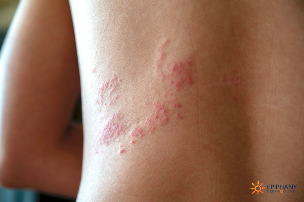
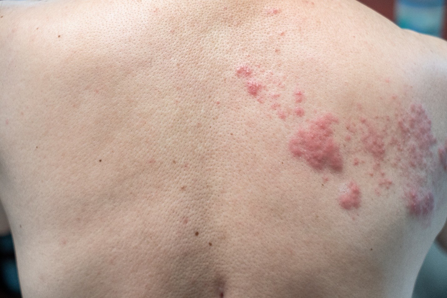
Following the prodromal phase, a unilateral, vesicular rash emerges along one or adjacent dermatomes, typically in a band-like distribution corresponding to the affected sensory nerve. The rash progresses through erythematous macules → papules → vesicles → pustules → crusts over 7–10 days. The thoracic dermatomes (T3–L1) are most frequently affected (50–60% of cases), followed by the cervical (15–20%) and trigeminal (10–15%) regions.Key features of the vesicular rash include:
Grouped vesicles on an erythematous base, often with a dew-drop-on-a-rosepetal appearance. Dermatomal confinement, though zoster sine herpete (pain without rash) occurs in ~10–20% of cases, particularly in older adults or immunocompromised patients. Posterior auricular or nasociliary branch involvement in trigeminal zoster, which carries a higher risk of ocular complications. The rash resolves within 2–4 weeks, but postherpetic complications may persist long after viral clearance. The dermatomal pattern reflects the segmental distribution of VZV reactivation from latent ganglia, with crossing of the midline (bilateral rash) rare except in severe immunosuppression.
Neurological Complications and Chronic Pain Mechanisms
The neurotropism of VZV underlies the chronic pain and neurological sequelae associated with shingles. Following viral reactivation, ganglionitis and neuronal damage disrupt normal nociceptive signaling, leading to peripheral and central sensitization. Key mechanisms include:- Peripheral Sensitization:
Nerve fiber degeneration: VZV infection damages Aδ and C-fibers, reducing their thresholds for activation. Neuropeptide release: Substance P and CGRP are upregulated, enhancing neurogenic inflammation and vasodilation, contributing to erythema and pain. Ion channel dysregulation: Nav1.7 and TRPV1 (heat/capsaicin receptors) are overexpressed, lowering pain thresholds. - Central Sensitization:
Spinal cord hyperexcitability: Persistent nociceptive input from damaged fibers leads to wind-up phenomenon in dorsal horn neurons, amplifying pain perception. Descending modulatory dysfunction: Disruption of serotonergic and noradrenergic pathways impairs endogenous pain inhibition. Postherpetic neuralgia (PHN), the most common complication, affects 10–20% of shingles cases and >50% of patients over 60 years old. It is defined as pain persisting ≥90 days post-rash resolution and may last months to years. Risk factors include:
Advanced age (incidence increases with each decade after 50). Severe acute pain during the prodromal phase. Ocular or widespread dermatomal involvement. Immunosuppression (e.g., HIV/AIDS, chemotherapy). Severe Complications and Systemic Involvement
While most shingles cases resolve without sequelae, certain complications may arise due to viral dissemination, immune-mediated damage, or secondary infections. These include:- Ocular Shingles (Herpes Zoster Ophthalmicus):
Involves the nasociliary branch of the trigeminal nerve (V1), with rash on the forehead, nose, or eyelids. Risk of corneal involvement (keratitis) in 50–75% of cases, potentially leading to blindness if untreated. Hutchinson’s sign (vesicles on the tip of the nose) predicts nasociliary involvement with 90% specificity. - Postherpetic Neuralgia (PHN):
Allodynia (pain from light touch) and hyperalgesia (exaggerated response to stimuli) dominate the clinical picture. Quality-of-life impairment: Chronic pain may lead to depression, sleep disturbances, and functional disability. - Meningoencephalitis and Myelitis:
VZV meningitis occurs in <1% of cases, presenting with fever, headache, and nuchal rigidity. Cranial nerve palsies (e.g., Ramsay Hunt syndrome, involving facial nerve [VII] and vestibulocochlear [VIII] nerves) may cause facial paralysis, hearing loss, and vertigo. - Disseminated Zoster:
Viremia in immunocompromised patients may lead to visceral involvement (pneumonia, hepatitis, encephalitis). Mortality risk approaches 15–20% in untreated HIV/AIDS patients with disseminated disease. - Zoster Paresis:
Motor weakness in the affected dermatome due to anterior horn cell involvement (rare, <1% of cases). Differential Diagnoses for Shingles-Like Rashes
Shingles must be distinguished from other vesicular, dermatomal, or painful rashes to ensure appropriate management. Below are key differential diagnoses, categorized by etiology:
Rule of thumb for shingles diagnosis:
"Unilateral, dermatomal, grouped vesicles in a patient with a history of varicella strongly suggest herpes zoster."Viral Causes: Herpes Simplex Virus (HSV) Zosteriformis: Bilateral or non-dermatomal distribution. Recurrent outbreaks in the same location (unlike VZV’s single-episode reactivation). PCR confirmation differentiates HSV from VZV. Enteroviral Exanthems (Coxsackievirus, Echovirus): Generalized rash (not dermatomal). Fever, myalgia, and oral ulcers (hand-foot-mouth disease). Disseminated Varicella: Generalized vesicular rash in immunocompromised hosts. - Bacterial Causes:
Impetigo (Staphylococcus aureus): Honey-crusted lesions (not vesicular). Purulent discharge and lymphadenopathy. Ecthyma (
Diagnostic Methods and Laboratory Techniques for Herpes Zoster (Shingles)
Accurate diagnosis of herpes zoster (shingles) relies on a combination of clinical assessment and laboratory confirmation, particularly in atypical or immunocompromised patients where presentation may differ. Diagnostic techniques range from rapid point-of-care tests to highly sensitive molecular assays, each with distinct roles in confirming active Varicella-Zoster Virus (VZV) infection. The selection of method depends on clinical context, resource availability, and the need for real-time results versus comprehensive laboratory analysis.Laboratory diagnostics for shingles prioritize direct detection of VZV or its nucleic acids, as well as serological evidence of exposure. While clinical diagnosis remains feasible in classic cases, laboratory confirmation is essential for immunocompromised individuals, suspected cases of disseminated disease, or situations requiring therapeutic intervention such as antiviral treatment.
Gold-Standard Diagnostic Tools for Shingles
The diagnosis of herpes zoster is supported by three primary laboratory techniques: polymerase chain reaction (PCR), viral culture, and serology. Each method offers distinct advantages in terms of sensitivity, specificity, and applicability in different clinical scenarios.
Gold-standard methods for VZV detection:Sensitivity, Specificity, and Limitations of Diagnostic Methods
PCR (Polymerase Chain Reaction): Highest sensitivity and specificity for active infection; detects viral DNA in clinical specimens. Viral Culture: Traditional method for isolating live virus, though less sensitive than PCR and slower in turnaround time. Serology (IgG/IgM): Useful for retrospective diagnosis or assessing immunity but not for active infection confirmation.
- PCR Testing
- Sensitivity: 70–95% (depending on sample type and timing; highest in vesicle fluid).
- Specificity: Near 100% when using VZV-specific primers.
- Limitations:
- False negatives may occur in late-stage lesions (crusted scabs) due to low viral load.
- Requires specialized equipment and trained personnel.
- Not suitable for point-of-care settings.
- Viral Culture
- Sensitivity: 50–70% (lower than PCR due to sample degradation during transport).
- Specificity: High, but cross-contamination risk exists.
- Limitations:
- Slow turnaround time (3–14 days).
- Requires cell culture facilities.
- Less sensitive in immunocompromised patients.
- Serology (IgG/IgM)
- Sensitivity for IgG: Near 100% in previously infected individuals (lifelong immunity marker).
- Specificity for IgM: Low due to cross-reactivity with other herpesviruses (e.g., HSV).
- Limitations:
- IgM may persist for months post-infection, complicating acute diagnosis.
- Not useful for distinguishing between varicella and zoster.
- False positives in immunocompromised patients with prior VZV exposure.
PCR Testing for Shingles: Sample Collection and Result Interpretation
PCR remains the most sensitive and specific method for detecting active VZV infection, particularly in early-stage lesions. The process involves collecting clinical specimens, extracting viral DNA, and amplifying target sequences for identification.Sample Collection for PCR
PCR Testing Protocol
- Specimen Types:
- Vesicle Fluid: Preferred sample (high viral load in early lesions). Collect using a sterile needle or swab.
- Skin Scrapings: From the base of unroofed vesicles (avoid crusts or scabs).
- Blood (for disseminated disease): Plasma or whole blood for VZV DNA detection.
- Cerebrospinal Fluid (CSF): In cases of suspected VZV meningitis or encephalitis.
- Sample Handling:
- Store specimens at 2–8°C if testing within 24 hours; otherwise, freeze at −70°C.
- Avoid contamination by using separate swabs/needles for each lesion.
- Label specimens clearly with patient details and lesion characteristics.
- DNA Extraction:
- Use commercial kits (e.g., QIAamp Viral RNA Mini Kit) to isolate viral DNA from clinical samples.
- Follow manufacturer instructions for lysis, binding, and elution steps.
- Amplification and Detection:
- Target VZV-specific genes (e.g., glycoprotein B, thymidine kinase, or open reading frame 21).
- Use real-time PCR for quantification (e.g., Roche LightCycler or TaqMan assays).
- Include positive (known VZV DNA) and negative (water or non-VZV sample) controls.
- Result Interpretation:
- Positive Result: Ct (cycle threshold) value < 35 indicates high viral load; values > 35 may represent low-level viremia.
- Negative Result: May occur if:
- Sample collected from healed or crusting lesions (low viral load).
- Improper sample handling (e.g., delayed transport, contamination).
- Early-stage infection with undetectable viral DNA.
- Ambiguous Results: Repeat testing with a second specimen or consider alternative methods (e.g., DFA).
Direct Fluorescent Antibody (DFA) Testing Protocol
DFA testing is a rapid, microscopy-based method for detecting VZV antigens in clinical specimens, offering results within hours. It is particularly useful in resource-limited settings or when PCR is unavailable.Required Reagents and Equipment
Staining Procedure
- Fluorescently Labeled Monoclonal Antibodies: Target VZV glycoproteins (e.g., gE, gB, or gH).
- Microscope: Equipped with ultraviolet (UV) light and a 40x objective lens.
- Slides and Coverslips: Clean, labeled glass slides for specimen preparation.
- Fixative: Acetone (for cell preservation) or commercial fixatives (e.g., Cytospin preparations).
- Positive and Negative Controls: Known VZV-positive and -negative specimens for validation.
Interpretation Criteria
- Sample Preparation:
- Collect vesicle fluid or skin scraping using a sterile swab or needle.
- Apply specimen to a clean slide and air-dry for 10–15 minutes.
- Fix with acetone for 10 minutes at room temperature, then air-dry again.
- Antibody Application:
- Apply 20–30 µL of fluorescently labeled VZV antibody to the fixed specimen.
- Incubate in a humid chamber for 30 minutes at 37°C.
- Wash slides three times with phosphate-buffered saline (PBS) for 5 minutes each.
- Microscopy Examination:
- Examine slides under UV light at 40x magnification.
- Identify green fluorescence indicating VZV antigen presence (compare to controls).
- Document the number of fluorescent cells per high-power field (HPF).
Positive DFA Result:
≥1 The varicella-zoster virus remains one of medicine’s most intriguing pathogens, demonstrating how a single infectious agent can adapt to human biology across decades, from asymptomatic latency to severe reactivation. Its clinical manifestations—ranging from mild rash to life-threatening complications—highlight the importance of early diagnosis, antiviral therapy, and preventive measures like vaccination. As global populations age and immunocompromised individuals increase, the challenge of controlling shingles persists, demanding continued research into viral latency mechanisms, immune evasion strategies, and novel therapeutic approaches. By understanding the full spectrum of VZV’s behavior, from molecular biology to epidemiological trends, healthcare systems can refine strategies to reduce transmission, improve patient outcomes, and ultimately curb the societal and economic impact of this pervasive herpesvirus.
FAQ
What virus causes shingles when it reactivates in the body?
Shingles (herpes zoster) is caused by the reactivation of the varicella-zoster virus (VZV), the same virus that causes chickenpox. After an initial chickenpox infection, the virus remains dormant in nerve cells and can reactivate years later, leading to shingles.
Which virus causes both shingles and chickenpox?
Both shingles and chickenpox are caused by the varicella-zoster virus (VZV). Chickenpox is the initial infection, while shingles occurs when the dormant virus reactivates in adults or older children.
Is shingles caused by a virus or a bacterial infection?
Shingles is caused by a virus, specifically the varicella-zoster virus (VZV), not by bacteria. Bacterial infections can sometimes complicate shingles (e.g., skin infections from scratching), but the rash itself is viral.
What virus causes herpes zoster (shingles)?
Herpes zoster (shingles) is caused by the varicella-zoster virus (VZV), which lies dormant in nerve tissues after a chickenpox infection and reactivates later in life.
Is shingles caused by the herpes virus?
Yes, shingles is caused by the herpes virus family, specifically the varicella-zoster virus (VZV), a type of herpesvirus. It is distinct from the herpes simplex viruses (HSV-1 and HSV-2) that cause cold sores or genital herpes.
Is shingles caused by the same virus that causes chickenpox?
Yes, shingles and chickenpox are caused by the same virus, the varicella-zoster virus (VZV). Chickenpox is the primary infection, while shingles is a reactivation of the virus in people who previously had chickenpox.
Leave a Comment
Comments are moderated before appearing. The data you submit is processed according to the Privacy Policy of Voltefac.