What Is Hip Bursitis Understanding Anatomy Symptoms And Treatment
Table of Contents
- Medical Definition and Anatomy of Hip Bursitis
- Anatomical Location and Structure of Hip Bursae
- Pathophysiology of Hip Bursitis: Inflammation Mechanisms
- Visual Representation: Hip Bursae and Bony Landmarks
- Comparative Anatomy: Hip Bursae vs. Other Joint Bursae
- Symptoms and Clinical Presentation of Hip Bursitis
- Characteristic Symptoms and Pain Patterns
- Physical Examination Techniques
- Patient Self-Assessment Symptom Checklist
- Causes and Risk Factors of Hip Bursitis
- Mechanical Causes of Hip Bursitis
- Systemic and Underlying Medical Conditions
- Population-Specific Risk Profiles
- Progression from Acute Irritation to Chronic Bursitis
- Diagnostic Methods and Imaging in Hip Bursitis
- Role of Imaging Modalities in Diagnosing Hip Bursitis
- Diagnostic Criteria for Trochanteric Bursitis
- Comparison of Imaging Features: Hip Bursitis vs. Other Pathologies
- Doppler Ultrasound in Acute Hip Bursitis: Interpretation Script
- FAQ
- What does hip bursitis pain feel like and where is it located?
- What are the most common symptoms of hip bursitis?
- What causes hip bursitis, and who is most at risk?
- What is hip bursitis called in medical terms?
- What are the best treatments for hip bursitis?
- What exercises help relieve or prevent hip bursitis?
Hip bursitis represents a common yet often misunderstood inflammatory condition affecting fluid-filled sacs—known as bursae—that cushion tendons, muscles, and bones in the hip joint. These bursae, particularly the trochanteric, ischiofemoral, and iliopsoas, act as shock absorbers during movement, but repetitive stress, trauma, or systemic factors can trigger their irritation and swelling. The resulting inflammation not only disrupts daily mobility but also mimics symptoms of other musculoskeletal disorders, complicating diagnosis. Understanding the precise anatomical triggers, clinical presentation, and diagnostic nuances of hip bursitis is critical for accurate identification and effective management, particularly in high-risk populations such as athletes, older adults, and individuals with occupational hazards.
The condition arises from a cascade of pathological changes at the cellular level, where mechanical stress or overuse leads to bursal thickening, fluid accumulation, and localized pain. Unlike joint-specific pathologies like labral tears or osteoarthritis, hip bursitis often presents with lateral hip pain radiating to the thigh or buttock, stiffness upon waking, or functional limitations during activities like stair climbing or prolonged sitting. Diagnostic challenges are further compounded by overlapping symptoms with conditions such as greater trochanteric pain syndrome (GTPS) or avascular necrosis, necessitating a systematic approach combining clinical examination, imaging, and patient-reported symptom triggers. By dissecting the interplay between anatomy, biomechanics, and systemic risk factors, clinicians can refine diagnostic accuracy and tailor interventions to restore function and alleviate discomfort.
Medical Definition and Anatomy of Hip Bursitis
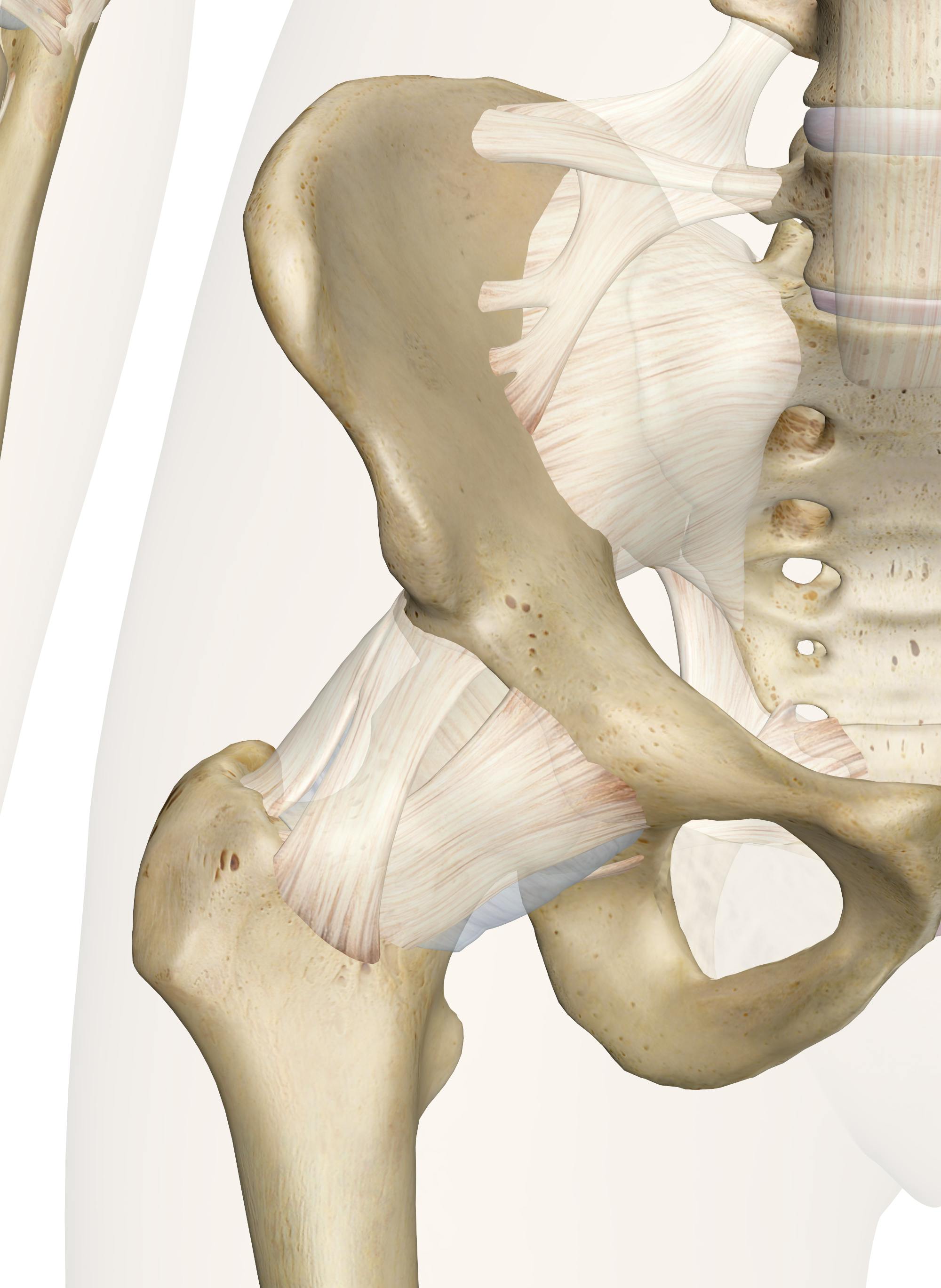
Hip bursitis refers to the inflammation of one or more bursae surrounding the hip joint, characterized by localized pain, swelling, and reduced mobility. The hip contains several bursae—fluid-filled sacs that reduce friction between tendons, muscles, and bony prominences—with the trochanteric, ischiofemoral, and iliopsoas bursae being the most clinically significant in cases of bursitis. These structures are strategically positioned to cushion repetitive movements, yet their irritation often stems from mechanical stress, overuse, or anatomical predispositions.
Anatomical Location and Structure of Hip Bursae
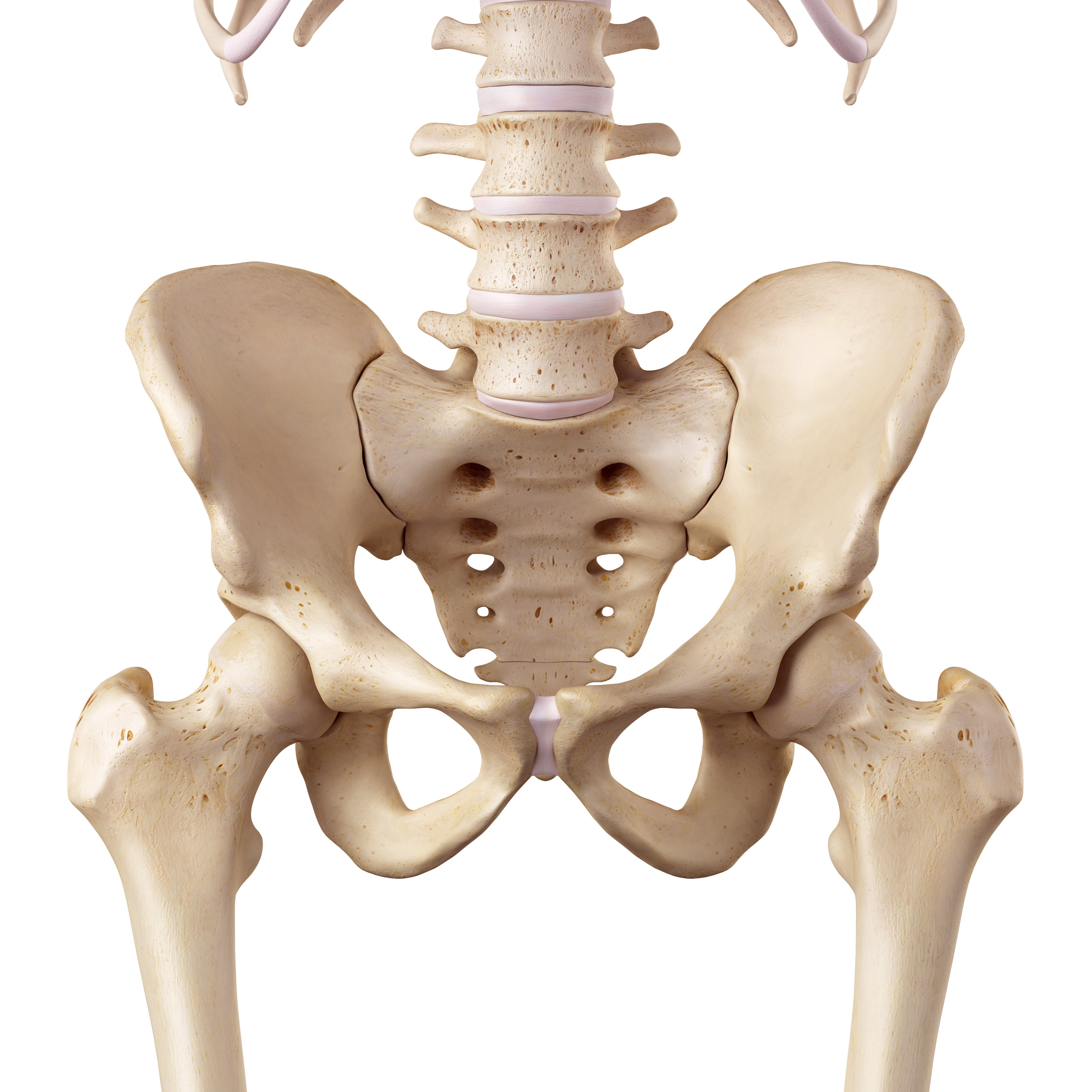
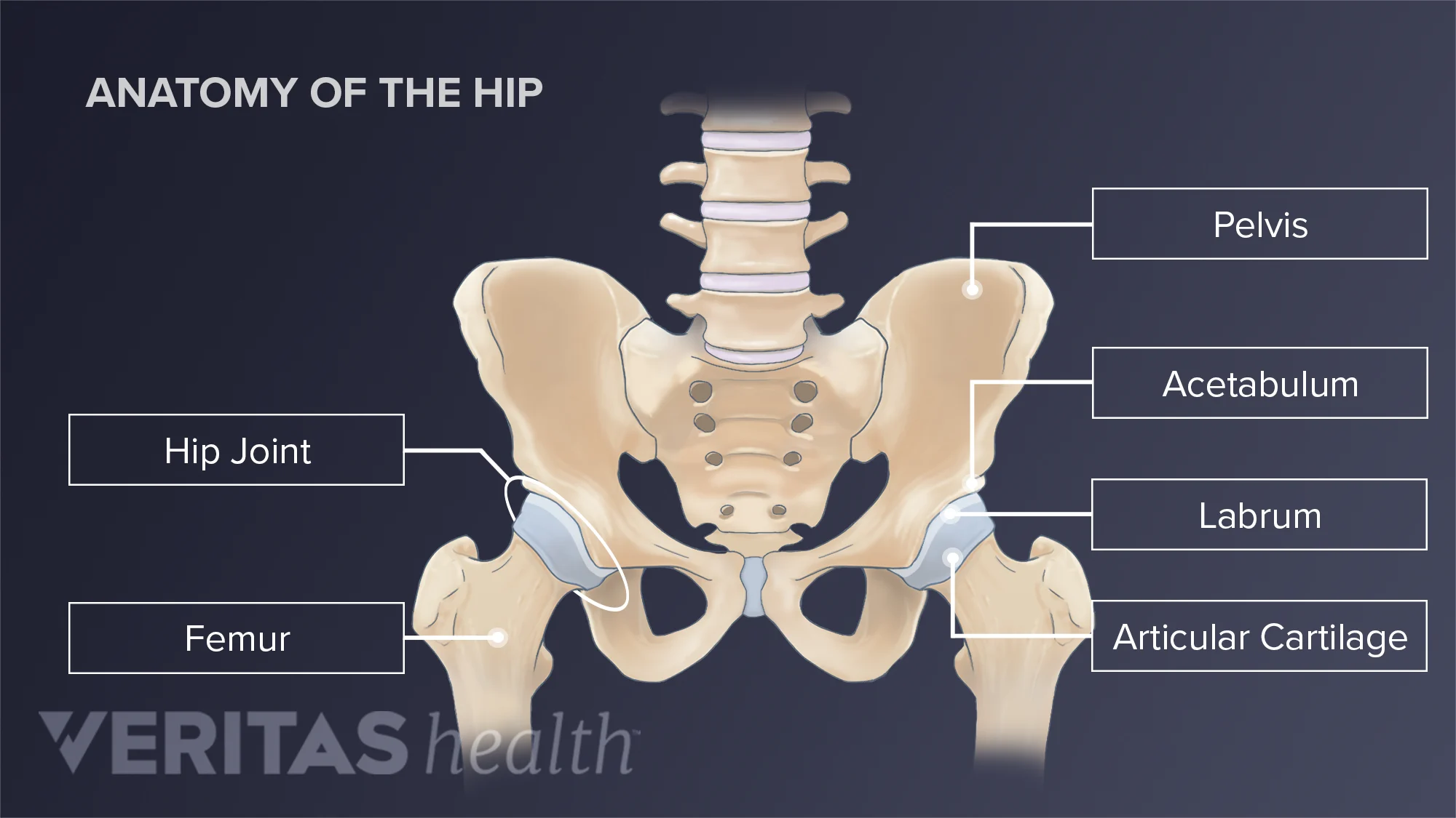
The hip’s bursae are situated in close proximity to major bony landmarks, tendons, and muscle groups, each serving a distinct biomechanical role. Below is a detailed breakdown of their positions and adjacent anatomical relationships:
Trochanteric Bursa
Iliopsoas Bursa
Ischiofemoral Bursa
Pathophysiology of Hip Bursitis: Inflammation Mechanisms
Inflammation in hip bursae arises from mechanical irritation, overuse, or trauma, leading to a cascade of cellular and structural changes. The primary triggers include:Mechanical Stress and Overuse
Trauma and Acute Injury
Cellular and Molecular Pathways
1. Initial Injury: Mechanical stress disrupts the bursal synovial lining, releasing pro-inflammatory cytokines (e.g., IL-1β, TNF-α).
2. Vascular Response: Increased capillary permeability leads to edema and leukocyte infiltration (neutrophils, macrophages).
3. Fibroblastic Proliferation: Chronic irritation stimulates fibroblast activation, thickening the bursal wall and reducing fluid mobility.
4. Calcification: Prolonged inflammation may deposit calcium hydroxyapatite crystals, further restricting motion.
Pathological Changes
Visual Representation: Hip Bursae and Bony Landmarks
Below is a descriptive table outlining the spatial relationships between hip bursae, bony structures, and associated tendons. This layout mirrors a labeled anatomical diagram, with arrows indicating common irritation sites.| Structure | Bony Landmark | Adjacent Tendons/Muscles | Common Irritation Sites |
|---|---|---|---|
| Trochanteric Bursa | Greater trochanter | Gluteus medius/minimus, IT band | Lateral hip (superficial bursa) |
| Deep bursa: Between greater trochanter and gluteus medius tendon | |||
| Iliopsoas Bursa | Femoral head/neck | Iliopsoas tendon (iliacus + psoas) | Anterior hip, near inguinal fold |
| Communicating variant: Adjacent to joint capsule | |||
| Ischiofemoral Bursa | Ischial tuberosity | Hamstring tendons (biceps femoris) | Posterior hip, near sciatic nerve pathway |
Comparative Anatomy: Hip Bursae vs. Other Joint Bursae
While bursae in all synovial joints share the function of reducing friction, the hip’s bursae exhibit unique anatomical and biomechanical features that distinguish hip bursitis from other joint pathologies.Shared Characteristics Across Joints
Distinct Features of Hip Bursae
| Feature | Hip Bursae | Shoulder (Subacromial Bursa) | Knee (Prepatellar Bursa) |
|---|---|---|---|
| Primary Function | Cushion repetitive hip motion | Protect rotator cuff during abduction | Absorb direct pressure (e.g., kneeling) |
| Mechanical Stress | High impact during gait/rotation | Compression in overhead activities | Shear forces from quadriceps contraction |
| Anatomical Constraints | Deep-seated (e.g., iliopsoas near joint capsule) | Superficial (subacromial space) | Superficial (patellar tendon pathway) |
| Clinical Presentation | Lateral/anteromedial hip pain | Shoulder impingement (pain arc) | Swelling over patella (baker’s cyst risk) |
| Unique Pathologies | Trochanteric bursitis (IT band friction) | Calcific tendinitis (rotator cuff) | Septic bursitis (post-traumatic infection) |
Example: A runner with trochanteric bursitis presents with lateral hip pain exacerbated by stair descent, whereas a weightlifter with subacromial bursitis reports pain during overhead presses—highlighting how joint-specific mechanics dictate bursal vulnerability.
Symptoms and Clinical Presentation of Hip Bursitis
Hip bursitis presents with a constellation of localized and functional symptoms that often mimic other musculoskeletal conditions, necessitating a structured clinical assessment. The characteristic pain patterns, physical examination findings, and patient-reported triggers collectively guide differential diagnosis and tailored management. Accurate identification of bursal involvement—whether trochanteric, ischiofemoral, or iliopsoas—refines diagnostic precision and informs therapeutic strategies.The clinical presentation of hip bursitis is dominated by pain, stiffness, and functional limitations, with variations depending on the specific bursa affected. Pain is typically mechanical in nature, exacerbated by activities that increase intra-bursal pressure or compress inflamed structures. Physical examination employs palpation, range-of-motion tests, and special maneuvers to isolate bursitis from conditions such as osteoarthritis, labral tears, or referred lumbar pathology. Below, the symptomatic features, diagnostic techniques, and differential considerations are detailed systematically.
Characteristic Symptoms and Pain Patterns
The primary symptom of hip bursitis is localized pain, often described as sharp, aching, or burning, with intensity correlating to bursal inflammation. Pain distribution varies by affected bursa:- Trochanteric bursitis (greater trochanteric pain syndrome, GTPS):
- Iliopsoas bursitis:
- Ischiofemoral bursitis:
Functional limitations frequently accompany pain, including:
Patient-reported triggers (to be assessed via history) include:
Physical Examination Techniques
A systematic physical examination distinguishes hip bursitis from other pathologies through palpation, range-of-motion tests, and special maneuvers. The following techniques are standardized for diagnostic accuracy:1. Palpation
- Iliopsoas bursitis:
- Ischiofemoral bursitis:
2. Range-of-Motion Tests
3. Special Maneuvers
- Ober’s Test:
- Resisted Straight Leg Raise (SLR):
- Fabere Test (Patrick’s Test):
4. Differential Diagnosis Considerations
Physical examination must exclude:
Patient Self-Assessment Symptom Checklist
To facilitate early recognition and self-monitoring, patients can use the following symptom checklist, rating severity on a 1–10 scale (1 = mild, 10 = disabling). Triggers should be documented to tailor activity modification.Pain Localization and Intensity
- Anterior hip/groin pain (iliopsoas bursitis):
- Posterolateral hip pain (ischiofemoral bursitis):
Functional Limitations
Associated Symptoms
Case Example: Trochanteric vs. Iliopsoas Bursitis
Causes and Risk Factors of Hip Bursitis
Hip bursitis arises from a combination of mechanical stress, systemic inflammation, and occupational or lifestyle-related factors. The primary etiology involves irritation or fluid accumulation within the hip bursae, often exacerbated by repetitive movements, direct trauma, or underlying musculoskeletal conditions. Understanding these contributors is essential for targeted prevention and intervention, particularly in high-risk populations such as athletes, older adults, and labor-intensive occupations.The development of hip bursitis follows a progressive pathological pathway influenced by biomechanical inefficiencies, tissue overload, and inadequate recovery. Below, the key causes and risk factors are categorized by mechanism, population-specific vulnerabilities, and a structured progression model illustrating how acute irritation evolves into chronic inflammation.
Mechanical Causes of Hip Bursitis
Mechanical factors account for the majority of hip bursitis cases, primarily through direct compression, friction, or repetitive strain on the bursae. The trochanteric bursa (most commonly affected) and iliopsoas bursa are particularly susceptible due to their proximity to bony prominences and high-mobility joints.Primary mechanical triggers include:
Real-world examples:
Systemic and Underlying Medical Conditions
Systemic inflammation or degenerative joint diseases predispose individuals to bursitis by altering tissue resilience and fluid dynamics. These conditions often coexist with mechanical stressors, amplifying bursal irritation.Key systemic contributors:
Pathophysiological link:
In systemic conditions, cytokine-mediated inflammation (e.g., TNF-α, IL-6) disrupts bursal membrane integrity, while reduced extracellular matrix turnover (e.g., collagen degradation in RA) compromises structural support. Mechanical stress then acts as a secondary trigger, converting subclinical irritation into symptomatic bursitis.
Population-Specific Risk Profiles
Risk factors for hip bursitis vary by demographic, with modifiable behaviors (e.g., ergonomics, activity modification) and non-modifiable traits (e.g., age, genetics) interacting to determine susceptibility.Athletes:
Older adults (≥65 years):
Occupational hazards:
Progression from Acute Irritation to Chronic Bursitis
The transition from reversible bursal irritation to chronic inflammation follows a biomechanical-inflammatory cascade, modulated by individual risk factors. Below is a structured flowchart outlining the stages:Stage 1: Initial MicrotraumaKey accelerants in progression:
Trigger: Single episode of overuse (e.g., marathon) or acute trauma (e.g., fall). Response: Localized bursal edema and mild synovial hyperplasia. Modifiable intervention: Rest, ice, and NSAIDs (if no systemic contraindications). Stage 2: Compensatory Adaptation
Trigger: Persistent mechanical stress (e.g., poor gait mechanics) or systemic inflammation (e.g., RA flare). Response: Mechanical: Fibroblastic proliferation and bursal thickening. Inflammatory: Neutrophil infiltration and cytokine release (IL-1β, PGE₂). Modifiable intervention: Corrective exercises (e.g., clamshells for gluteal activation) and activity modification. Stage 3: Chronic Inflammation
Trigger: Unresolved Stage 2 + repetitive cycles of irritation/recovery. Response: Structural: Bursal wall fibrosis and adhesions to adjacent tendons (e.g., gluteus medius). Systemic: Persistent low-grade inflammation (elevated CRP, ESR). Modifiable intervention: Physical therapy (e.g., manual therapy for tight IT band), corticosteroid injections (short-term), or lifestyle changes (weight loss, ergonomic adjustments). Stage 4: Degenerative Changes
Trigger: Long-standing chronic bursitis (>6 months) with inadequate management. Response: Mechanical: Heterotopic ossification or tendon calcification (e.g., "calcific tendinitis"). Functional: Reduced hip range of motion and compensatory gait patterns (e.g., Trendelenburg limp). Modifiable intervention: Surgical bursectomy (last resort) or targeted rehabilitation for secondary impairments (e.g., hip flexor tightness).
Diagnostic Methods and Imaging in Hip Bursitis
Imaging plays a critical role in confirming hip bursitis, excluding alternative diagnoses, and guiding targeted treatment. While clinical examination remains foundational, advanced imaging modalities—such as X-rays, ultrasound, and MRI—provide objective evidence of bursal inflammation, structural abnormalities, and differential pathologies. These techniques ensure accurate diagnosis, particularly in cases where symptoms overlap with greater trochanteric pain syndrome (GTPS), labral tears, or avascular necrosis (AVN). The selection of imaging modality depends on clinical suspicion, cost-effectiveness, and the need for soft-tissue or bony detail.The diagnostic approach begins with non-invasive, cost-effective imaging to rule out fractures, osteoarthritis, or other bony pathologies before progressing to more detailed soft-tissue assessments. Ultrasound and MRI offer superior visualization of bursal anatomy and inflammatory changes, while Doppler techniques can quantify vascular activity in acute cases. Below, the role of each modality is detailed, followed by diagnostic criteria for trochanteric bursitis and a comparative analysis of imaging features across common hip pathologies.
Role of Imaging Modalities in Diagnosing Hip Bursitis
X-raysX-rays serve as the first-line imaging modality in evaluating hip pain, primarily to exclude bony abnormalities that may mimic or coexist with bursitis. Key findings include:
Limitation: X-rays lack sensitivity for soft-tissue inflammation and cannot directly visualize bursae. Negative findings do not rule out bursitis but guide further imaging.Ultrasound
Ultrasound is the preferred modality for diagnosing trochanteric bursitis due to its real-time capability, lack of ionizing radiation, and ability to assess dynamic structures. Key ultrasound features include:
Doppler ultrasound enhances diagnostic accuracy by detecting increased vascularity in acute bursitis, where hyperemia correlates with inflammation. This modality is particularly useful in differentiating acute vs. chronic bursitis and ruling out septic bursitis (where vascularity is markedly elevated).
MRI
MRI provides high-resolution images of soft tissues, making it invaluable for complex cases where ultrasound is inconclusive or other pathologies (e.g., labral tears, AVN) are suspected. Key MRI findings in hip bursitis include:
MRI vs. Ultrasound: While MRI offers superior detail for deep structures, ultrasound is often sufficient for trochanteric bursitis due to its accessibility and lower cost. MRI is reserved for cases with atypical presentations or suspected coexisting pathologies.
Diagnostic Criteria for Trochanteric Bursitis
Trochanteric bursitis is diagnosed based on a combination of clinical, ultrasound, and MRI findings, with the goal of distinguishing it from greater trochanteric pain syndrome (GTPS), which encompasses multiple etiologies (e.g., gluteal tendinopathy, snapping hip syndrome). The following criteria integrate clinical examination with imaging:Clinical Criteria:
Ultrasound Criteria:
MRI Criteria:
Differentiating GTPS from Trochanteric Bursitis:
GTPS is an umbrella term that includes trochanteric bursitis but also encompasses gluteal tendinopathy, snapping hip syndrome, and other soft-tissue pathologies. Imaging helps refine the diagnosis:
Pure trochanteric bursitis: Isolated bursal inflammation on ultrasound/MRI. GTPS with bursitis: Bursal changes + gluteal tendon degeneration or tears. Snapping hip syndrome: Dynamic ultrasound may show iliotibial band (ITB) or gluteus maximus snapping over the trochanter.
Comparison of Imaging Features: Hip Bursitis vs. Other Pathologies
The following table summarizes key imaging differences between trochanteric bursitis and common hip pathologies, aiding in differential diagnosis:| Feature | Trochanteric Bursitis | Greater Trochanteric Pain Syndrome (GTPS) | Labral Tear | Avascular Necrosis (AVN) |
|---|---|---|---|---|
| Location of Abnormality | Superficial to greater trochanter (bursa) | Gluteal tendons, ITB, or bursa (multifocal) | Intra-articular (labrum, acetabulum) | Subchondral bone (femoral head) |
| X-ray Findings | Normal or degenerative changes | Normal or trochanteric calcifications | Joint space narrowing, osteophytes | Subchondral lucency, crescent sign, collapse |
| Ultrasound Findings | Bursal fluid/thickening, Doppler hyperemia | Gluteal tendon tears, ITB snapping, bursitis | Labral irregularity, joint effusion | Normal (unless secondary synovitis) |
| MRI Findings | T2/STIR hyperintensity in bursa, enhancement | Gluteal tendon edema/tears, bursitis | Labral contrast enhancement, marrow edema | Subchondral low T1, high T2 signal, collapse |
| Doppler Ultrasound (Acute) | Marked hypervascularity | Variable (depends on tendinopathy) | Synovial hyperemia (if inflammatory) | Usually absent (unless reactive) |
Doppler Ultrasound in Acute Hip Bursitis: Interpretation Script
Doppler ultrasound is instrumental in quantifying inflammatory activity in acute bursitis, where increased blood flow correlates with neutrophil infiltration and cytokine release. Below is a step-by-step interpretation protocol for identifying inflammatory changes:1. Patient Positioning and Probe Selection:
2. Identification of the Bursa:
Hip bursitis underscores the delicate balance between mechanical stress and physiological resilience in the musculoskeletal system, where even minor irritations can escalate into chronic discomfort if left unaddressed. From the trochanteric bursa’s vulnerability to lateral hip compression in runners to the iliopsoas bursa’s susceptibility in dancers performing repetitive hip flexion, each anatomical site presents unique diagnostic and therapeutic considerations. Advances in imaging—such as Doppler ultrasound for identifying inflammatory vascularity or MRI for differentiating soft-tissue involvement—have revolutionized precision diagnostics, while early intervention strategies, including activity modification, physical therapy, and targeted injections, can mitigate progression to debilitating conditions. Ultimately, recognizing hip bursitis as a preventable and treatable disorder empowers patients and clinicians alike to intervene proactively, ensuring optimal recovery and long-term hip health.
FAQ
What does hip bursitis pain feel like and where is it located?
Hip bursitis pain is usually a sharp, aching, or burning sensation on the outside of the hip, near the upper thigh or buttock. It often worsens when walking, climbing stairs, or lying on the affected side. Some people describe it as a deep, throbbing discomfort that may radiate down the thigh.
What are the most common symptoms of hip bursitis?
Common symptoms include pain or tenderness over the hip joint, swelling or stiffness, limited range of motion, and discomfort when pressing on the hip. Activities like sitting for long periods, squatting, or getting up from a chair may also trigger pain. Some people experience warmth or redness in the affected area.
What causes hip bursitis, and who is most at risk?
Hip bursitis is usually caused by inflammation of the bursae (fluid-filled sacs) due to repetitive motion, overuse, or pressure on the hip. Risk factors include activities like running or cycling, prolonged sitting, arthritis, or conditions that cause hip irritation (e.g., bone spurs). Poor posture or muscle weakness can also contribute.
What is hip bursitis called in medical terms?
Hip bursitis is also called greater trochanteric bursitis when it affects the bursa on the outer hip (near the greater trochanter). Other types include ischial bursitis (near the tailbone) or iliopsoas bursitis (front of the hip).
What are the best treatments for hip bursitis?
Treatment typically involves rest, ice, and over-the-counter pain relievers (like NSAIDs). Physical therapy (e.g., stretching and strengthening exercises) and cortisone injections can reduce inflammation. Severe cases may require activity modification or surgery if other treatments fail.
What exercises help relieve or prevent hip bursitis?
Gentle exercises like hip stretches (e.g., butterfly stretch), clamshells, and glute bridges strengthen supporting muscles and improve mobility. Avoid high-impact activities; low-impact options like swimming or cycling (with proper form) may help. Always warm up and cool down to prevent flare-ups.
Leave a Comment
Comments are moderated before appearing. The data you submit is processed according to the Privacy Policy of Voltefac.