What The Excretory System Functions And Mechanisms
Table of Contents
- The Excretory System: Definition, Core Functions, and Structural Breakdown
- Key Functions of the Excretory System
- Major Components of the Excretory System: Organ Functions and Associated Pathologies
- Kidney Function: Blood Filtration and Waste Excretion Process
- Key Organs and Their Mechanisms in Excretion
- Nephron Structure and Renal Filtration Mechanisms
- Liver’s Dual Role in Excretion: Bile Production and Detoxification
- Skin Excretion and Thermoregulation via Sweat Glands
- Lesser-Known Excretory Organs and Their Contributions
- Physiological Processes and Pathways in Excretion
- Urine Formation Process
- Excretion Pathways for Water-Soluble vs. Fat-Soluble Wastes
- Excretion Routes and Chemical Transformations of Common Wastes
- Diseases and Dysfunctions of the Excretory System
- Common Excretory System Disorders and Their Impact on Homeostasis
- Case Study: Progression of Chronic Kidney Disease (CKD) and Systemic Effects
- Evolutionary and Comparative Perspectives on Excretory Systems
- Comparative Excretory Mechanisms in Humans, Fish, and Insects
- Evolution of Excretory Efficiency: Mammals vs. Reptiles
- Species-Specific Solutions to Excretory Challenges
- Experimental and Educational Tools for Excretory System Instruction
- Simulation of Kidney Filtration Using a DIY Lab Setup
- Fill-in-the-Blank Template for Excretory System Anatomy
- Quiz-Style True/False Statements on Excretory Processes
- FAQ
- what's the excretory system?
- what's the excretory system function?
- what's the endocrine system?
- what the endocrine system does?
- what's the urinary system?
- what's the endocrine system made up of?
The excretory system serves as the body’s intricate waste management network, ensuring survival through precise regulation of fluid balance, toxin elimination, and metabolic byproduct clearance. Beyond its foundational role in homeostasis, this system integrates complex physiological pathways—from the kidneys’ ultrafiltration processes to the liver’s detoxification cascades—demonstrating nature’s efficiency in balancing chemical equilibrium. By examining its anatomical components, cellular mechanisms, and evolutionary adaptations, we uncover how this system adapts to environmental demands while safeguarding internal stability. This exploration bridges scientific rigor with practical insights, revealing why disruptions in excretion can trigger systemic disorders.
From the nephron’s selective filtration to the skin’s thermoregulatory sweating, each organ contributes uniquely to waste processing, often operating in tandem with other systems like the endocrine or respiratory networks. Comparative analysis further illuminates how species optimize excretion—whether through urea synthesis in mammals or uric acid conservation in desert animals—highlighting evolutionary trade-offs between efficiency and survival. Understanding these processes not only demystifies physiological functions but also underscores the fragility of homeostasis when excretory pathways falter, as seen in diseases like chronic kidney disease or cystic fibrosis.
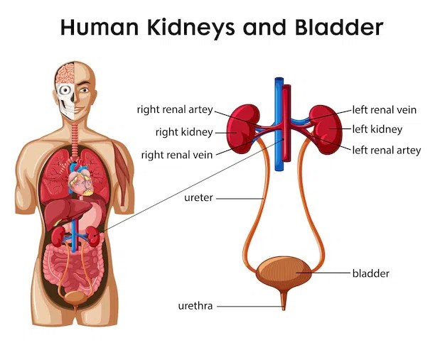
The Excretory System: Definition, Core Functions, and Structural Breakdown
The excretory system is a complex network of organs and structures responsible for eliminating metabolic waste, maintaining internal chemical balance, and regulating physiological processes essential for survival. Its primary role revolves around homeostasis, ensuring the body’s internal environment remains stable despite external fluctuations. Through filtration, secretion, and excretion, the system prevents toxic accumulation while preserving vital substances like water, electrolytes, and nutrients.
The excretory system operates through a coordinated interplay of organs, each specializing in removing specific waste products while contributing to fluid and electrolyte equilibrium. Below, a structured breakdown highlights its key functions: waste removal (e.g., urea, carbon dioxide), fluid balance (via osmosis and hormone regulation), and toxin regulation (detoxification and pH adjustment). The kidneys, liver, lungs, skin, and intestines serve as the primary components, each with distinct yet interconnected roles in sustaining physiological stability.
Key Functions of the Excretory System
The excretory system performs three overarching functions critical to survival:1. Waste Removal
Elimination of metabolic byproducts generated during cellular respiration, protein breakdown, and other biochemical processes. Failure to excrete these wastes—such as urea (from protein metabolism) or bilirubin (from hemoglobin degradation)—leads to systemic toxicity.
2. Fluid and Electrolyte Balance
Regulation of water, sodium, potassium, and other ions to maintain osmotic pressure, blood volume, and pH levels. Hormones like antidiuretic hormone (ADH) and aldosterone play pivotal roles in this process.
3. Toxin Regulation and Detoxification
Neutralization or removal of exogenous toxins (e.g., drugs, heavy metals) and endogenous waste products (e.g., ammonia) to prevent cellular damage. The liver and kidneys are particularly vital in this function, converting harmful substances into less toxic forms or excreting them directly.
Major Components of the Excretory System: Organ Functions and Associated Pathologies
The following table summarizes the primary organs of the excretory system, their functions, example waste products, and associated diseases when dysfunction occurs.| Organ | Primary Function | Example Waste Product | Associated Disease |
|---|---|---|---|
| Kidneys | Filtration of blood to remove metabolic wastes, regulate fluid/electrolyte balance, and secrete hormones (e.g., erythropoietin). | Urea, creatinine, excess water, potassium | Chronic Kidney Disease (CKD), nephrolithiasis (kidney stones), renal failure |
| Liver | Detoxification of blood via metabolism of drugs/toxins, synthesis of bile for fat emulsification, and conversion of ammonia to urea. | Bilirubin, ammonia (converted to urea), excess hormones | Hepatitis, cirrhosis, hepatic encephalopathy (ammonia toxicity) |
| Lungs | Excretion of carbon dioxide (a waste product of cellular respiration) and regulation of blood pH through respiratory adjustments. | Carbon dioxide (CO₂), water vapor | Chronic Obstructive Pulmonary Disease (COPD), respiratory acidosis/alkalosis |
| Skin | Excretion of sweat (containing water, electrolytes, and urea) and thermoregulation via perspiration. | Urea, lactic acid, excess salts | Cystic fibrosis (impaired sweat gland function), hyperhidrosis (excessive sweating) |
| Intestines | Absorption of water and electrolytes from digested food, excretion of solid waste (feces), and elimination of bile pigments (e.g., stercobilin). | Stercobilin, undigested food residues, excess cholesterol | Constipation, diarrhea, colorectal cancer, diverticulitis |
Kidney Function: Blood Filtration and Waste Excretion Process
The kidneys are the primary filtration units of the excretory system, processing approximately 120–150 liters of blood daily to produce urine. The process occurs in three main stages: glomerular filtration, tubular reabsorption, and tubular secretion. Below is a step-by-step text-based flow diagram describing the pathway:1. Glomerular Filtration
Blood enters the glomerulus (a network of capillaries) under high pressure, forcing water, ions, glucose, and small waste molecules (e.g., urea) into the Bowman’s capsule to form glomerular filtrate. Large molecules (e.g., proteins, blood cells) remain in the bloodstream due to the glomerular filtration barrier.
2. Tubular Reabsorption
The filtrate travels through the proximal convoluted tubule, where ~65% of water, sodium, and nutrients (e.g., glucose, amino acids) are reabsorbed into the blood via active and passive transport. The loop of Henle further refines concentration gradients to regulate water and electrolyte balance.
3. Tubular Secretion
In the distal convoluted tubule (DCT) and collecting duct, additional waste products (e.g., potassium, hydrogen ions, drugs) are actively secreted into the filtrate. Antidiuretic hormone (ADH) and aldosterone modulate water and sodium reabsorption here, determining urine concentration.
4. Urine Formation and Excretion
The processed filtrate, now urine, flows into the renal pelvis, descends through the ureters, and is stored in the bladder before voluntary expulsion via the urethra. The kidneys also secrete erythropoietin (EPO) to stimulate red blood cell production and renin to regulate blood pressure.
Key Physiological Principle:
"The kidneys maintain homeostasis by balancing filtration, reabsorption, and secretion, ensuring only waste and excess substances are excreted while preserving essential solutes."
Key Organs and Their Mechanisms in Excretion
The excretory system relies on specialized organs that perform distinct yet interconnected functions to eliminate metabolic waste, maintain homeostasis, and regulate fluid balance. Among these, the kidneys, liver, and skin play central roles, while lesser-known contributors such as salivary glands and the large intestine also participate in waste processing. Each organ employs unique cellular and biochemical mechanisms to ensure efficient excretion, often integrating filtration, secretion, and metabolic conversion processes.The kidneys serve as the primary filtration units, processing blood to remove excess solutes and water, while the liver detoxifies harmful substances and synthesizes bile for fat emulsification. Meanwhile, the skin acts as a secondary excretory pathway, releasing waste through sweat while aiding thermoregulation. Below, the structural and functional intricacies of these organs—along with their cellular-level operations—are examined in detail.
Nephron Structure and Renal Filtration Mechanisms
The nephron, the functional unit of the kidney, consists of a glomerulus, proximal convoluted tubule (PCT), loop of Henle, distal convoluted tubule (DCT), and collecting duct. These components work in tandem to perform filtration, reabsorption, and secretion, ensuring precise regulation of blood composition.The glomerulus, a network of capillaries, applies hydrostatic pressure to filter plasma into Bowman’s capsule, producing glomerular filtrate containing water, glucose, ions, and waste products like urea and creatinine. This process is governed by the filtration fraction (typically 20% of renal plasma flow) and regulated by glomerular filtration rate (GFR), influenced by afferent/efferent arteriolar resistance and oncotic pressure.
In the proximal convoluted tubule (PCT), 65% of filtered water, all glucose, and essential ions (e.g., Na⁺, K⁺, HCO₃⁻) are reabsorbed via symporters (e.g., Na⁺-glucose cotransporter) and antiporters (e.g., Na⁺/H⁺ exchanger). The loop of Henle establishes a concentration gradient via the countercurrent multiplier system, where descending limbs passively reabsorb water, while ascending limbs actively transport Na⁺ and Cl⁻. The distal convoluted tubule (DCT) and collecting duct fine-tune electrolyte balance through aldosterone (Na⁺ reabsorption) and antidiuretic hormone (ADH-mediated water reabsorption).
Secretion occurs primarily in the PCT and DCT, where organic acids (e.g., creatinine, uric acid) and bases (e.g., ammonia) are actively transported from peritubular capillaries into the tubule lumen. This ensures removal of substances not efficiently filtered at the glomerulus.
Liver’s Dual Role in Excretion: Bile Production and Detoxification
The liver performs excretion through bile synthesis and detoxification, with urea synthesis being a critical metabolic pathway. Bile, produced by hepatocytes, contains bile salts (derived from cholesterol), bilirubin (a breakdown product of hemoglobin), and phospholipids. It emulsifies dietary fats in the small intestine, facilitating absorption while eliminating cholesterol and waste pigments.For urea synthesis, ammonia—a toxic byproduct of amino acid catabolism—undergoes a three-step process in the urea cycle (occurring in hepatocytes and mitochondria):
-
Ammonia (NH₃) + CO₂ + ATP → Carbamoyl phosphate
Catalyzed by carbamoyl phosphate synthetase I (CPS-I), this reaction consumes 2 ATP molecules, incorporating NH₃ and CO₂ into carbamoyl phosphate. -
Carbamoyl phosphate + Ornithine → Citrulline
Ornithine transcarbamoylase (OTC) transfers the carbamoyl group to ornithine, forming citrulline, which diffuses into the cytoplasm. -
Citrulline + Aspartate + 3 ATP → Urea + Fumarate
Two enzymes, argininosuccinate synthetase and argininosuccinase, convert citrulline to argininosuccinate, then to arginine. Arginase finally hydrolyzes arginine into urea (excreted via kidneys) and ornithine (recycled).
Additionally, the liver detoxifies drugs and toxins via Phase I (oxidation, reduction, hydrolysis) and Phase II (conjugation) reactions, converting hydrophobic compounds into water-soluble metabolites (e.g., glucuronides, sulfates) for renal excretion.
Skin Excretion and Thermoregulation via Sweat Glands
The skin contributes to excretion through eccrine glands, which secrete sweat—a hypotonic fluid composed of:Sweat excretion is regulated by the sympathetic nervous system, with acetylcholine stimulating eccrine glands (unlike apocrine glands, which respond to adrenaline). The primary function is thermoregulation, as evaporative cooling reduces body temperature by up to 10% of metabolic heat. Additionally, sweat eliminates 0.5–1.0 L/day of waste (e.g., urea, ammonia), though its role in detoxification is secondary to renal function. Chronic sweating disorders (e.g., hyperhidrosis) or impaired gland function (e.g., cystic fibrosis) disrupt electrolyte balance and thermoregulation.Sweat composition varies with diet, hydration, and activity. For example, high-protein diets increase urea excretion, while intense exercise elevates lactate and K⁺ concentrations. The skin’s excretory capacity is limited compared to kidneys but is critical in maintaining homeostasis during heat stress or prolonged physical exertion.
Lesser-Known Excretory Organs and Their Contributions
Beyond the kidneys, liver, and skin, three additional organs play specialized roles in waste elimination:-
Salivary Glands
Produce saliva containing lysozyme (antibacterial enzyme), amylase (digestive enzyme), and mucus, but also excrete urea, uric acid, and ammonia via diffusion. While not a primary excretory pathway, salivary glands contribute to oral detoxification and maintain oral pH. Dysfunction (e.g., xerostomia) can impair waste clearance and increase oral microbial load. -
Large Intestine
Absorbs water and electrolytes from indigestible food residues, but also excretes bile salts, cholesterol, and dead bacteria via feces. The colon’s microbiota metabolizes undigested proteins into indoles, skatoles, and ammonia, which are partially reabsorbed or excreted. Constipation disrupts this process, increasing toxin reabsorption and systemic load. -
Lungs
Excrete carbon dioxide (CO₂) as a byproduct of cellular respiration, with ~200 mL of CO₂ produced daily by an average adult. Additionally, they remove volatile organic compounds (e.g., acetone from fat metabolism) and water vapor during exhalation. Pulmonary excretion is passive, driven by alveolar gas exchange, but critical for maintaining acid-base balance via CO₂ elimination.

Physiological Processes and Pathways in Excretion
The excretory system relies on precise physiological mechanisms to filter, process, and eliminate metabolic wastes while maintaining homeostasis. Urine formation, waste solubility, and autonomic regulation represent critical pathways that integrate renal, hepatic, and neural functions. These processes ensure the removal of harmful byproducts while conserving essential substances through selective reabsorption and feedback-driven adjustments."Excretion is not merely waste disposal but a finely tuned balance between filtration, selective retention, and adaptive responses to physiological demands."
Urine Formation Process
Urine formation occurs in three sequential stages—glomerular filtration, tubular reabsorption, and tubular secretion—each governed by distinct anatomical structures. The glomerulus initiates filtration under high hydrostatic pressure, while the loop of Henle establishes osmotic gradients for water reabsorption. The collecting ducts then finalize urine concentration through hormonal regulation.-
Glomerular Filtration
The glomerulus, a network of capillaries within the renal corpuscle, filters plasma under glomerular hydrostatic pressure (GHP, ~55 mmHg) while opposing forces—colloid osmotic pressure (COP, ~30 mmHg) and Bowman’s capsule pressure (~15 mmHg)—determine net filtration pressure (NFP = GHP − COP − Bowman’s pressure). This process yields glomerular filtrate, which contains water, glucose, ions, and small solutes but excludes blood cells and large proteins (e.g., albumin). The filtration rate averages 125 mL/min in healthy adults, producing ~180 L of filtrate daily. -
Tubular Reabsorption in the Proximal Convoluted Tubule (PCT)
The PCT reabsorbs ~65% of filtered water, Na⁺, Cl⁻, and glucose via active transport (e.g., Na⁺/K⁺ ATPase) and co-transport mechanisms (e.g., SGLT2 for glucose). Organic solutes like amino acids are reclaimed through secondary active transport, while paracellular pathways allow passive movement of ions. The PCT’s extensive microvilli and high mitochondrial density support these energy-dependent processes. -
Loop of Henle and Countercurrent Multiplier System
The descending limb is permeable to water but impermeable to solutes, allowing osmotic water reabsorption as filtrate passes through the hypertonic medulla (up to 1,200 mOsm/L). The ascending limb actively pumps Na⁺, Cl⁻, and K⁺ into the interstitium via Na⁺/K⁺/2Cl⁻ cotransporters (NKCC2), creating a diluting segment while maintaining the medullary osmotic gradient. This gradient enables the collecting ducts to concentrate urine via antidiuretic hormone (ADH)-mediated aquaporin-2 insertion. -
Selective Secretion and Final Adjustments in Distal Tubule/Collecting Duct
The distal convoluted tubule (DCT) and collecting duct refine urine composition through:
- Selective secretion of H⁺ (via H⁺-ATPase), K⁺, and organic acids (e.g., creatinine) into the tubule lumen.
- ADH-regulated water reabsorption in the collecting duct, where aquaporin-2 channels insert in response to plasma osmolality detected by osmoreceptors in the hypothalamus.
- Aldosterone enhances Na⁺ reabsorption and K⁺ secretion in the DCT, while atrial natriuretic peptide (ANP) promotes Na⁺ excretion during volume overload.
-
Final Urine Composition and Volume Regulation
The end product is ~1–2 L of concentrated urine/day, with osmolality ranging from 50 mOsm/L (dilute) to 1,200 mOsm/L (concentrated). Key regulatory hormones include:
- ADH (vasopressin): Increases water permeability in collecting ducts.
- Aldosterone: Enhances Na⁺/K⁺ exchange in DCT.
- ANP: Inhibits Na⁺ reabsorption in the collecting duct.
Excretion Pathways for Water-Soluble vs. Fat-Soluble Wastes
The solubility of metabolic wastes dictates their excretion route, as hydrophilic compounds rely on aqueous transport, while lipophilic toxins require bile salts, carrier proteins, or enzymatic modification for elimination. The liver plays a pivotal role in converting fat-soluble wastes into water-soluble conjugates via Phase I (oxidation, reduction) and Phase II (conjugation) reactions."Fat-soluble toxins (e.g., steroids, bilirubin) are sequestered in bile or bound to plasma proteins to prevent reabsorption in the renal tubules."
-
Water-Soluble Wastes (Renal Excretion Pathway)
Hydrophilic wastes, such as urea, creatinine, and excess ions (e.g., NH₄⁺, HCO₃⁻), are filtered by the glomerulus and excreted via:
- Passive glomerular filtration (e.g., urea, inulin).
- Active tubular secretion (e.g., organic cations/anions via OATs/OCTs in the PCT).
- pH-dependent reabsorption/secretion (e.g., NH₄⁺ excretion to buffer acidity). Renal clearance ensures these wastes avoid enterohepatic recirculation, as they are not reabsorbed by lipid membranes.
-
Fat-Soluble Wastes (Hepatic and Biliary Excretion Pathway)
Lipophilic compounds (e.g., bilirubin, steroid hormones, drug metabolites) require:
- Liver conjugation (e.g., bilirubin → bilirubin diglucuronide via UDP-glucuronosyltransferase).
- Bile salt-mediated solubilization (e.g., cholesterol derivatives emulsified in micelles).
- Enterohepatic circulation for some toxins (e.g., bile acids reabsorbed in the ileum via ASBT transporters). Failure to conjugate fat-soluble wastes (e.g., in Gilbert’s syndrome) leads to jaundice or cholestasis.
-
Carrier-Mediated Excretion for Toxins
Certain toxins (e.g., heavy metals, lipophilic drugs) bind to:
- Plasma proteins (e.g., albumin for bilirubin) to prevent filtration.
- Organic anion transporters (OATs) in the kidney for active secretion.
- Multidrug resistance proteins (MRPs) in the liver/bile ducts. Example: Bilirubin is transported by MRP2 into bile, while digoxin is secreted via P-glycoprotein (MDR1) in the kidney.
Excretion Routes and Chemical Transformations of Common Wastes
The following table summarizes the origin, excretion pathway, and biochemical modifications of key metabolic byproducts. These transformations ensure compatibility with aqueous excretion systems while minimizing toxicity.| Substance | Source | Excretion Route | Chemical Transformation | ||||||||||||||||||||||||||||||
|---|---|---|---|---|---|---|---|---|---|---|---|---|---|---|---|---|---|---|---|---|---|---|---|---|---|---|---|---|---|---|---|---|---|
| Urea | Protein catabolism (liver via ornithine cycle) | Renal (glomerular filtration + tubular secretion) | No modification; excreted as is (osmotic diuretic) | ||||||||||||||||||||||||||||||
| Bilirubin | Heme breakdown (RBCs → spleen/liver) | Biliary (conjugated form) → fecal excretion; small fraction renal | Unconjugated (lipophilic) → conjugated with glucuronic acid (water-soluble) | ||||||||||||||||||||||||||||||
| Carbon Dioxide (CO₂) | Cellular respiration (Krebs cycle) | Pulmonary (exhaled as CO₂) + minor renal (HCO₃⁻) | CO₂ + H₂O → H₂CO₃ → H⁺ + HCO₃⁻ (buffered in blood) | ||||||||||||||||||||||||||||||
| Excess Ions (e.g., K⁺, H⁺, PO₄³⁻) | Dietary intake or metabolic imbalance | Renal (selective reabsorption/secretion) |
|

Leave a Comment
Comments are moderated before appearing. The data you submit is processed according to the Privacy Policy of Voltefac.