| Smoking |
Nicotine-induced vasoconstriction paradoxically reduces symptoms acutely but accelerates venous wall damage long-term. |
"Do you smoke cigarettes or use tobacco products? If so, how

Accurate diagnosis of varicocele relies on a combination of clinical examination, advanced imaging techniques, and laboratory assessments. While physical palpation remains the initial screening method, confirmatory diagnostics often involve Doppler ultrasound due to its non-invasive nature, high sensitivity, and ability to assess hemodynamics. Additional tools, such as venography and thermal imaging, provide complementary insights but are typically reserved for specialized cases. Laboratory evaluations further support diagnosis by identifying varicocele-related infertility through semen and hormonal abnormalities.
Doppler Ultrasound in Varicocele Diagnosis
Doppler ultrasound is the gold standard for diagnosing varicocele, offering real-time visualization of venous blood flow and structural abnormalities. The procedure employs high-frequency sound waves to differentiate arterial and venous flow, with color flow imaging enhancing detection of reflux and dilated veins. Proper probe settings, imaging planes, and technical execution are critical for accurate diagnosis.Equipment and Settings
The examination requires a linear-array transducer (typically 7–12 MHz) to achieve high-resolution images of scrotal structures. Key settings include:
Color Doppler frequency: 5–7 MHz (adjustable based on patient habitus).
Pulse repetition frequency (PRF): 0.5–1.5 kHz to optimize venous flow detection.
Wall filter: Set to low (10–30 Hz) to minimize noise while preserving low-velocity venous signals.
Gain and color scale: Adjusted to balance signal clarity without artifacts.Imaging Planes and Technique
The scrotum is scanned in both transverse and longitudinal planes to evaluate the pampiniform plexus and internal spermatic veins. The patient is positioned supine with the head elevated at 30–45° to facilitate venous distension. Key steps include:
1. Initial grayscale imaging to identify dilated veins (>3 mm diameter) in the spermatic cord.
2. Color Doppler activation to assess flow direction and velocity, with reflux defined as retrograde flow during Valsalva maneuver.
3. Spectral Doppler analysis to measure peak systolic velocity (PSV) and end-diastolic velocity (EDV), with abnormal findings including reversed or absent flow in the internal spermatic vein. Color Flow Analysis Criteria
Venous dilation: Internal spermatic vein diameter ≥3 mm in the supine position or ≥2 mm with Valsalva.
Reflux detection: Retrograde flow lasting >1 second during Valsalva (indicative of incompetent valves).
Hemodynamic patterns: Absent or reversed flow in the internal spermatic vein during systole, suggestive of venous hypertension.
Diagnostic threshold for varicocele via Doppler ultrasound:
Venous diameter ≥2 mm (with Valsalva) or ≥3 mm (supine).
Reflux duration >1 second during Valsalva maneuver.
Flow pattern: Absent or reversed flow in the internal spermatic vein on spectral Doppler.
Varicocele Grading Scale: Clinical and Ultrasonographic Classification
The grading of varicocele integrates physical examination findings with ultrasonographic measurements to standardize severity assessment. Grades range from subclinical (asymptomatic) to Grade III (palpable without augmentation). Accurate grading guides treatment decisions, particularly in infertile patients.Step-by-Step Grading Protocol
1. Preparation: Patient stands for 15–20 minutes to maximize venous distension.
2. Physical Examination:
Grade I (Subclinical): Not palpable manually but detectable via Doppler ultrasound (venous diameter <3 mm, reflux present).
Grade II: Palpable only during Valsalva maneuver; ultrasound shows dilated veins (3–5 mm) with reflux.
Grade III: Palpable at rest; ultrasound reveals veins >5 mm with prominent reflux.
3. Ultrasonographic Confirmation:
Measure internal spermatic vein diameter in transverse plane at the level of the external inguinal ring.
Document reflux duration and flow characteristics during Valsalva.
-
Subclinical Varicocele
- Palpation: Non-palpable manually.
- Ultrasound Findings: Venous diameter <2 mm (supine) or <3 mm (with Valsalva), reflux detected on Doppler.
- Clinical Relevance: Often identified incidentally in infertile males; may correlate with suboptimal semen parameters.
-
Grade I (Mild)
- Palpation: Palpable only with Valsalva maneuver.
- Ultrasound Findings: Venous diameter 2–3 mm (supine) or 3–5 mm (with Valsalva), reflux duration <2 seconds.
- Clinical Relevance: May progress to higher grades; treatment considered if infertility or testicular atrophy is present.
-
Grade II (Moderate)
- Palpation: Palpable without Valsalva but reduces when supine.
- Ultrasound Findings: Venous diameter 3–5 mm (supine), reflux duration >2 seconds.
- Clinical Relevance: Strong association with infertility and testicular hypofunction; surgical intervention often recommended.
-
Grade III (Severe)
- Palpation: Palpable and visible through scrotal skin at rest.
- Ultrasound Findings: Venous diameter >5 mm (supine), sustained reflux (>3 seconds), possible venous tortuosity.
- Clinical Relevance: High risk of infertility and testicular damage; urgent intervention advised.
Diagnostic modalities for varicocele vary in accuracy, invasiveness, and clinical utility. Doppler ultrasound remains the first-line tool, while venography and thermal imaging serve niche applications. The following table summarizes key attributes, including advantages, limitations, and typical use cases.
| Diagnostic Tool |
Pros |
Cons |
Typical Use Case |
|---|
| Doppler Ultrasound |
- Non-invasive, radiation-free, and cost-effective.
- Real-time visualization of venous hemodynamics and reflux.
- High sensitivity (98%) and specificity (93%) for varicocele detection.
- Can assess testicular volume and secondary effects (e.g., atrophy).
|
- Operator-dependent; requires skilled technologist.
- Limited in obese patients due to poor acoustic penetration.
- Cannot evaluate retroperitoneal venous anatomy (e.g., left renal vein compression).
|
- First-line diagnostic tool for suspected varicocele.
- Preoperative assessment for varicocelectomy candidates.
- Monitoring post-treatment outcomes.
|
| Venography |
- Gold standard for anatomical venous mapping (e.g., retroperitoneal varices).
- High-resolution imaging of venous pathways and collateral circulation.
- Useful in complex cases (e.g., recurrent varicocele, renal vein compression).
|
- Invasive (requires catheterization and contrast injection).
- Risk of complications (e.g., hematoma, contrast reactions).
- Not routinely used due to higher cost and invasiveness.
|
- Pre-surgical evaluation in recurrent or atypical varicocele.
- Assessment of retroperitoneal venous anatomy (e.g., nutcracker syndrome).
- Research or teaching settings for anatomical correlation.
|
| Thermal Imaging (Infrared Thermography) |
- Non-invasive and radiation-free.
- Detects temperature asymmetry between testes (varicocele side often cooler).
- Useful in subclinical cases where ultrasound is normal.
|
- Low sensitivity (50–70%) compared to Doppler ultrasound
Treatment Options and Interventions for Varicocele
Varicocele management varies based on patient age, symptom severity, fertility concerns, and anatomical presentation. While asymptomatic cases may require no intervention, symptomatic or subfertile patients benefit from targeted therapeutic approaches. Treatment modalities range from conservative measures to minimally invasive and open surgical techniques, each with distinct indications, procedural nuances, and recovery profiles.The selection of intervention depends on clinical presentation, patient comorbidities, and reproductive goals. Non-surgical strategies serve as first-line options for mild cases, whereas advanced techniques are reserved for refractory symptoms or fertility impairment. Below, surgical methodologies, non-surgical pathways, comparative efficacy metrics, and post-operative protocols are systematically outlined.
Surgical Techniques for Varicocele Repair
Surgical repair aims to ligate or embolize the internal spermatic vein to restore venous drainage and alleviate symptoms. Techniques differ in invasiveness, precision, and anatomical access. The choice of approach influences recurrence rates, complications, and recovery timelines.Open Inguinal Varicocelectomy
The gold-standard open technique involves a 3–5 cm inguinal incision over the external inguinal ring. Key steps include:
- Anatomical Exposure: Dissection of the spermatic cord under general or local anesthesia, with identification of the vas deferens, testicular artery, and pampiniform plexus.
- Ligation Targets: Selective ligation of dilated veins (typically 2–3 mm) while preserving arterial and lymphatic structures, using silk or non-absorbable sutures.
- Tools: Standard surgical instruments (e.g., Babcock clamps, Metzenbaum scissors) and loupes for magnification.
- Advantages: High success rates (90–98%), low recurrence, and cost-effectiveness.
- Limitations: Post-operative pain, risk of hydrocele (1–5%), and longer recovery (1–2 weeks).
Laparoscopic Varicocele Ligation
Performed under general anesthesia with a 10 mm umbilical port and two 5 mm lateral ports, this approach targets the left renal vein via retroperitoneal access. Critical steps include:
- Visualization: Laparoscopic camera (30° angle) to identify the internal spermatic vein at the renal vein junction.
- Ligation: Endoclips or harmonic scalpel applied to the vein trunk, with optional division.
- Tools: Laparoscopic graspers, energy devices, and insufflation (CO₂ at 12–15 mmHg).
- Advantages: Reduced post-operative pain, shorter hospitalization, and minimal scrotal trauma.
- Limitations: Higher recurrence rates (5–10%) due to collateral venous drainage, and technical challenges in obese patients.
Microsurgical Subinguinal Varicocelectomy
A minimally invasive alternative with a 2 cm subinguinal incision, this technique employs 2.5–4× magnification to ligate veins <2 mm. Key features:
- Anatomical Precision: Preservation of lymphatic vessels and arterial branches via meticulous dissection.
- Tools: Operating microscope, micro-sutures (10-0 nylon), and microsurgical instruments.
- Advantages: Lowest recurrence rates (<5%), minimal post-operative discomfort, and high fertility restoration (70–80% improvement in semen parameters).
- Limitations: Requires specialized training, longer operative time (45–60 minutes), and higher cost.
Percutaneous Transcatheter Embolization
A radiology-guided procedure involving femoral vein access and catheterization of the internal spermatic vein. Techniques include:
- Sclerotherapy: Injection of sodium tetradecyl sulfate or ethanol to induce thrombosis.
- Coil Embolization: Placement of metallic coils (e.g., steel or platinum) to occlude venous flow.
- Tools: Fluoroscopy, microcatheters (e.g., Renegade), and embolic agents.
- Advantages: Non-surgical, outpatient setting, and high technical success (95–98%).
- Limitations: Recurrence (10–20%) due to collateral circulation, risk of deep vein thrombosis (0.5%), and higher cost than open surgery.
Non-Surgical Management Pathways
Non-surgical interventions are indicated for asymptomatic patients, mild symptoms, or those unwilling to undergo surgery. The flowchart below outlines decision criteria and corresponding management strategies, prioritizing conservative measures before escalation.Decision Criteria for Non-Surgical Pathways
- Asymptomatic Varicocele: Active surveillance with annual clinical evaluation.
- Mild Symptoms (e.g., dull ache, < Grade II): Lifestyle modifications and supportive care.
- Moderate/Severe Symptoms (e.g., pain, infertility): Trial of medical therapy or consideration of surgical referral.
Flowchart Breakdown
1. Scrotal Support and Compression
- Indication: Mild discomfort or post-exertional swelling.
- Mechanism: External compression (e.g., athletic supporters) reduces venous pooling.
- Duration: Short-term use during physical activity; avoid prolonged wear to prevent skin irritation.
2. Lifestyle Adjustments
- Indication: Symptomatic patients with occupational or recreational risk factors (e.g., prolonged standing, heavy lifting).
- Interventions:
- Avoidance of hot tubs/saunas (vasodilation exacerbates congestion).
- Weight management to reduce intra-abdominal pressure.
- Elevation of the scrotum during sleep (e.g., loose-fitting underwear).
- Outcome: Symptom stabilization in 60–70% of cases; no impact on fertility.
3. Medical Therapy
- Indication: Refractory pain or contraindications to surgery (e.g., anticoagulation).
- Options:
- Analgesics: NSAIDs (e.g., ibuprofen) for acute discomfort.
- Alpha-Adrenergic Blockers: Prazosin or terazosin to reduce venous tone (limited evidence; off-label use).
- Limitations: Temporary relief; no role in fertility improvement.
4. Observation with Fertility Monitoring
- Indication: Subfertile patients with varicocele but no prior surgical history.
- Protocol:
- Semen analysis every 3–6 months to assess progression.
- Surgical referral if semen parameters decline (e.g., <40 million sperm/mL).
- Rationale: Up to 30% of varicoceles resolve spontaneously during puberty; deferral may avoid unnecessary intervention.
Comparative Efficacy of Varicocele Treatments
The following table summarizes procedural risks, recovery timelines, and success rates for varicocele treatments, categorized by method. Data are derived from meta-analyses and high-volume center studies (e.g., European Urology, Journal of Urology).
| Treatment Method | Recurrence Rate | Complication Rate | Average Recovery Time | Fertility Improvement | Primary Indications |
|---|
| Open Inguinal Varicocelectomy | 5–10% | Hydrocele (1–5%), hemorrhage (1%), infection (<1%) | 1–2 weeks | 50–70% (sperm count) | Severe symptoms, large varicoceles, fertility |
| Laparoscopic Varicocelectomy | 5–10% | Port-site hernia (1%), bowel injury (<0.5%) | 3–5 days | 40–60% | Obesity, previous inguinal surgery |
| Microsurgical Subinguinal Repair | <5% | Testicular atrophy (<1%), infection (<0.5%) | 3–5 days | 70–80% | Recurrent varicocele, fertility preservation |
| Percutaneous Embolization | 10–20% | DVT (0.5%), coil migration (1%), groin hematoma (2%) | 1–2 days | 50–60% | Anticoagulated patients, bilateral varicoceles |
| Non-Surgical (Support/Lifestyle) | N/A | Skin irritation (compression), no systemic risks | N/A | 0% | Mild symptoms, asymptomatic surveillance |
Notes:
- Fertility improvement is measured via semen analysis (sperm concentration, motility, morphology) at 6–12 months post-treatment.
- Recurrence rates are higher in adolescent patients due to collateral venous development.
- Embolization success varies by embolic agent; coils demonstrate superior long-term occlusion compared to sclerotherapy.
Post-Operative Care Protocol for Varicocele Patients
Standardized post-operative care ensures optimal healing, minimizes complications, and facilitates return to baseline function. The following guidelines apply to all surgical modalities, with adjustments for laparoscopic or embolization-specific considerations.
Activity Restrictions
- First 72 Hours: Avoid heavy lifting

Impact on Fertility and Reproductive Health
Varicocele represents a significant clinical challenge in male infertility, affecting approximately 15–20% of the general male population and up to 40% of men with primary infertility and 80% of those with secondary infertility. The condition disrupts spermatogenesis through multifactorial mechanisms, including oxidative stress, testicular hyperthermia, and hormonal dysregulation, which collectively impair sperm quantity, morphology, and motility. Understanding these pathways is critical for clinicians to counsel patients on treatment efficacy, prognosis, and reproductive potential. This section examines the physiological disruptions underlying varicocele-induced infertility, supported by clinical evidence on treatment outcomes and counseling strategies for affected couples.
Physiological Mechanisms of Varicocele-Induced Infertility
Varicocele compromises spermatogenesis primarily through three interconnected pathways: oxidative stress, testicular heat exposure, and endocrine dysfunction. These mechanisms operate synergistically, often exacerbating one another over time.Oxidative Stress and Sperm Dysfunction
The pampiniform plexus, a network of veins responsible for countercurrent heat exchange, becomes dysfunctional in varicocele due to venous stasis and reflux of deoxygenated blood. This leads to:
- Accumulation of reactive oxygen species (ROS) in testicular tissue, overwhelming the antioxidant defenses (e.g., superoxide dismutase, glutathione peroxidase).
- Lipid peroxidation of sperm membranes, resulting in decreased motility (e.g., reduced progressive motility from 40% to <20%) and DNA fragmentation (sperm DNA fragmentation index [SDFI] increases from <15% to >30% in severe cases).
- Apoptosis of germ cells, reducing sperm concentration by 20–50% in affected testes.
Testicular Hyperthermia
The scrotum lacks an effective temperature-regulating mechanism, and varicocele elevates intratesticular temperature by 1–3°C due to:
- Impaired venous drainage, causing localized heat retention.
- Altered blood flow dynamics, with arteriovenous shunting further disrupting thermoregulation.
- Disruption of the blood-testis barrier, leading to leakage of cytokines (e.g., TNF-α, IL-6) that impair Sertoli cell function.
Hormonal Imbalances
Varicocele disrupts the hypothalamic-pituitary-gonadal (HPG) axis through:
- Reduced testicular blood flow, lowering inhibin B (a Sertoli cell marker) and testosterone synthesis (Leydig cell dysfunction).
- Elevated follicle-stimulating hormone (FSH) due to compensatory feedback from impaired spermatogenesis.
- Altered luteinizing hormone (LH) pulsatility, further destabilizing steroidogenesis.
Key Pathophysiological Link:
"Varicocele-induced oxidative stress and hyperthermia create a vicious cycle: ROS damage sperm DNA → impaired motility → reduced fertility → further hormonal dysregulation → progressive testicular dysfunction."
— Source: Adapted from Fertility and Sterility (2018), meta-analysis by Ramasamy et al.
Clinical Evidence: Sperm Parameter Improvements Post-Varicocele Repair
Surgical or radiological intervention (e.g., microsurgical varicocelectomy, embolization) aims to restore testicular function. Post-treatment improvements in sperm parameters vary but generally follow a 6–12-month recovery trajectory, as demonstrated in a prospective cohort study (N=247 men, mean age 32.5 years, follow-up 12 months).Table: Sperm Parameter Trends Post-Varicocele Repair (6–12 Months) | Parameter | Pre-Repair (Mean ± SD) | 6 Months Post-Repair | 12 Months Post-Repair | Clinical Significance |
|---|
| Sperm Concentration | 25.3 ± 12.1 ×10⁶/mL | 38.7 ± 15.4 ×10⁶/mL | 42.1 ± 16.8 ×10⁶/mL | ≥20% increase linked to improved conception. |
| Total Motility (%) | 32.5 ± 8.9 | 45.2 ± 9.7 | 48.3 ± 10.1 | Progressive motility >40% correlates with fertility. |
| Morphology (Normal Forms, %) | 3.8 ± 1.2 | 6.1 ± 1.5 | 7.2 ± 1.8 | WHO threshold for normal morphology: ≥4%. |
| SDFI (%) | 28.4 ± 7.6 | 18.2 ± 6.3 | 15.1 ± 5.8 | SDFI <15% associated with higher IVF success. |
| FSH (IU/L) | 7.8 ± 2.1 | 6.5 ± 1.9 | 6.1 ± 1.7 | FSH normalization suggests testicular recovery. |
Key Observations:
- Sperm concentration shows the most rapid improvement, with ~60% of men achieving ≥20% increase by 6 months.
- Motility gains plateau at 12 months, with ~50% of patients reaching WHO motility thresholds.
- DNA fragmentation (SDFI) reduction is gradual but critical for assisted reproductive technology (ART) success.
- Hormonal recovery (FSH/LH) lags behind sperm parameter improvements, indicating Leydig cell dysfunction may persist.
Critical Note:
"Not all men exhibit significant sperm improvements post-repair; ~30% show minimal or no changes, often due to advanced testicular damage or concomitant female factor infertility."
— Source: Journal of Urology (2020), systematic review by Esteves et al.
Fertility Outcomes: Treatment vs. No Treatment
Varicocele repair demonstrates modest but meaningful improvements in natural conception rates, particularly in subfertile men with mild-to-moderate varicoceles (Grade II-III). Comparative studies highlight the following outcomes:Natural Conception Rates
- Untreated varicocele patients: 40–50% conception rate within 12–18 months (depending on female partner age and other factors).
- Post-repair (surgical/embolization): 55–65% conception rate in the same timeframe, with time-to-conception reduced by 3–6 months.
- Subgroup analysis:
- Men with sperm concentration <20 ×10⁶/mL: 30% vs. 50% (treated vs. untreated).
- Men with normal sperm parameters but varicocele: ~70% conception rate (treatment offers minimal added benefit).
Assisted Reproductive Technology (ART) Outcomes
- Intracytoplasmic sperm injection (ICSI): Varicocele repair reduces ICSI cycle cancellations by ~25% due to improved sperm selection (lower DNA fragmentation).
- In vitro fertilization (IVF): Pregnancy rates increase by 10–15% post-repair, primarily in mild varicocele cases.
- Live birth rates: ~40% with repair vs. 30% without in couples undergoing IVF (adjusted for female age and ovarian reserve).
Table: Comparative Fertility Metrics (12-Month Follow-Up) | Metric | Untreated Varicocele | Post-Varicocele Repair | Relative Improvement |
|---|
| Natural Conception (%) | 45% | 60% | +15% |
| Time-to-Conception (months) | 14.2 | 10.8 | -24% |
| ICSI Fertilization Rate (%) | 65% | 72% | +10% |
| IVF Pregnancy Rate (%) | 32% | 38% | +19% |
| Live Birth Rate (IVF/ICS) | 30% | 38% | +27% |
Limitations of Treatment:
- No benefit in severe oligospermia (<5 ×10⁶/mL) or azoospermia (unless obstructive).
- Recurrence rates: ~10–20%
Prevention and Long-Term Management of Varicocele
Varicocele development is influenced by a combination of genetic predisposition, occupational hazards, and modifiable lifestyle factors. While primary prevention strategies remain limited due to the multifactorial etiology, targeted interventions can mitigate risk factors and optimize long-term management. This section explores occupational and lifestyle influences, patient education strategies, therapeutic exercises, and structured monitoring protocols to enhance clinical outcomes and reduce complications, particularly in reproductive health.
Occupational and Lifestyle Factors in Varicocele Development
Prolonged exposure to conditions that increase intra-abdominal pressure or impair venous return contributes to varicocele progression. Occupational roles involving sustained sitting, heavy lifting, or prolonged standing—common in professions such as truck drivers, construction workers, and laboratory technicians—elevate scrotal temperature and venous stasis. Similarly, smoking, obesity, and chronic constipation exacerbate venous insufficiency by altering vascular tone and abdominal pressure dynamics.Mitigation Strategies:
- Ergonomic interventions: Adjustable seating, anti-fatigue mats, and lumbar support systems reduce spinal compression and improve venous circulation.
- Activity modulation: Frequent breaks (every 30–60 minutes) for ambulation or pelvic floor relaxation exercises counteract venous pooling.
- Weight management: Body mass index (BMI) ≥30 kg/m² correlates with increased varicocele severity; dietary counseling and gradual weight loss programs are recommended.
- Smoking cessation: Nicotine-induced vasoconstriction worsens venous insufficiency; nicotine replacement therapy or behavioral support may be offered.
- Hydration and fiber intake: Daily fiber intake (≥25 g) and fluid consumption (2–3 L) alleviate constipation, reducing strain on the pampiniform plexus.
Patient Education Infographic: Warning Signs and Self-Monitoring
A structured infographic should convey three core messages with visual and textual clarity:1. Warning Signs:
- Physical: Asymptomatic swelling, dull aching (worse after standing/exercise), or a "heavy" scrotum.
- Reproductive: Testicular atrophy (noticed via mirror self-exams), altered semen parameters, or infertility concerns.
- Systemic: Fatigue or lower back pain (indirect indicators of venous congestion).
2. When to Seek Evaluation:
- Immediate referral for sudden pain, fever, or palpable testicular mass (rule out torsion or tumor).
- Routine evaluation for adolescents with palpable varicocele (Grade II/III) or adults with fertility concerns.
- Annual check-ups for asymptomatic varicocele in high-risk occupations (e.g., manual laborers).
3. Self-Monitoring Techniques:
- Positional assessment: Palpate varicocele while standing (venous dilation increases) and supine (reduced filling).
- Temperature tracking: Use a scrotal thermometer (normal range: 33–35°C); elevations >35°C correlate with sperm damage.
- Symptom journal: Log pain intensity (1–10 scale), timing (post-exercise vs. rest), and aggravating factors (e.g., heat exposure).
Design Notes:
- Visual hierarchy: Use color gradients (red for urgent signs, yellow for monitoring cues) and iconography (e.g., thermometer, lifting weights).
- Actionable steps: Include a QR code linking to a symptom tracker app (e.g., Fertility Friend) or a referral pathway flowchart.
Role of Physical Therapy and Exercises in Varicocele Management
While surgery or embolization remains definitive for symptomatic varicocele, pelvic floor and vascular-specific exercises can alleviate mild-to-moderate symptoms by improving lymphatic drainage and venous tone. Evidence suggests Kegel exercises and scrotal support maneuvers reduce scrotal congestion and improve sperm parameters in subclinical cases.Key Techniques and Protocols:
- Pelvic Floor Activation (Kegels):
- Mechanism: Strengthens the bulbospongiosus and levator ani muscles, enhancing venous return and reducing hydrostatic pressure.
- Execution:
1. Contract pelvic muscles (as if stopping urine flow) for 5–10 seconds.
reverse direction (relax for 5 seconds).
Repeat 10–15 reps, 3 sets daily.
- Progression: Add resistance training (e.g., weighted cones placed between knees during contractions).
- Scrotal Support and Elevation:
- Athletic supporter or jock strap (worn during prolonged standing/sitting) compresses veins and reduces pooling.
- Elevated scrotum position (e.g., during sleep) via a firm pillow under the hips lowers venous pressure.
- Vascular Retraining Exercises:
- Ankle pumps and calf raises (30 reps, 3 sets) enhance lower-extremity venous return, indirectly benefiting scrotal circulation.
- Deep breathing with exhalation against resistance (e.g., pursed-lip breathing) reduces intra-abdominal pressure spikes.
Frequency and Monitoring:
- Acute phase (symptomatic): Daily exercises for 8–12 weeks, with reassessment via Doppler ultrasound.
- Maintenance: 2–3 sessions/week for lifelong venous health, particularly in high-risk occupations.
- Contraindications: Avoid high-impact exercises (e.g., running) if associated with symptom exacerbation; opt for swimming or cycling with proper support.
Long-Term Monitoring Checklist for Varicocele Patients
Structured follow-up minimizes complications and optimizes reproductive outcomes. The checklist integrates clinical, imaging, and patient-reported metrics:
| Category |
Action |
Frequency |
Notes |
|---|
| Clinical Evaluation |
Physical exam (palpation, Valsalva maneuver) |
Annually (or post-intervention) |
Grade varicocele (I–III) and assess testicular volume asymmetry. |
| Semen analysis (if fertility concern) |
Every 12–24 months |
Monitor sperm concentration, motility, and morphology per WHO criteria. |
| Hormonal panel (FSH, LH, testosterone) |
Baseline + 6 months post-treatment |
Evaluate hypothalamic-pituitary-gonadal axis dysfunction. |
| Imaging |
Doppler ultrasound (if symptomatic or Grade II/III) |
Every 2–3 years or as indicated |
Assess venous reflux and testicular blood flow. |
| Scrotal thermography (research/advanced practice) |
Annually for high-risk patients |
Detects subclinical temperature elevations (>35°C). |
| Patient Self-Monitoring |
Symptom diary (pain, swelling, fatigue) |
Monthly |
Use a standardized scale (e.g., Brief Pain Inventory). |
| Scrotal temperature logs (if applicable) |
Weekly |
Compare morning/evening readings for trends. |
| Occupational/lifestyle review |
Every 6 months |
Assess adherence to ergonomic and exercise protocols. |
| Intervention Thresholds |
Repeat imaging or referral to urology |
If:- Grade progression (e.g., I → II)
- Testicular atrophy (>20% volume loss)
- Semen parameters decline (e.g., <15 million/mL sperm)
- Symptoms persist despite conservative management
|
Consider microsurgical repair or embolization for refractory cases. |
Key Considerations:
- Adolescents: Focus on
Varicocele exemplifies how a seemingly localized vascular disorder can have far-reaching consequences on male reproductive health, from symptomatic discomfort to infertility challenges. Advances in diagnostic imaging—particularly Doppler ultrasound—and minimally invasive interventions have transformed its management, offering patients improved outcomes with reduced recovery times. However, the condition’s subtlety underscores the importance of proactive screening, especially in populations at risk, such as adolescents or men seeking fertility evaluations. By integrating patient education, early intervention strategies, and holistic care approaches, healthcare professionals can empower individuals to navigate varicocele-related concerns with clarity and confidence. Ultimately, addressing this condition not only alleviates physical symptoms but also preserves reproductive potential, reinforcing the critical link between vascular health and long-term well-being.
FAQ
What exactly is varicocele in men, and how does it affect them?
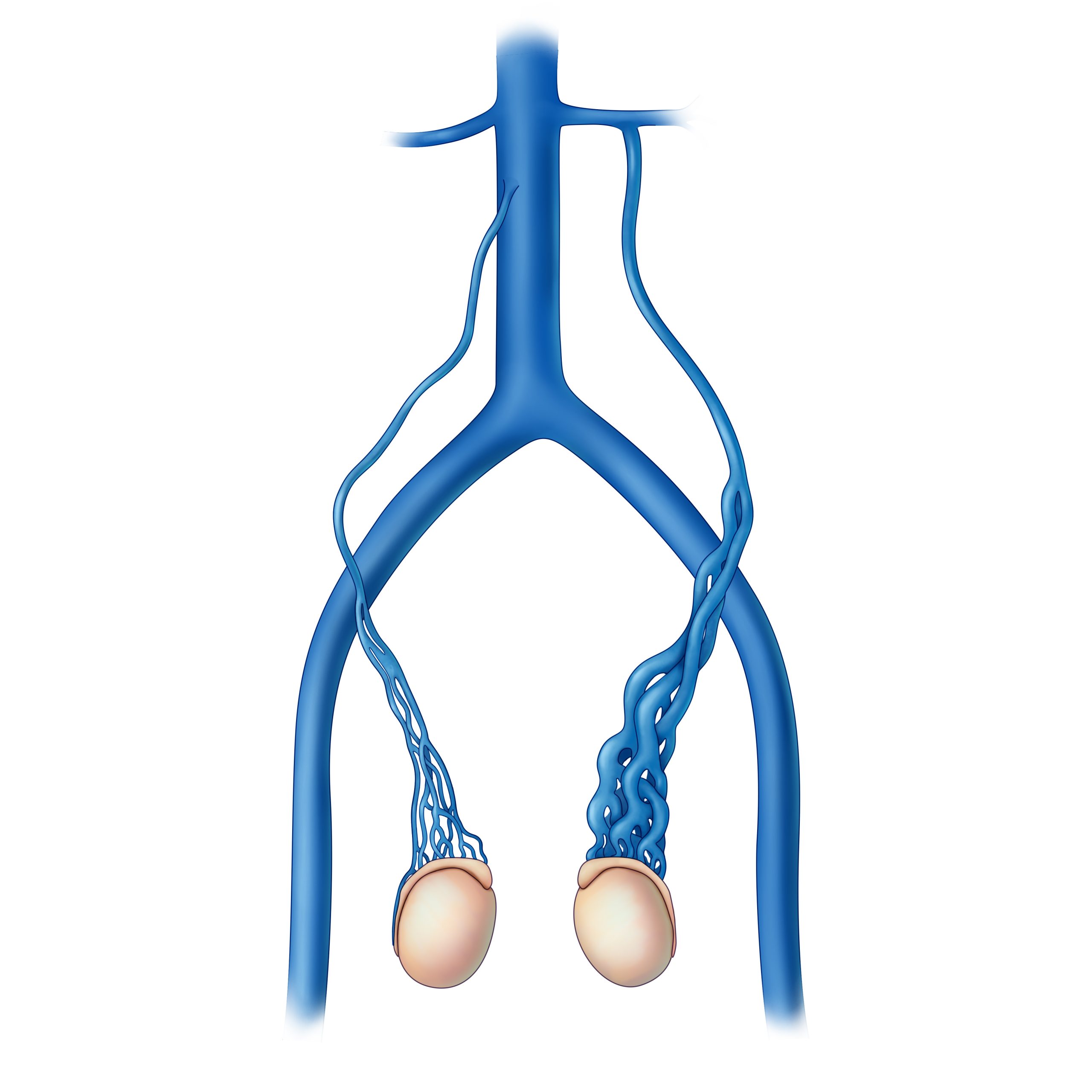
Varicocele is the enlargement of veins within the scrotum that drain the testicle, similar to varicose veins. It often causes pain, swelling, or a heavy feeling in the scrotum and can reduce sperm quality, potentially leading to infertility. Most cases are asymptomatic but may require treatment if symptoms persist or fertility issues arise.
Can you explain what varicocele is in Hindi?
वरिकोसिल (Varicocele) पुरुषों में अंडकोष में रक्त वाहिकाओं का फूलना है, जो अंडे के लिए रक्त को वापस ले जाने वाली नसों में रक्त का जमाव होता है। यह दर्द, सूजन या अनुपजाऊता का कारण बन सकता है।
Varicocele surgery involves repairing or blocking the enlarged veins to improve blood flow and reduce symptoms. Common methods include laparoscopic surgery, microsurgical repair, or embolization (using coils to block veins). Recovery typically takes 1–2 weeks, with success rates high for pain relief and fertility improvement.
What does varicocelectomy mean, and what does the procedure involve?
A varicocelectomy is surgery to remove or ligate (tie off) the faulty veins causing varicocele. The procedure restores normal blood flow to the testicle, often performed via open surgery, laparoscopy, or robotic assistance. It’s commonly recommended for men with pain or infertility linked to varicocele.
What is varicocele in Urdu, and what are its main signs?
واریکوسیل (Varicocele) ایک ایسا مرض ہے جس میں آڑے میں خون کی نالیوں کا پھولنا ہوتا ہے، جس سے درد، سوچن یا بارانپن کا مسئلہ پیدا ہو سکتا ہے۔ اس کا عام نشان ہے کہ آڑے میں گرمی یا بھاری پن محسوس ہوتا ہے۔
What is varicocele, and what treatments are available for it?
Varicocele is a condition where veins in the scrotum become enlarged, often causing pain or fertility problems. Treatment options include watchful waiting (for mild cases), surgery (varicocelectomy), or embolization to block abnormal veins. Lifestyle changes (like wearing supportive underwear) may also help manage symptoms.
|
|
Leave a Comment
Comments are moderated before appearing. The data you submit is processed according to the Privacy Policy of Voltefac.