What Is Dysentery Medical Insights And Global Impact
Table of Contents
- Medical Definition and Classification of Dysentery
- Pathogen-Specific Characteristics and Comparative Analysis
- Pathophysiological Mechanisms of Dysentery
- Flowchart: Progression from Infection to Systemic Complications
- Symptoms and Clinical Presentation of Dysentery
- Acute Phase: Core Symptomatic Spectrum
- Chronic Phase: Persistent and Atypical Presentations
- Red Flags Requiring Immediate Medical Intervention
- Differential Diagnosis: Dysentery vs. Other Diarrheal Diseases
- Diagnostic Approaches and Tools for Dysentery
- Stool Sample Collection, Transport, and Preservation Protocols
- Laboratory Diagnostic Tests: Sensitivity, Specificity, and Limitations
- Non-Invasive Diagnostic Methods in Resource-Limited Settings
- Treatment Protocols and Management Strategies for Dysentery
- Antimicrobial Therapy for Bacterial Dysentery
- Oral Rehydration Therapy (ORT) for Dehydration Management
- Epidemiology and Public Health Impact of Dysentery
- Global Burden and High-Risk Populations
- Notable Outbreaks and Control Measures
- Transmission Dynamics: Waterborne and Foodborne Pathways
- Public Health Intervention Strategy to Reduce Dysentery Incidence
- FAQ
- What is dysentery disease and what are its key characteristics?
- What does dysentery mean in terms of health conditions?
- What is dysentery called today in medical terms?
- What causes dysentery, and how is it transmitted?
- What is dysentery, and how do you get infected with it?
- What’s the difference between dysentery and regular diarrhea?
Dysentery remains a critical global health challenge, characterized by severe gastrointestinal distress with potentially life-threatening complications. As a preventable yet persistent infectious disease, it disproportionately affects vulnerable populations in regions with inadequate sanitation, underscoring the urgent need for targeted medical and public health interventions. This condition manifests in distinct forms—bacillary and amoebic—each driven by distinct pathogens that exploit weaknesses in mucosal defenses, leading to inflammation, fluid loss, and systemic deterioration when untreated. Understanding its pathophysiology, clinical spectrum, and epidemiologic patterns is essential for clinicians, epidemiologists, and policymakers to mitigate its burden through evidence-based diagnostics, therapeutics, and preventive strategies.
The disease’s dual classification—bacillary, caused by Shigella species, and amoebic, attributable to Entamoeba histolytica—reflects divergent transmission dynamics, symptom profiles, and diagnostic complexities. While bacillary dysentery often spreads through fecal-oral routes in crowded settings, amoebic dysentery thrives in tropical climates with poor hygiene, complicating differential diagnoses in endemic regions. Symptom severity varies from acute bloody diarrhea and tenesmus to chronic malnutrition and extraintestinal abscesses, demanding a nuanced approach that integrates clinical acumen with laboratory precision. This overview synthesizes medical definitions, diagnostic workflows, and management protocols to equip practitioners with actionable insights for early intervention and outbreak containment.
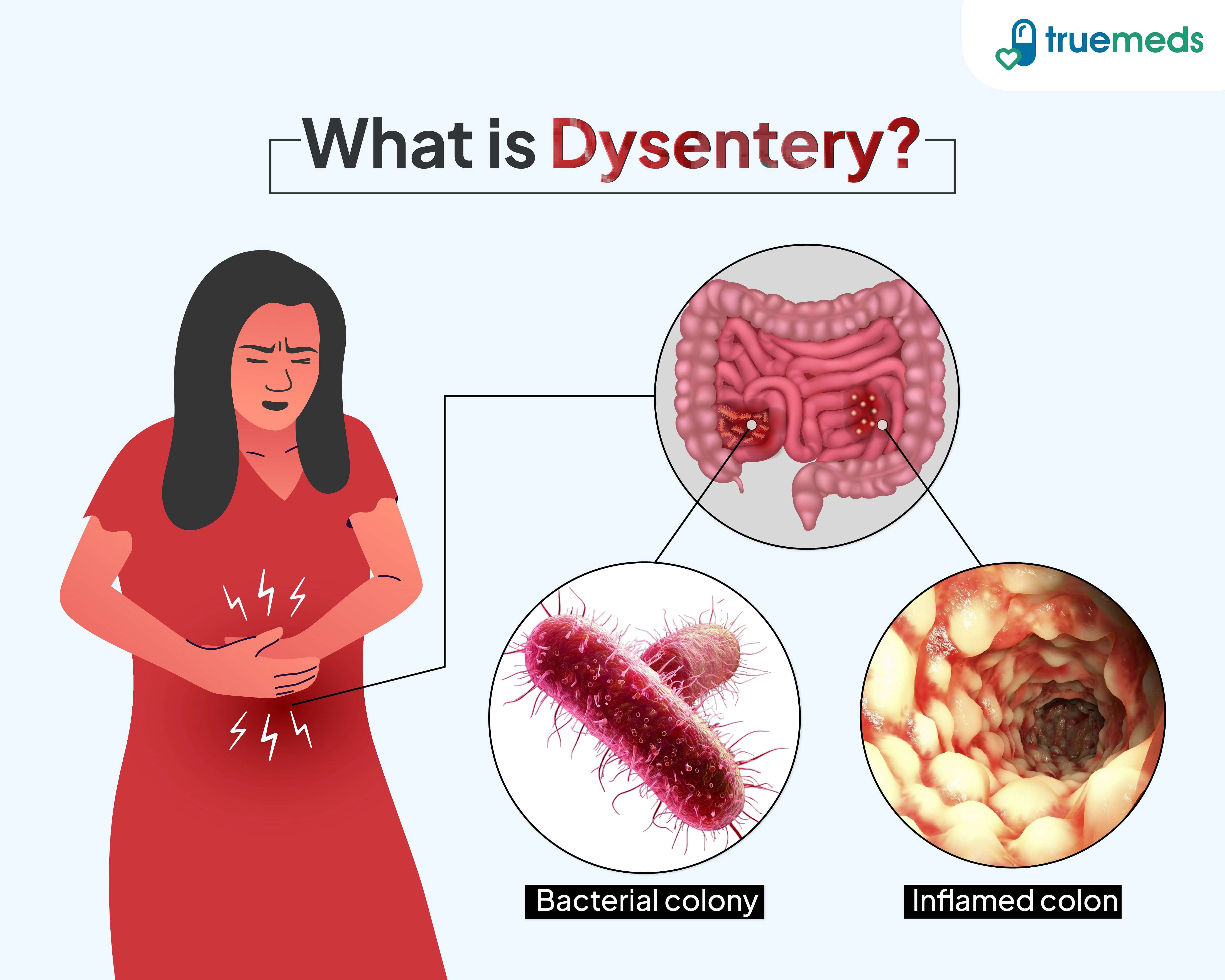
Medical Definition and Classification of Dysentery
Dysentery represents a group of acute intestinal infections characterized by inflammation of the colon, severe diarrhea with blood or mucus, and systemic symptoms such as fever and abdominal pain. The term encompasses two primary forms—bacillary dysentery, caused by bacterial pathogens, and amoebic dysentery, resulting from a parasitic infection. Both forms exhibit distinct clinical, epidemiological, and pathological features, necessitating precise differentiation for accurate diagnosis and targeted therapeutic intervention.The classification of dysentery is fundamentally rooted in its etiologic agents and pathophysiological mechanisms. Bacillary dysentery is predominantly attributed to Shigella species, while amoebic dysentery arises from Entamoeba histolytica. These distinctions influence transmission routes, symptom severity, and diagnostic approaches, underscoring the need for a structured comparative analysis to elucidate their unique clinical presentations.
Pathogen-Specific Characteristics and Comparative Analysis
Dysentery’s two primary forms—bacillary and amoebic—differ significantly in their causative agents, modes of transmission, clinical manifestations, and diagnostic methodologies. Below is a comparative table summarizing these distinctions:| Pathogen | Transmission Route | Symptoms | Diagnostic Methods |
|---|---|---|---|
| Bacillary Dysentery(Shigella spp.: S. dysenteriae, S. flexneri, S. boydii, S. sonnei) |
|
|
|
| Amoebic Dysentery(Entamoeba histolytica) |
|
|
|
Pathophysiological Mechanisms of Dysentery
The clinical manifestations of dysentery arise from a cascade of inflammatory and cytotoxic processes within the gastrointestinal mucosa, culminating in fluid/electrolyte imbalances and systemic complications. The pathophysiological progression involves three interconnected phases: mucosal invasion, inflammatory response, and secondary complications.1. Mucosal Invasion and Cytotoxicity
Critical Pathogen-Virulence Factor Interactions:2. Inflammatory Response and Mucosal Damage
Shigella: Shiga toxin (in S. dysenteriae type 1) disrupts protein synthesis in host cells, exacerbating mucosal damage. E. histolytica: Galactose/N-acetylglucosamine-inhibitable lectin (Gal/GalNAc) mediates adhesion and cytotoxicity.
Both pathogens trigger a robust inflammatory cascade:
3. Fluid and Electrolyte Imbalance
The destruction of colonic epithelial cells impairs ion transport mechanisms (e.g., Na⁺/K⁺ ATPases, chloride channels), leading to:
4. Systemic Complications
Prolonged or severe dysentery may progress to life-threatening conditions:
Flowchart: Progression from Infection to Systemic Complications
The clinical trajectory of dysentery follows a predictable yet variable path, influenced by pathogen virulence, host immunity, and environmental factors. Below is a structured flowchartSymptoms and Clinical Presentation of Dysentery
Dysentery presents a heterogeneous spectrum of gastrointestinal symptoms that vary in severity, duration, and systemic impact depending on the causative pathogen, host immune status, and environmental factors. The clinical manifestation ranges from mild, self-limiting diarrhea to life-threatening complications, particularly in vulnerable populations. Understanding these variations is critical for accurate diagnosis, appropriate management, and prevention of severe outcomes. Below, the symptomatology is categorized into acute and chronic phases, with emphasis on distinguishing features and high-risk presentations.Acute Phase: Core Symptomatic Spectrum
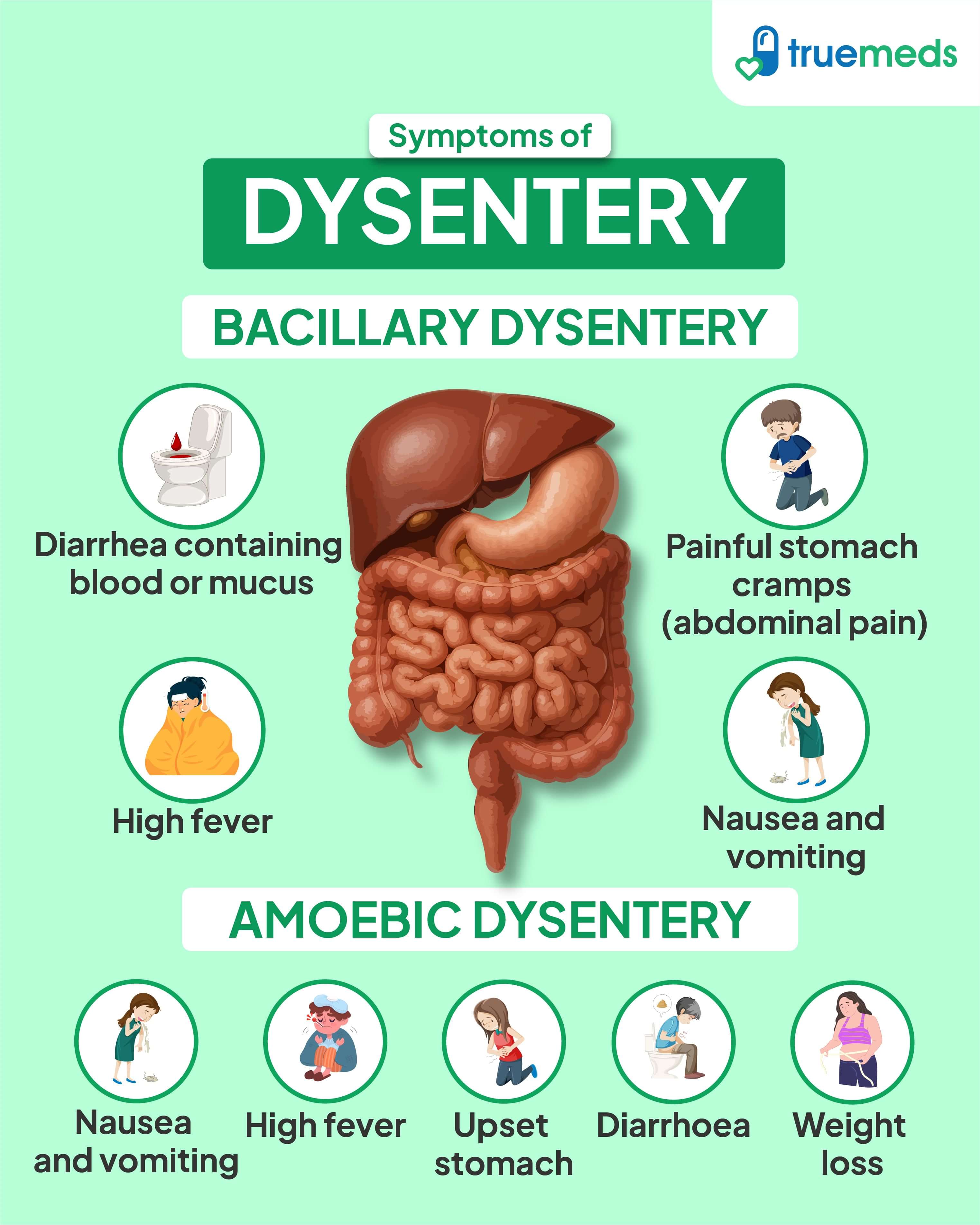
The acute phase of dysentery typically manifests within 12–72 hours of pathogen exposure and is characterized by a constellation of symptoms reflecting colonic inflammation and systemic toxicity. The severity of symptoms correlates with the pathogen’s virulence (e.g., Shigella spp. vs. Entamoeba histolytica) and host factors such as age, malnutrition, and comorbid conditions.Gastrointestinal Symptoms:
Systemic Manifestations:
Duration: Acute symptoms typically resolve within 3–7 days in uncomplicated cases, though relapses may occur with Entamoeba histolytica or Shigella.
Chronic Phase: Persistent and Atypical Presentations
Inadequate treatment, immunosuppression, or recurrent reinfection can transition dysentery into a chronic phase, defined by symptoms persisting beyond 4 weeks. Chronic dysentery is more commonly associated with Entamoeba histolytica and Balantidium coli, though bacterial causes (e.g., Salmonella in HIV/AIDS) may also contribute.Key Features:
Atypical Presentations:
Red Flags Requiring Immediate Medical Intervention
Certain clinical signs indicate severe disease progression or complications necessitating urgent evaluation and intervention. Recognition of these "red flags" is critical to prevent morbidity and mortality.High-risk symptoms demanding emergency care:
- Fever >39°C with signs of sepsis (e.g., altered mental status, hypotension, tachypnea), suggesting systemic invasion (e.g., Shigella bacteremia or amoebic abscess rupture).
- Persistent vomiting with inability to tolerate oral fluids, leading to dehydration (sunken eyes, dry mucous membranes, absent urine output) and risk of shock.
- Hematochezia with hemodynamic instability (e.g., systolic BP <90 mmHg, tachycardia >120 bpm), indicating massive lower GI bleeding or toxic megacolon.
- Neurological deterioration (e.g., confusion, seizures) in immunocompromised patients, raising suspicion of amoebic meningoencephalitis or HUS.
- Abdominal distension with absent bowel sounds, suggestive of toxic megacolon (a surgical emergency with perforation risk).
- Microangiopathic hemolytic anemia + thrombocytopenia + AKI, consistent with Shiga toxin–mediated HUS, particularly in children.
Differential Diagnosis: Dysentery vs. Other Diarrheal Diseases
Dysentery must be distinguished from other diarrheal illnesses to guide targeted therapy. Below is a comparative analysis of key features:| Feature | Dysentery (Shigella/Entamoeba) | Cholera | E. coli Gastroenteritis (ETEC/STEC) | |||||||||||||||||||||||||||||||||||||||||||||||||||||||||||||||||||||||||||||||||||||||||||||||||||||||
|---|---|---|---|---|---|---|---|---|---|---|---|---|---|---|---|---|---|---|---|---|---|---|---|---|---|---|---|---|---|---|---|---|---|---|---|---|---|---|---|---|---|---|---|---|---|---|---|---|---|---|---|---|---|---|---|---|---|---|---|---|---|---|---|---|---|---|---|---|---|---|---|---|---|---|---|---|---|---|---|---|---|---|---|---|---|---|---|---|---|---|---|---|---|---|---|---|---|---|---|---|---|---|---|---|---|---|
| Primary site of infection | Colon (invasive/proctitis) | Small intestine (secretory) | Small intestine (ETEC) or colon (STEC) | |||||||||||||||||||||||||||||||||||||||||||||||||||||||||||||||||||||||||||||||||||||||||||||||||||||||
| Stool characteristics | Small volume, bloody/mucousy ("dysenteric"), tenesmus | Large volume, watery ("rice-water"), odorless | Watery (ETEC) or bloody (STEC, e.g., O157:H7) | |||||||||||||||||||||||||||||||||||||||||||||||||||||||||||||||||||||||||||||||||||||||||||||||||||||||
| Fever | Common (38.5–40°C), especially bacterial causes | Absent or mild (<38°C) | Variable (ETEC: rare; STEC: possible) | |||||||||||||||||||||||||||||||||||||||||||||||||||||||||||||||||||||||||||||||||||||||||||||||||||||||
| Systemic toxicity | High (sepsis, HUS, abscesses) | Low (unless severe dehydration) | Moderate (HUS in STEC; rare in ETEC) | |||||||||||||||||||||||||||||||||||||||||||||||||||||||||||||||||||||||||||||||||||||||||||||||||||||||
| Dehydration onset | Rapid (hours to days) | Very rapid (<24 hours) | Moderate (12–72 hours) | |||||||||||||||||||||||||||||||||||||||||||||||||||||||||||||||||||||||||||||||||||||||||||||||||||||||
| Complications | Toxic megacolon, HUS, abscesses, strictures | Hypovolemic shock, metabolic acidosis | HUS (STEC), hemolytic anemia, TTP | |||||||||||||||||||||||||||||||||||||||||||||||||||||||||||||||||||||||||||||||||||||||||||||||||||||||
| Treatment focus | Antimicrobials (e.g., azithromycin, metronidazole), supportive care | IV fluids + electrolytes (antibiotics rarely indicated) | Supportive;
Diagnostic Approaches and Tools for DysenteryDiagnostic accuracy in dysentery relies on a structured, evidence-based approach that balances sensitivity, specificity, and resource availability. Early and precise identification of the causative pathogen—whether bacterial (Shigella, Salmonella, Campylobacter), parasitic (Entamoeba histolytica), or viral (norovirus, rotavirus)—guides targeted therapy and infection control measures. The diagnostic workflow integrates stool sample handling, laboratory testing, and non-invasive rapid assays, with selection influenced by clinical presentation, geographic risk, and healthcare infrastructure.The diagnostic process begins with patient history and symptom assessment, followed by systematic stool collection and preservation to ensure pathogen viability. Laboratory techniques range from traditional microscopy to advanced molecular assays, each with distinct advantages and limitations in accuracy and cost. Non-invasive methods, such as rapid antigen tests and multiplex PCR panels, play a critical role in low-resource settings, though their performance varies by pathogen and setting. Clinicians must navigate these options using a decision-tree framework that incorporates symptom duration, travel history, and patient-specific risk factors to optimize diagnostic yield. Stool Sample Collection, Transport, and Preservation ProtocolsProper stool specimen collection and handling are fundamental to maintaining pathogen integrity for accurate diagnostic testing. Contamination, improper storage, or delayed processing can lead to false-negative results, particularly for fastidious organisms like Shigella or Entamoeba histolytica. The following protocols ensure specimen viability while minimizing degradation:- Collection Requirements: - Transport Conditions: - Preservation Methods: Critical Note: Delayed or improperly preserved specimens may yield false-negative cultures for Shigella (viability declines within 24 hours at room temperature) and missed parasitic cysts due to autolysis. Laboratory Diagnostic Tests: Sensitivity, Specificity, and LimitationsLaboratory confirmation of dysentery employs a tiered approach, combining microscopy, culture, antigen detection, and molecular methods. Each technique varies in turnaround time, cost, and diagnostic performance. Below is a comparative analysis formatted for clinical decision-making:
Key Limitation: Culture-based methods may fail to detect carriage states (e.g., asymptomatic Shigella shedding) or fastidious pathogens (e.g., Cyclospora), while antigen tests often miss low-level infections. PCR overcomes these gaps but requires specialized infrastructure. Non-Invasive Diagnostic Methods in Resource-Limited SettingsNon-invasive rapid diagnostic tests (RDTs) and multiplex PCR panels address critical gaps in low-resource settings, where laboratory infrastructure is limited. These tools enable point-of-care (POC) testing, reducing reliance on centralized labs and accelerating treatment. However, their performance varies by pathogen, and clinicians must interpret results cautiously to avoid misdiagnosis.- Rapid Antigen Key Considerations for Antimicrobial Selection: Standardized Dosage Regimens:
Oral Rehydration Therapy (ORT) for Dehydration ManagementDehydration is the leading cause of mortality in dysentery, particularly in children and malnourished individuals. Oral rehydration therapy (ORT) restores electrolyte and fluid balance while minimizing gastrointestinal losses. The WHO recommends low-osmolarity ORS (245 mOsm/L) to reduce stool output and vomiting compared to high-osmolarity solutions. Homemade alternatives (e.g., rice-based ORS) may be used in resource-limited settings but require careful preparation to avoid contamination.Comparison of ORT Solutions:
Epidemiology and Public Health Impact of DysenteryDysentery remains a significant global health challenge, disproportionately affecting vulnerable populations in regions with limited access to clean water, sanitation, and healthcare. The disease exhibits marked geographic and demographic disparities, with high-risk groups including children under five years old, refugees, internally displaced persons, and communities in low-resource settings. Seasonal fluctuations and epidemic outbreaks further exacerbate transmission, particularly in tropical and subtropical climates where environmental conditions favor pathogen survival. Understanding these patterns is critical for targeted public health interventions, including infrastructure improvements, vaccination strategies, and community-based education.The burden of dysentery extends beyond individual morbidity, straining healthcare systems and economic productivity in endemic areas. Shigellosis, caused by Shigella spp., and amoebic dysentery, driven by Entamoeba histolytica, are the most prevalent forms, with Shigella sonnei and Shigella flexneri accounting for the majority of bacterial cases. Waterborne and foodborne transmission routes dominate epidemiology, with fecal-oral contamination serving as the primary vector. Below, the global distribution, key outbreaks, transmission dynamics, and evidence-based mitigation strategies are examined. Global Burden and High-Risk PopulationsDysentery incidence varies by region, with the highest disease burden observed in sub-Saharan Africa, South Asia, and Latin America, where an estimated 1.7 billion cases of diarrheal illness—including dysentery—occur annually, leading to 525,000 deaths (WHO, 2023). Children under five are most vulnerable, accounting for ~40% of severe cases, while refugee and conflict-affected populations face elevated risks due to overcrowding, poor hygiene, and disrupted healthcare access. Seasonal trends correlate with monsoon seasons in South Asia and rainy seasons in tropical regions, where flooding contaminates water sources and increases pathogen exposure.Key high-risk groups include: Blockquote: Notable Outbreaks and Control MeasuresHistorical and recent outbreaks highlight dysentery’s epidemic potential, particularly in settings with weakened surveillance. Below is a summary of key events, categorized by pathogen and geographic context:
Transmission Dynamics: Waterborne and Foodborne PathwaysDysentery spreads primarily through fecal-oral transmission, with water and food serving as the dominant vectors. Waterborne transmission accounts for ~80% of cases in endemic regions, while foodborne routes contribute significantly in urban and peri-urban settings.Waterborne Transmission: Foodborne Transmission: Blockquote: Public Health Intervention Strategy to Reduce Dysentery IncidenceA multi-pronged approach combining sanitation infrastructure, vaccination, and community engagement is essential to curb dysentery transmission. Below is a structured intervention framework:1. Sanitation and Water Safety Infrastructure Dysentery exemplifies the intersection of infectious disease pathology and public health systemic failures, where timely diagnosis and treatment can avert severe morbidity and mortality. From the microscopic invasion of intestinal mucosa by Shigella or Entamoeba to the macroscopic consequences of dehydration and sepsis, the disease’s progression underscores the fragility of gastrointestinal integrity in the face of pathogen exposure. Advances in rapid diagnostics, antimicrobial stewardship, and community-based sanitation initiatives offer promising avenues to curb its spread, particularly in high-risk populations. As global health efforts intensify—through vaccination trials, improved water infrastructure, and targeted education campaigns—the fight against dysentery serves as a litmus test for equitable healthcare access and the resilience of vulnerable communities against preventable infectious threats. FAQWhat is dysentery disease and what are its key characteristics?Dysentery is an infectious intestinal disease causing severe diarrhea, often with blood or mucus in the stool, along with abdominal pain, fever, and dehydration. It’s typically spread through contaminated food or water and can be caused by bacteria (like Shigella or E. coli) or parasites (like Entamoeba histolytica). Without treatment, it can lead to serious complications, especially in children or those with weak immune systems. What does dysentery mean in terms of health conditions?Dysentery refers to an inflammatory disorder of the intestines marked by frequent, watery or bloody stools, cramping, and systemic symptoms like fever and chills. The term historically described severe diarrhea with mucus or blood, though modern medicine distinguishes between bacterial (e.g., shigellosis) and amoebic forms. It’s a major cause of morbidity in regions with poor sanitation. What is dysentery called today in medical terms?Today, dysentery is often specified by its cause: bacillary dysentery (from bacteria like Shigella) or amoebic dysentery (from the parasite Entamoeba histolytica). The general term "dysentery" is still used colloquially, but doctors prefer the precise microbial name for diagnosis and treatment. What causes dysentery, and how is it transmitted?Dysentery is caused by bacteria (Shigella, E. coli, Salmonella) or parasites (Entamoeba histolytica), usually spread through ingestion of fecal-contaminated food, water, or hands. Poor hygiene, unsafe drinking water, and close contact with infected individuals are primary transmission routes. Outbreaks often occur in areas with inadequate sanitation. What is dysentery, and how do you get infected with it?Dysentery is an infection of the intestines leading to severe diarrhea, often with blood, abdominal pain, and fever. You get it by consuming food or water contaminated with feces containing dysentery-causing bacteria or parasites, or through poor handwashing after contact with infected stool. Travelers and people in crowded or unsanitary conditions are at higher risk. What’s the difference between dysentery and regular diarrhea?Dysentery is a severe form of diarrhea characterized by bloody or mucus-filled stools, accompanied by high fever, intense abdominal cramps, and systemic illness. Regular diarrhea is usually watery and milder, without blood or systemic symptoms, though both can cause dehydration. Dysentery often requires medical treatment, while mild diarrhea may resolve with fluids and rest. |

Leave a Comment
Comments are moderated before appearing. The data you submit is processed according to the Privacy Policy of Voltefac.