| Interdisciplinary Collaboration |
Historical Development and Milestones in Pathology
Pathology has evolved from ancient observational practices into a precision-driven medical science, integrating anatomical, physiological, and molecular insights. The discipline’s trajectory reflects broader advancements in medicine, technology, and scientific methodology, with each era introducing transformative paradigms that redefined disease understanding. Key milestones—spanning from early autopsy traditions to contemporary genomic and artificial intelligence applications—illustrate how pathology shifted from descriptive morphology to predictive, systems-based diagnostics. This progression underscores the interplay between empirical discovery, theoretical innovation, and technological adaptation, shaping pathology’s role as both a diagnostic and investigative cornerstone in modern healthcare.
Ancient and Pre-Modern Foundations: Autopsy and Early Observations
The origins of pathology trace back to ancient civilizations where systematic examinations of deceased individuals provided foundational insights into disease mechanisms. In ancient Egypt (c. 1600 BCE), the Edwin Smith Papyrus—one of the earliest known medical texts—detailed clinical observations and postmortem findings, including descriptions of traumatic injuries and organ pathologies. Similarly, Hippocrates (460–370 BCE) emphasized the importance of autopsy in his Corpus Hippocraticum, advocating for empirical evidence to distinguish natural diseases from supernatural explanations. During the Middle Ages, Islamic scholars such as Ibn Sina (Avicenna, 980–1037 CE) expanded upon these traditions, documenting autopsies in his Canon of Medicine and linking anatomical abnormalities to physiological dysfunctions. However, progress stagnated in Europe until the Renaissance, when artists like Leonardo da Vinci (1452–1519) and anatomists such as Andreas Vesalius (1514–1564) revived systematic dissection, laying the groundwork for modern anatomical pathology.The 16th–17th centuries marked a turning point with the advent of microscopy, pioneered by Antonie van Leeuwenhoek (1632–1723), who observed bacteria and blood cells, though their pathological significance remained unclear. Meanwhile, Giovanni Morgagni (1682–1771), often called the "father of pathological anatomy," formalized the concept of localizing disease to specific organs through his 1761 work De Sedibus et Causis Morborum (On the Seats and Causes of Diseases). Morgagni’s systematic correlation of gross anatomical findings with clinical symptoms established pathology as a scientific discipline, shifting focus from speculative theories to evidence-based diagnostics.
19th Century: Cellular Pathology and the Germ Theory Revolution
The 19th century witnessed two revolutionary frameworks that fundamentally altered pathology: cell theory and germ theory. These paradigms shifted the field from macroscopic observations to microscopic and infectious disease etiologies, respectively.Cellular Pathology
The foundational work of Theodor Schwann (1810–1882) and Matthias Schleiden (1804–1881) in the 1830s–1840s established that all living organisms are composed of cells, a principle later refined by Rudolf Virchow (1821–1902). Virchow’s 1858 declaration "Omnis cellula e cellula" ("Every cell arises from another cell") dismantled the prevailing theory of spontaneous generation and introduced the concept of pathological cellular changes as the basis of disease. His contributions extended beyond theory: Virchow founded the Berlin Pathological Institute (1849), pioneered histopathology (microscopic examination of tissues), and linked chronic inflammation to cancer—a precursor to modern tumor immunology. His multidisciplinary approach—integrating clinical medicine, politics, and social pathology—demonstrated pathology’s role in public health, exemplified by his investigations into typhus epidemics in 19th-century Prussia. Germ Theory and Infectious Disease Pathology
While Louis Pasteur (1822–1895) laid the groundwork for germ theory with his work on fermentation and vaccination, Robert Koch (1843–1910) provided the definitive pathological framework. Koch’s postulates (1876–1890)—a series of criteria to establish the causative relationship between a microorganism and a disease—enabled the identification of pathogens such as Bacillus anthracis (1876), Mycobacterium tuberculosis (1882), and Vibrio cholerae (1883). His discoveries not only validated the germ theory but also introduced bacterial staining techniques (e.g., carbol fuchsin for tuberculosis) and culture methods, which remain cornerstones of microbiological pathology. Koch’s work also highlighted the host-pathogen interaction, a concept later expanded by Paul Ehrlich (1854–1915) with his side-chain theory of immunity, foreshadowing modern immunopathology.
Critical Discoveries and Their Implications-
Cell Theory (1838–1858)
Contribution: Virchow’s Omnis cellula e cellula and the principle that diseases arise from abnormal cellular processes. Implication: Shifted pathology from organ-based to cellular-level diagnostics, enabling histopathology as the gold standard for tissue analysis. This framework underpins modern oncology (studying cancer as a cellular disorder) and regenerative medicine.
-
Germ Theory (1860s–1890s)
Contribution: Koch’s postulates and Pasteur’s microbial discoveries established infectious agents as primary causes of disease. Implication: Revolutionized infectious disease pathology, leading to antimicrobial therapies, vaccines, and the field of clinical microbiology. It also introduced epidemiological pathology, linking microbial spread to public health policies.
-
Autopsy and Anatomical Correlation (16th–19th centuries)
Contribution: Morgagni’s systematic autopsies and Virchow’s integration of clinical-pathological correlations. Implication: Established pathology as a bridge between clinical medicine and basic science, enabling evidence-based diagnostics and the development of surgical pathology and forensic pathology.
Early 20th Century: Specialization and Technological Innovations
The early 1900s saw pathology diversify into specialized subfields, driven by technological advancements and the need to address emerging diseases. Key developments included:Histochemistry and Cytology
The introduction of staining techniques such as hematoxylin and eosin (H&E, 1870s) by Franz Nissl and Camillo Golgi allowed for detailed tissue differentiation, while George Papanicolaou’s (1883–1962) cytological smears (1920s) enabled early cancer detection via Pap tests. Histochemistry—the study of chemical components in tissues—gained traction with Paul Ehrlich’s (1854–1915) vital dyes and later enzymatic staining (e.g., alkaline phosphatase for bone and placental tissues), which became essential for neuropathology and oncological diagnostics. Immunopathology and Serology
The 1920s–1940s marked the rise of immunological techniques, including precipitation reactions and agglutination tests, which identified antibodies in sera. Karl Landsteiner’s (1868–1943) discovery of blood groups (1901) laid the foundation for transfusion medicine, while Albert Coons’ (1912–1978) fluorescent antibody technique (1941) enabled direct visualization of antigens in tissues, a precursor to modern immunohistochemistry (IHC). These innovations were pivotal in diagnosing autoimmune diseases (e.g., rheumatoid arthritis) and infectious agents (e.g., syphilis via VDRL tests). Electron Microscopy and Ultrastructural Pathology
The 1950s introduced electron microscopy (EM), with Albert Claude (1899–1983) and Christian de Duve (1917–2013) identifying cell organelles (e.g., lysosomes, mitochondria) and their pathological alterations. EM revealed viral structures (e.g., polio virus, 1954) and ultrastructural changes in diseases like amyloid deposits in Alzheimer’s, bridging the gap between light microscopy and molecular pathology

Methods and Techniques in Pathology
Pathology relies on a diverse array of laboratory techniques to diagnose diseases, understand mechanisms of illness, and guide therapeutic decisions. These methods range from traditional histological staining to cutting-edge molecular and imaging technologies, each offering unique insights into tissue architecture, cellular abnormalities, and disease biomarkers. Advances in pathology techniques have transformed diagnostic accuracy, enabling early detection, personalized medicine, and integration with genomic and proteomic data. Below, the key methodologies are categorized, compared, and contextualized within their procedural workflows and emerging innovations.
Categorized Laboratory Techniques in Pathology
Pathology techniques are broadly classified based on their analytical focus: morphological assessment, molecular characterization, and functional imaging. Each category serves distinct diagnostic or research purposes, often employed in combination for comprehensive evaluations.
-
Histopathology
The gold standard for examining tissue architecture and cellular morphology, histopathology involves slicing fixed tissues into thin sections (3–5 µm) and staining them for microscopic evaluation. Applications include cancer diagnosis, inflammatory disease assessment, and organ-specific pathology (e.g., liver cirrhosis, neurodegenerative disorders).
Histopathology remains the cornerstone of surgical pathology, providing definitive diagnoses for ~70% of cancer cases worldwide.
-
Cytopathology
Focuses on individual cells or small clusters, typically obtained via fine-needle aspiration (FNA), Pap smears, or body fluids. Used for detecting malignancies (e.g., thyroid nodules, gynecological cancers) and infectious agents (e.g., Papillomavirus in cervical cytology).
Cytopathology’s liquid-based preparation (e.g., ThinPrep) reduces obscuring blood/clot artifacts, improving diagnostic sensitivity.
-
Immunohistochemistry (IHC)
Detects specific antigens in tissue sections using antibody-based staining, enabling identification of protein expression patterns (e.g., Ki-67 for proliferation, ER/PR in breast cancer). IHC bridges morphology with molecular pathology, critical for subclassifying tumors (e.g., HER2 status in gastric cancer).
-
In Situ Hybridization (ISH)
Localizes nucleic acids (DNA/RNA) within tissue sections, including fluorescence (FISH) for gene fusions (e.g., ALK in lung cancer) or chromogenic (CISH) for amplifications (e.g., HER2 gene copies). Chromogenic ISH is particularly useful in formalin-fixed paraffin-embedded (FFPE) samples.
-
Molecular Pathology
Encompasses techniques like PCR, NGS (next-generation sequencing), and microarray analysis to detect genetic mutations, epigenetic alterations, or microbial DNA/RNA. Used for hereditary cancer syndromes (e.g., BRCA1/2 testing) and infectious disease diagnostics (e.g., COVID-19 PCR).
-
Flow Cytometry
Quantifies and characterizes cellular populations based on surface/ intracellular markers (e.g., CD markers in hematologic malignancies). Essential for diagnosing leukemias/lymphomas and monitoring minimal residual disease (MRD).
-
Special Stains
Highlight specific tissue components: Prussian blue (iron), Masson’s trichrome (collagen), GMS/PAS (fungal/cryptococcal cell walls), and PSA (amyloid deposits). These stains complement routine H&E staining for targeted diagnoses.
-
Electron Microscopy (EM)
Provides ultrastructural details (e.g., viral particles, mitochondrial abnormalities) at nanometer resolution. Used in rare diseases (e.g., Fabry disease) or when light microscopy is inconclusive.
Comparison of Traditional Staining vs. Advanced Imaging Modalities
Traditional staining methods, particularly hematoxylin and eosin (H&E), have been the backbone of pathology for over a century. However, advanced imaging modalities offer higher throughput, quantitative data, and integration with digital workflows. Below is a comparative analysis of key techniques:
| Feature |
Traditional Staining (H&E) |
Advanced Imaging (Digital Pathology/Multiplex IF) |
|---|
| Principle |
Chemical dyes bind to nucleic acids (hematoxylin) and proteins (eosin), providing contrast via light microscopy. |
Fluorescent/colorimetric markers or machine learning algorithms analyze tissue sections digitally, enabling multiplexing or AI-assisted diagnostics. |
| Applications |
General tissue morphology, tumor grading, inflammatory patterns. |
- Multiplex immunofluorescence (mIF): Simultaneous detection of 5+ proteins (e.g., PD-L1, CD8, Ki-67 in TME analysis).
- Digital pathology: Whole-slide imaging (WSI) for telepathology, AI-driven detection (e.g., prostate cancer Gleason scoring).
- Spatial transcriptomics: Mapping RNA expression in tissue context (e.g., Visium by 10x Genomics).
|
| Limitations |
- Subjective interpretation (inter-observer variability).
- Limited to 1–2 stains per slide.
- Destruction of tissue after staining.
|
- High cost and technical complexity.
- Photobleaching (fluorescent methods).
- Data overload requiring specialized software (e.g., QuPath, HALO).
|
| Advantages |
- Widely standardized; low infrastructure requirements.
- Proven diagnostic accuracy for routine cases.
|
- Quantitative and objective (e.g., AI quantifies cell densities).
- Preservation of tissue for additional testing.
- Enables longitudinal studies (e.g., tracking tumor evolution).
|
| Emerging Integration |
Hybrid approaches: H&E slides scanned for digital archives, with AI flagging suspicious regions for pathologist review. |
Combining mIF with spatial transcriptomics to correlate protein expression with gene activity in situ. |
Step-by-Step Tissue Sample Processing from Fixation to Slide Preparation
Proper tissue processing is critical to preserve morphology and antigenicity for downstream analysis. Deviations in this workflow can lead to artifacts, diagnostic errors, or failed molecular testing. Below is a standardized protocol with common pitfalls:
-
Fixation
- Purpose: Cross-links proteins and nucleic acids to halt autolysis and maintain structural integrity.
- Method: Submerge tissue in 10% neutral-buffered formalin (NBF) at a 1:10 tissue-to-fixative ratio for 6–24 hours (optimal: 6–12 hours for small biopsies; up to 48 hours for large specimens).
- Pitfalls:
- Overfixation: >48 hours causes hard, brittle tissue, complicating sectioning and increasing background in IHC.
- Underfixation: <6 hours leads to poor morphology and antigen loss.
- Improper formalin: Acidic or unbuffered formalin causes tissue shrinkage and nuclear pyknosis.
- Alternatives: For molecular testing, PAXgene (RNA preservation) or ethanol-based fixatives (e.g., HistoChoice) may be used.
-
Trimming and Orientation
- Purpose: Remove excess tissue and orient the specimen for optimal sectioning (e.g., perpendicular to long axis for uniform slices).
- Method: Use a scalpel under a dissecting microscope. Ink margins (e.g., black for deep
Pathology in Disease Diagnosis
Pathological examination serves as the cornerstone of precise disease diagnosis, bridging clinical suspicion with definitive therapeutic decisions. Through systematic analysis of tissue, fluids, and cellular morphology, pathologists distinguish between benign and malignant conditions, identify infectious agents, and uncover genetic aberrations that drive disease progression. The integration of macroscopic, microscopic, immunohistochemical, and molecular techniques ensures comprehensive diagnostic accuracy, directly influencing patient stratification and treatment selection.Pathological diagnostics rely on a structured workflow that begins with specimen acquisition—such as biopsies, cytological smears, or body fluids—and progresses through gross examination, histological processing, and advanced molecular profiling. Key differentiators between benign and malignant lesions include cellular atypia, mitotic activity, invasion patterns, and molecular signatures. For instance, while benign tumors exhibit organized architecture and low proliferative indices, malignant neoplasms demonstrate pleomorphism, high nuclear-to-cytoplasmic ratios, and genomic instability. Below, the diagnostic pathway for breast cancer illustrates how pathology guides clinical decision-making from initial presentation to final management.
Differentiation Between Benign and Malignant Conditions Through Pathological Examination
The distinction between benign and malignant pathologies hinges on histomorphological criteria, immunohistochemical markers, and molecular diagnostics. Pathologists evaluate several parameters to classify lesions:- Cellular Architecture: Benign tumors maintain tissue-of-origin structure (e.g., glandular patterns in adenomas), whereas malignant tumors exhibit disorganized, infiltrative growth.
- Nuclear Features: Malignant cells display atypia (irregular nuclear shapes, prominent nucleoli) and hyperchromasia (dark-staining nuclei due to increased DNA content).
- Mitotic Activity: High mitotic rates (>20 mitoses/10 high-power fields) correlate with malignancy, as seen in carcinomas and sarcomas.
- Invasion and Metastasis: Malignant tumors breach basement membranes (e.g., ductal carcinoma in situ progressing to invasive ductal carcinoma) and may present with distant metastases.
- Immunophenotyping: Markers like Ki-67 (proliferation index), p53 (tumor suppressor mutation), and HER2 (oncogene amplification) refine diagnostic specificity.
Example: In prostate cancer, Gleason grading evaluates architectural patterns (grades 1–5) to predict aggressiveness, while PSA (prostate-specific antigen) immunohistochemistry confirms epithelial origin. Molecular tests for ERG gene rearrangements further distinguish between indolent and aggressive subtypes.
Diagnostic Pathway for Breast Cancer: A Structured Flowchart
The following flowchart outlines the pathological evaluation of breast cancer, from initial clinical suspicion to definitive reporting, emphasizing key decision points:-
Initial Suspicion
- Clinical presentation: palpable mass, mammographic abnormality (e.g., spiculated lesion), or nipple discharge.
- Imaging-guided biopsy (core needle or vacuum-assisted) or surgical excision (lumpectomy/wire-localized biopsy).
-
Gross Examination
- Tumor size, margins, and relationship to surrounding tissue documented.
- Sampling of multiple regions if heterogeneous (e.g., invasive vs. in situ components).
-
Histological Assessment
- H&E staining identifies:
- Ductal carcinoma in situ (DCIS): malignant cells confined to ducts.
- Invasive ductal carcinoma (IDC): stromal invasion with desmoplastic response.
- Lobular carcinoma: single-file infiltration.
- Grading (Notching system or Bloom-Richardson): nuclear atypia, tubule formation, mitotic count.
-
Immunohistochemistry (IHC) and Molecular Profiling
- Hormone receptors (ER/PR) and HER2 status guide endocrine/anti-HER2 therapy.
- Multigene panels (e.g., Oncotype DX, PAM50) assess recurrence risk for ER+ tumors.
- Basal-like subtype (triple-negative) identified by CK5/6, EGFR, or BRCA mutations.
-
Final Report and Staging
- TNM classification (e.g., T2N0M0 for 2–5 cm tumor, no nodes, no metastasis).
- Recommendations: surgery (lumpectomy/mastectomy), adjuvant chemotherapy, or targeted therapy (e.g., trastuzumab for HER2+).
Critical Insight: Pathological staging (e.g., pT3N1) is superior to clinical staging for treatment planning, as it accounts for microscopic tumor extent (e.g., lymphovascular invasion).
Case Studies Demonstrating Pathology’s Impact on Patient Management
Pathological findings have revolutionized treatment paradigms in five landmark cases, each highlighting the discipline’s role in precision medicine:
-
Lynch Syndrome Detection via Microsatellite Instability (MSI) Testing
- Case: A 45-year-old woman with stage II colorectal cancer (CRC) underwent MSI testing, revealing high instability (MSI-H).
- Impact: Identified Lynch syndrome (germline MLH1/PMS2 mutations), prompting:
- Prophylactic colectomy to prevent metachronous tumors.
- Screening for endometrial and ovarian cancers in family members.
- Immunotherapy eligibility (e.g., pembrolizumab for MSI-H metastatic CRC).
-
ALK Rearrangement in Non-Small Cell Lung Cancer (NSCLC)
- Case: A never-smoker with advanced NSCLC tested positive for EML4-ALK fusion via FISH or NGS.
- Impact:
- Targeted therapy with crizotinib or alectinib replaced chemotherapy, achieving 70% objective response rates.
- Avoided toxicities of platinum-based regimens.
-
BRCA1/2 Mutations in Triple-Negative Breast Cancer
- Case: A 32-year-old with BRCA1-mutated triple-negative breast cancer (TNBC) underwent germline testing.
- Impact:
- Eligibility for PARP inhibitors (olaparib) in metastatic setting.
- Risk-reducing salpingo-oophorectomy to lower ovarian cancer risk.
- Family cascade testing identified 12 at-risk relatives.
-
Diffuse Large B-Cell Lymphoma (DLBCL) Subtype Classification
- Case: A patient with DLBCL underwent Hans algorithm (CD10, Bcl-6, MUM1) and cell-of-origin (COO) profiling.
- Impact:
- Germinal center B-cell (GCB) subtype responded better to R-CHOP (rituximab + cyclophosphamide).
- Non-GCB subtype required DA-EPOCH (dose-adjusted etoposide) for improved outcomes.
-
Prostate Cancer: Gleason Grade Upgrading and Active Surveillance
- Case: A 62-year-old with Gleason 3+3=6 prostate cancer on initial biopsy was upgraded to Gleason 4+3=7 on radical prostatectomy.
- Impact:
- Avoided overtreatment in low-risk patients (ISUP Grade Group 1).
- High-risk patients (Grade Group ≥3) received ADT (androgen deprivation therapy) or radiation.
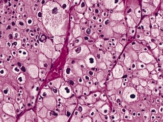
Molecular Pathology and Personalized Medicine: Descriptive Examples
Ethical and Regulatory Considerations in Pathology
Pathology operates at the intersection of medical science, patient care, and legal frameworks, necessitating rigorous adherence to ethical principles and regulatory standards. Ethical dilemmas arise from the dual role of pathologists as diagnosticians and stewards of patient data, particularly in areas such as tissue banking, incidental genomic findings, and genetic privacy. Concurrently, regulatory compliance ensures the integrity of diagnostic processes, public safety, and the prevention of medical errors. This section examines the ethical challenges faced by pathologists, key regulatory frameworks governing laboratory practices, and the legal responsibilities of pathologists in forensic and clinical contexts. Additionally, it explores the critical role of pathology in public health emergencies, where the tension between rapid diagnostics and diagnostic accuracy must be carefully managed.
Ethical Dilemmas in Pathology
Pathologists encounter complex ethical challenges that stem from their involvement in patient care, research, and public health. These dilemmas often involve conflicts between individual patient rights, societal benefits, and professional obligations. Key areas of concern include informed consent for tissue banking, incidental findings in genetic testing, and genetic privacy.Pathologists frequently rely on biobanked tissues for research, diagnostic validation, and educational purposes. Obtaining informed consent for such use presents challenges, particularly when tissues are collected for clinical purposes but later repurposed for research. The ethical tension arises from balancing the potential benefits of research against the autonomy of donors, who may not fully understand the long-term implications of their tissue contributions. Incidental findings—unexpected genetic or molecular abnormalities detected during routine testing—pose additional dilemmas. Pathologists must determine whether to disclose such findings to patients, especially when they lack clinical relevance or may cause psychological distress. Guidelines from organizations like the American College of Medical Genetics and Genomics (ACMG) recommend disclosure of actionable findings but leave room for interpretation in ambiguous cases. Genetic privacy is another critical ethical concern, given the sensitive nature of genomic data. Pathologists must ensure that genetic information is protected from unauthorized access or misuse, while also complying with laws such as the Genetic Information Nondiscrimination Act (GINA) in the U.S., which prohibits discrimination based on genetic information in employment and health insurance. However, emerging technologies like whole-genome sequencing and multi-omics analyses complicate privacy safeguards, as they generate vast datasets that may inadvertently reveal non-medical traits (e.g., ancestry, carrier status for non-disease genes).
Ethical principles in pathology must align with autonomy, beneficence, non-maleficence, and justice, while navigating the evolving landscape of genomic medicine and data sharing.
Key Regulations Governing Pathology Practices
Pathology laboratories operate under a multi-layered regulatory framework designed to ensure accuracy, safety, and quality in diagnostic processes. In the U.S., the Clinical Laboratory Improvement Amendments (CLIA) of 1988 establish federal standards for laboratory testing, including proficiency testing, personnel qualifications, and quality control measures. Compliance with CLIA is mandatory for all laboratories performing patient testing, and non-compliance can result in sanctions or loss of accreditation.The College of American Pathologists (CAP) provides voluntary accreditation that exceeds CLIA requirements, focusing on quality assurance, laboratory management, and continuous improvement. CAP accreditation is widely recognized and often required for participation in Medicare/Medicaid programs. Key CAP standards include:
- Laboratory accreditation programs for anatomic and clinical pathology.
- Inspection protocols evaluating equipment calibration, reagent validation, and error reporting.
- Proficiency testing to assess laboratory performance against national benchmarks.
Additional regulations address specificity in testing, such as the Food and Drug Administration (FDA) oversight of in vitro diagnostic (IVD) devices, including molecular assays and immunohistochemistry stains. The FDA classifies IVDs into three categories (I, II, or III) based on risk, with higher-risk devices (e.g., next-generation sequencing panels) requiring premarket approval (PMA) or 510(k) clearance. International standards, such as those from the International Organization for Standardization (ISO 15189), provide frameworks for medical laboratory quality and competence, ensuring global harmonization of best practices. Compliance with these regulations mitigates risks of misdiagnosis, false positives/negatives, and legal liabilities, while fostering public trust in pathology services.
Pathologists’ Legal Responsibilities in Clinical and Forensic Contexts
Pathologists assume legal responsibilities that extend beyond diagnostic accuracy, particularly in forensic pathology, medical malpractice cases, and expert testimony. Their role in courtroom proceedings requires adherence to scientific rigor, ethical integrity, and professional standards. Below is a structured overview of pathologists’ legal obligations across key domains:
| Legal Context |
Responsibilities |
Key Considerations |
|---|
| Forensic Pathology |
Conducting autopsies and determining cause and manner of death. |
Adherence to medical examiner/coroner protocols, chain of custody for evidence, and avoidance of confirmation bias in interpretations. |
| Providing expert testimony in criminal or civil cases. |
Compliance with Daubert criteria (relevance, reliability, scientific validity) and transparency in uncertainty quantification (e.g., "reasonable medical certainty"). |
| Handling mass fatality incidents (e.g., disasters, pandemics). |
Coordination with public health agencies, use of standardized identification methods, and documentation for legal proceedings. |
| Medical Malpractice |
Ensuring diagnostic accuracy and timely reporting of critical findings. |
Documentation of differential diagnoses, consultation processes, and follow-up actions (e.g., repeat testing, specialist referrals). |
| Defending against claims of negligence or misdiagnosis. |
Demonstration of standard of care compliance, use of evidence-based guidelines, and peer-reviewed literature to justify decisions. |
| Expert Witness and Consultation |
Providing objective, unbiased opinions in legal disputes. |
Avoidance of conflicts of interest, clear distinction between clinical opinions and legal conclusions, and compliance with courtroom ethics. |
| Assisting in medical licensing board investigations or insurance fraud cases. |
Maintaining confidentiality while fulfilling legal obligations, and ensuring scientific validity of interpretations. |
In medical malpractice cases, pathologists may face litigation if their diagnoses contribute to patient harm (e.g., delayed cancer detection, misinterpreted cytology). Courts often evaluate whether the pathologist’s actions met the "standard of care" expected from a reasonably competent professional in the field. Documentation is critical; incomplete or contradictory records can undermine a pathologist’s defense. Forensic pathologists, meanwhile, must navigate legal scrutiny in cases involving homicide, suicide, or accidental deaths, where their findings directly influence criminal prosecutions or civil settlements.
Pathologists in legal contexts must balance scientific objectivity with legal expectations, ensuring that their testimony is both accurate and admissible under relevant jurisprudence.
Pathology’s Role in Public Health Crises and Diagnostic Trade-offs
Pathology plays a pivotal role in public health crises, particularly during pandemics, bioterrorism events, or emerging infectious disease outbreaks. The COVID-19 pandemic exemplified the dual pressures of rapid diagnostics and diagnostic accuracy, as laboratories adapted to high-volume testing while maintaining quality assurance. Key challenges included:
- Scaling up testing capacity without compromising specificity/sensitivity of assays (e.g., false negatives in early PCR testing).
- Balancing speed and precision in autopsy protocols for deceased patients with suspected infectious diseases.
- Ensuring equitable access to diagnostics while managing supply chain disruptions (e.g., reagent shortages).
During such crises, pathologists collaborate with public health agencies (e.g., CDC, WHO) to:
- Validate rapid diagnostic tests (e.g., antigen tests,
Future Trends and Innovations in Pathology
Pathology stands at the precipice of a transformative era, driven by exponential advancements in computational biology, genomics, and digital technologies. The integration of artificial intelligence (AI), next-generation sequencing (NGS), and single-cell genomics is redefining diagnostic precision, workflow efficiency, and therapeutic personalization. Concurrently, emerging global health challenges—such as antimicrobial resistance (AMR) and zoonotic diseases—demand adaptive pathological frameworks that merge cutting-edge technology with robust regulatory compliance. This section explores the evolving landscape of pathology, focusing on AI-driven diagnostics, genomic innovations, and the conceptualization of a unified digital pathology ecosystem.The convergence of high-throughput data generation and computational analysis is reshaping pathological diagnostics from reactive to predictive and preventive. AI and machine learning (ML) algorithms now assist in automating image analysis, while NGS and single-cell genomics unlock granular insights into disease mechanisms. Meanwhile, digital pathology workflows must address interoperability, data security, and global standardization to ensure seamless integration across healthcare systems. The following sections delineate these innovations, their technical underpinnings, and their implications for future disease management.
Integration of Artificial Intelligence and Machine Learning in Pathological Image Analysis
AI and ML are revolutionizing histopathological and cytopathological diagnostics by enhancing accuracy, reducing inter-observer variability, and accelerating turnaround times. Deep learning models, particularly convolutional neural networks (CNNs), have demonstrated superior performance in classifying tissue slides, detecting malignancies, and quantifying biomarkers—tasks traditionally reliant on pathologist expertise.Key Applications and Advancements
The adoption of AI in pathology is structured around three primary domains: diagnostic assistance, quality control, and predictive analytics. For instance, Google’s DeepMind Health and IBM Watson for Oncology have pioneered AI-driven slide analysis, achieving diagnostic concordance rates exceeding 90% for certain cancers. Meanwhile, platforms like Aperio’s ImageScope and Leica’s Aperio AT2 integrate ML to automate tissue microarrays (TMAs) and digital slide archiving, enabling large-scale retrospective studies.
"AI in pathology is not a replacement for pathologists but an augmentation tool, enhancing diagnostic confidence and reducing cognitive bias through data-driven insights."
— American Society for Clinical Pathology (ASCP) White Paper, 2023
Technical Workflow and Challenges
AI-driven image analysis follows a structured pipeline:
1. Preprocessing: Staining normalization (e.g., H&E) and artifact removal via algorithms like CLAM (Deep Learning-based Cell Detection).
2. Feature Extraction: Use of ResNet, U-Net, or Vision Transformers (ViT) to identify cellular morphology and spatial patterns.
3. Classification: Ensemble models combining supervised (e.g., random forests) and unsupervised (e.g., clustering) learning for multi-class diagnostics.
4. Validation: Integration with pathologist-in-the-loop (PITL) systems for real-time feedback and error correction.Challenges persist in generalizability across datasets, interpretability of AI decisions, and regulatory approval for clinical deployment. The FDA’s 2021 Safer Technologies Program for AI/ML-based software as a medical device (SaMD) outlines rigorous validation requirements, including clinical performance benchmarks and adversarial testing.
Next-Generation Sequencing and Single-Cell Genomics in Pathological Diagnostics
Next-generation sequencing (NGS) has transitioned from research tool to clinical staple, enabling comprehensive genomic profiling for cancer, infectious diseases, and genetic disorders. Single-cell genomics further refines this capability by dissecting cellular heterogeneity, uncovering clonal evolution, and identifying rare subpopulations critical to disease progression.Clinical Impact of NGS in Pathology
NGS platforms like Illumina’s NovaSeq, Thermo Fisher’s Ion GeneStudio, and PacBio’s Sequel II facilitate targeted (e.g., Oncomine, FoundationOne) and whole-exome sequencing (WES) at reduced costs. Key applications include:
- Oncology: Detection of actionable mutations (e.g., EGFR, BRAF, KRAS) in lung, colorectal, and breast cancers, guiding targeted therapies.
- Infectious Diseases: Metagenomic sequencing (e.g., Illumina’s DRAGEN) for rapid pathogen identification in sepsis or unexplained fevers, reducing empirical antibiotic use.
- Hereditary Disorders: Panel-based testing for Lynch syndrome, familial hypercholesterolemia, and neuromuscular diseases via GeneDx or Invitae.
Single-Cell Genomics: Unraveling Cellular Complexity
Technologies such as 10x Genomics’ Chromium, Drop-Seq, and scRNA-seq allow resolution of intra-tumoral heterogeneity, immune microenvironments, and drug resistance mechanisms. For example:
- Lung Adenocarcinoma: Single-cell RNA sequencing (scRNA-seq) revealed two distinct fibroblast subsets associated with poor prognosis, enabling targeted fibroblast-modulating therapies.
- Autoimmune Diseases: Identification of exhausted T-cell subsets in rheumatoid arthritis via CyTOF mass cytometry, informing checkpoint inhibitor strategies.
Future Directions
Emerging trends include:
- Spatial Transcriptomics: Combining Visium (10x Genomics) with imaging to map gene expression in tissue context (e.g., Human Protein Atlas integration).
- Long-Read Sequencing: PacBio’s HiFi reads improve structural variant detection in neurodegenerative diseases (e.g., Alzheimer’s APP duplications).
- Multi-Omics Integration: Fusion of epigenomics (e.g., ATAC-seq), proteomics (e.g., SOMAscan), and metabolomics for holistic disease stratification.
Conceptual Framework for a Digital Pathology Workflow
A digital pathology (DP) workflow integrates imaging, data analytics, and clinical decision support into a cohesive, interoperable system. The framework must address data standardization, security, and scalability while ensuring compliance with HL7 FHIR, DICOM, and IHE profiles.Core Components of the Digital Pathology Ecosystem
1. Image Acquisition and Digitization
- Whole Slide Imaging (WSI): High-resolution scanners (e.g., Hamamatsu NanoZoomer, 3DHISTECH Pannoramic) capture slides at 40x or 60x magnification (0.25–0.5 µm/pixel).
- Multispectral Imaging: Leica’s Aperio Versa enables immunofluorescence and multiplex IHC for co-localization studies.
2. Data Storage and Management
- Cloud-Based Solutions: Google Health, AWS HealthLake, and Microsoft Azure Pathology offer HIPAA-compliant storage with AI-optimized retrieval.
- Federated Learning: Decentralized model training (e.g., IBM’s Federated Pathology Network) preserves data privacy while improving algorithm robustness.
3. Interoperability Standards
- DICOM-SR: Structured reporting for pathology findings.
- HL7 FHIR: Pathology-specific PathologyReport and DiagnosticReport resources for EHR integration.
- GA4GH Standards: Data Repository Service (DRS), Workflow Service (WES), and Genomics API for cross-platform compatibility.
4. Analytical Pipeline
- AI/ML Modules: NVIDIA Clara and Intel’s OpenVINO accelerate slide analysis on edge devices.
- Quantitative Pathology: QuPath, HALO, and StrataQuest enable cell segmentation, texture analysis, and digital biomarkers (e.g., mitotic index, stromal density).
5. Clinical Integration and Reporting
- Natural Language Processing (NLP): PathAI’s report generation auto-populates structured data from unstructured notes.
- Telepathology: Real-time consultation via Zoom for Healthcare or PathX bridges remote experts.
Challenges and Solutions | Challenge | Solution |
|---|
| Data silos | Standardized APIs (e.g., IHE Pathology Integration Profile) |
| Regulatory hurdles | FDA’s Digital Pathology Assessment Framework (2022) |
| Workforce adaptation | AI-assisted training (e.g., PathAI’s virtual mentor system) |
| Cybersecurity risks | Blockchain for audit trails (e.g., MedRec by MIT) |
Pathology’s Evolution in Response to Emerging Diseases
Pathology must adapt to antimicrobial resistance (AMR) and zoonotic pathogens through rapid diagnostics, surveillance, and adaptive research. The COVID-19 pandemic demonstrated the critical role of pathology in molecular epidemiology, variant tracking, and post-mortem investigations.Antimicrobial Resistance (AMR) and Pathogen Surveillance
- Molecular Diagnostics: PCR-based AMR genotyping (e.g., Xpert CARBA
Pathology stands at the intersection of science, medicine, and technology, where every tissue sample and genetic sequence holds the potential to alter diagnoses, treatments, and public health strategies. By synthesizing historical advancements with emerging tools like AI-assisted imaging and single-cell genomics, the discipline ensures its relevance in an era demanding faster, more accurate, and patient-centered healthcare solutions. As its methods refine and ethical frameworks evolve, pathology will not only deepen our comprehension of disease but also pioneer the next generation of precision medicine—solidifying its legacy as the backbone of modern clinical practice.
FAQ
What exactly is a pathology test and what does it involve?
A pathology test is a medical examination that analyzes body fluids, tissues, or cells to diagnose diseases, monitor health, or detect abnormalities. Common types include blood tests, biopsies, urine tests, and cytology smears, which are analyzed in a lab by pathologists or automated systems.
What does pathology collection refer to in medical settings?
Pathology collection refers to the process of obtaining and preparing biological specimens (like blood, tissue, or swabs) for laboratory testing. Proper collection ensures accurate results, often requiring sterile techniques, correct containers, and adherence to chain-of-custody protocols for legal or diagnostic purposes.
Who or what is a pathology collector, and what do they do?
A pathology collector is a healthcare professional (often a phlebotomist, nurse, or medical assistant) trained to safely obtain and handle specimens for pathology testing. Their role includes drawing blood, collecting tissue samples, or processing swabs while following strict guidelines to maintain sample integrity.
What does the term "pathology" mean in medicine?
Pathology is the branch of medicine that studies disease—its causes, mechanisms, development, and effects on the body. It combines laboratory analysis (e.g., microscopy, molecular testing) with clinical correlation to diagnose conditions like cancer, infections, or genetic disorders.
What is pathology as the study of?
Pathology is the study of disease at the cellular, tissue, and organ levels, including how abnormalities arise from infections, genetic mutations, inflammation, or trauma. It bridges clinical medicine and laboratory science to explain symptoms, guide treatments, and advance medical research.
What do pathology results tell you about your health?
Pathology results provide objective data about your body’s condition, such as abnormal cell growth (e.g., cancer), infection markers (e.g., high white blood cells), or metabolic imbalances (e.g., diabetes). They help doctors diagnose diseases, assess treatment effectiveness, or monitor chronic conditions.
|
Leave a Comment
Comments are moderated before appearing. The data you submit is processed according to the Privacy Policy of Voltefac.