| Grade III (Severe) |
Complete (>50% fibers torn, possible tendon avulsion) |
- Immediate, sharp pain followed by loss of function (e.g., inability to weight-bear in Achilles tendon rupture).
- Rapid swelling, ecchymosis, and palpable gap in muscle belly.
- Active contraction impossible (e.g., no knee extension in quadriceps rupture).
- Possible compartment syndrome (e.g., in severe calf strains).
|
3–6 months (or longer with complications) |
- Surgical repair indicated for:
- Complete tendon ruptures (e.g., Achilles, patellar tendon).
- Muscle belly avulsions (e.g., rectus femoris, sartorius).
- Compartment syndrome requiring fasciotomy.
- Post-op rehabilitation:
- 6 weeks immobilization (cast/brace).
- Progressive loading with electrical stimulation for denervated fibers.
- Scar tissue management via manual therapy and low-load stretching.
- Non-surgical management (for select cases):
- Extended immobilization (8–12 weeks) with early ROM exercises.
- High-risk patients (e.g., elderly, comorbidities) may require orthotic support.
|
- Complete sarcomere fragmentation with muscle fiber necrosis.
<
Common Causes and Risk Factors for Muscle Strains
Muscle strains result from excessive mechanical stress applied to muscle fibers beyond their physiological limits, often due to a combination of external forces and intrinsic vulnerabilities. Understanding the underlying causes and risk factors is essential for prevention, particularly in high-risk populations such as athletes, manual laborers, and individuals engaging in repetitive or high-intensity activities. These factors can be categorized into activity-specific triggers and modifiable or non-modifiable physiological conditions, each contributing uniquely to strain susceptibility. Biomechanical principles, such as the force-velocity relationship and stretch-shortening cycle efficiency, further explain how improper loading patterns disrupt muscle integrity, leading to microtears or complete ruptures.
Activity-Specific Causes of Muscle Strains
Muscle strains frequently occur in contexts where sudden, uncontrolled movements or prolonged static loads exceed muscle capacity. The following scenarios illustrate how different activities increase strain risk, categorized by biomechanical demands and environmental constraints.Sports and Athletic Activities
Athletes experience strains due to explosive movements, high-speed changes in direction, or eccentric contractions (lengthening under load). Common examples include:
- Ballistic movements: Sprinting, jumping, or throwing (e.g., hamstring strains in sprinters during acceleration or deceleration phases).
- Plyometric exercises: Landing from jumps with improper knee alignment (e.g., Achilles tendinopathy progressing to gastrocnemius strains in volleyball players).
- Repetitive overhead motions: Serving in tennis or spiking in badminton, which overload the rotator cuff and deltoid muscles.
- Contact sports collisions: Direct trauma to muscles (e.g., quadriceps strains in rugby players during tackles or football linemen during blocking).
Manual Labor and Occupational Hazards
Workplace strains arise from sustained postures, awkward lifting techniques, or vibrations transmitted through tools. Key examples include:
- Heavy lifting with improper form: Bending at the waist instead of using leg muscles (e.g., lumbar strains in construction workers lifting materials).
- Repetitive motions: Assembly line tasks or prolonged gripping (e.g., forearm strains in factory workers operating machinery).
- Vibration exposure: Operating power tools (e.g., carpal tunnel syndrome exacerbating forearm muscle strains in mechanics).
- Slip-and-fall incidents: Sudden muscle activation to break a fall (e.g., calf strains in warehouse employees on uneven surfaces).
Sudden or Uncontrolled Movements
Unanticipated physical demands often overwhelm muscle groups unprepared for rapid force generation. Notable scenarios include:
- Slipping on wet surfaces: Abrupt hip flexion to regain balance (e.g., adductor strains in office workers).
- Car accidents: Seatbelt tension causing pectoral or latissimus dorsi strains during sudden deceleration.
- Falling asleep in awkward positions: Compression of nerves and muscles (e.g., sciatic nerve irritation leading to gluteal strains).
- Defensive reactions: Sudden arm extension to block an object or person (e.g., biceps strains in self-defense situations).
Everyday Activities and Household Tasks
Even low-impact movements can strain muscles if performed incorrectly or with underlying weaknesses. Examples include:
- Gardening: Digging or pulling weeds with extended arm positions (e.g., supraspinatus strains in golfers or gardeners).
- Carrying groceries: Uneven load distribution causing oblique or trapezius strains.
- DIY projects: Using tools requiring excessive wrist flexion (e.g., flexor carpi radialis strains in painters).
- Sleeping positions: Prolonged side-sleeping compressing the piriformis muscle (e.g., piriformis syndrome mimicking sciatica).
Intrinsic Risk Factors for Muscle Strains
Intrinsic factors stem from an individual’s anatomical, physiological, or pathological conditions, often reducing muscle elasticity, strength, or recovery capacity. These factors are less modifiable but can be mitigated through targeted interventions such as conditioning programs or medical management.Age-Related Muscle Decline
Muscle mass and connective tissue resilience decline with age due to:
- Reduced collagen synthesis: Leading to less pliable muscle tendons (e.g., older adults experiencing calf strains during walking).
- Neuromuscular coordination deficits: Slower reaction times increasing fall-related strains (e.g., hip flexor strains in elderly individuals).
- Sarcopenia: Age-related muscle atrophy reducing force production (e.g., quadriceps weakness predisposing to knee strains).
Muscle Imbalances and Weaknesses
Asymmetrical muscle development or chronic underuse disrupts joint stability and force distribution. Common imbalances include:
- Overdeveloped agonists: Dominant muscle groups compensating for weaker antagonists (e.g., tight hip flexors leading to hamstring strains in runners).
- Postural deviations: Forward head posture weakening deep neck flexors (e.g., upper trapezius strains in office workers).
- Single-joint vs. multi-joint muscle dominance: Isolated quadriceps strength without hamstring development (e.g., patellar tendon strains in soccer players).
Poor Flexibility and Joint Mobility
Restricted range of motion (ROM) increases strain risk by forcing muscles to work outside optimal lengths. Key limitations include:
- Reduced dynamic ROM: Stiff hip flexors limiting stride length (e.g., groin strains in dancers).
- Joint hypomobility: Arthritic stiffness reducing shoulder mobility (e.g., rotator cuff strains in swimmers).
- Adhesions or scar tissue: Post-injury fibrosis reducing muscle extensibility (e.g., quadriceps strains in recovering ACL patients).
Pre-Existing Medical Conditions
Underlying pathologies alter muscle metabolism, blood flow, or structural integrity. Notable conditions include:
- Arthritis: Joint inflammation reducing muscle efficiency (e.g., rheumatoid arthritis predisposing to biceps strains).
- Diabetes: Peripheral neuropathy impairing proprioception (e.g., foot muscle strains in diabetic patients).
- Neurological disorders: Spinal cord injuries or stroke affecting motor control (e.g., spasticity-induced muscle overuse strains).
Extrinsic Risk Factors for Muscle Strains
Extrinsic factors originate from external environments, equipment, or behavioral patterns that increase mechanical stress on muscles. These are often preventable through education, ergonomic adjustments, or proper preparation.Inadequate Warm-Up and Cool-Down
Failure to prepare muscles for physical activity reduces blood flow, oxygen delivery, and neural activation. Critical deficiencies include:
- Absence of dynamic stretching: Skipping leg swings or arm circles before sprinting (e.g., increased hamstring strain risk).
- Static stretching without activation: Holding stretches without subsequent movement (e.g., reduced quadriceps responsiveness in weightlifters).
- Cold muscle temperature: Performing high-intensity exercise in cold environments (e.g., 2–3× higher strain incidence in outdoor winter sports).
Improper Equipment and Footwear
Suboptimal gear alters biomechanics, redistributing forces to vulnerable muscle groups. Common issues include:
- Ill-fitting shoes: High heels increasing calf muscle strain risk by altering ankle dorsiflexion.
- Worn-out cleats: Reduced traction causing compensatory muscle activation (e.g., ankle sprains leading to peroneal strains in football players).
- Overly rigid or flexible equipment: Ski boots restricting knee ROM (e.g., quadriceps strains in downhill skiing).
Environmental and Workplace Conditions
External stressors exacerbate strain risk by introducing unpredictable or excessive loads. Key factors include:
- Vibration exposure: Prolonged use of power tools increasing forearm muscle fatigue (e.g., carpal tunnel syndrome progression).
- Poor lighting or slippery surfaces: Reducing reaction time for balance corrections (e.g., wrist strains in factory workers).
- Temperature extremes: Heat causing dehydration and muscle cramping (e.g., heat-related calf strains in construction workers).
Fatigue and Dehydration
Physiological depletion impairs muscle contractility, recovery, and injury resistance. Mechanisms include:
- Glycogen depletion: Reducing ATP availability for muscle contractions (e.g., delayed-onset muscle soreness progressing to strains in endurance athletes).
- Electrolyte imbalances: Low sodium or potassium levels increasing cramp susceptibility (e.g., quadriceps strains in marathon runners).
- Central nervous system fatigue: Slower motor unit recruitment (e.g., reduced gluteal activation leading to hamstring strains in cyclists).
Biomechanical Explanations for Exacerbated Risk
The interplay between fatigue, dehydration, and pre-existing conditions alters muscle function through:
- Reduced tendon stiffness: Dehydration lowers tendon elasticity, increasing strain risk during eccentric contractions (e.g., Achilles tendinopathy).
- Altered motor control: Fatigue-induced recruitment of type II (fast-twitch) fibers, which are more prone to injury (e.g., lateral gastrocnemius strains in sprinters).
- Increased joint laxity: Conditions like Ehlers-Danlos syndrome reducing connective tissue strength (e.g., multi-muscle strains in hypermobile individuals).
Critical Warning: The cumulative effect of intrinsic and extrinsic factors often leads to compensatory movement patterns, where secondary muscle groups overwork to compensate for primary weaknesses. For example, tight hip flexors may cause the hamstrings to overstretch
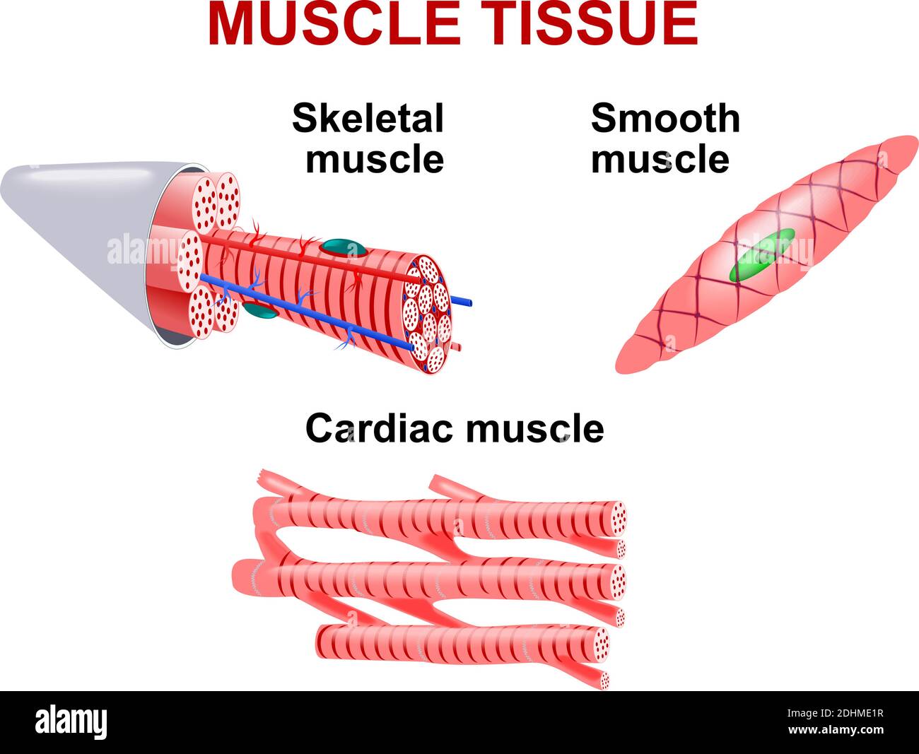
Symptoms and Diagnostic Procedures in Muscle Strains
Muscle strains present with a spectrum of clinical manifestations that evolve over time, often correlating with the severity of muscle fiber disruption and associated inflammatory response. Accurate symptom recognition and systematic diagnostic evaluation are critical to distinguishing muscle strains from other musculoskeletal injuries, guiding appropriate management, and preventing chronic dysfunction. Diagnostic procedures range from non-invasive clinical assessments to advanced imaging, each with distinct advantages, limitations, and contraindications. This section organizes symptoms by temporal progression and compares diagnostic modalities based on their clinical utility, cost, and invasiveness, culminating in a structured approach to differential diagnosis.
Symptoms of Muscle Strains by Timeframe
The presentation of muscle strain symptoms varies depending on the acute, subacute, and chronic phases of injury. Below is a structured breakdown of immediate, delayed, and chronic manifestations, categorized by time since onset.
| Timeframe |
Immediate Symptoms (0–24 hours) |
Delayed Symptoms (24–72 hours) |
Chronic Symptoms (>72 hours to weeks/months) |
|---|
| Pain Intensity and Quality |
- Sudden, sharp pain at the moment of injury, often described as a "tearing" or "popping" sensation.
- Localized tenderness upon palpation, exacerbated by passive or active movement.
- Pain may subside temporarily if the muscle is rested but recurs with provocation.
|
- Pain intensifies due to inflammation, often described as a dull ache or throbbing sensation.
- Pain may radiate to adjacent structures (e.g., hamstring strain radiating to the knee).
- Swelling and warmth develop, contributing to increased pain with movement.
|
- Persistent dull ache, particularly during prolonged activity or at rest.
- Pain may become intermittent, triggered by specific movements (e.g., stair climbing for calf strains).
- Stiffness or muscle tightness, especially after inactivity (e.g., morning stiffness).
|
| Functional Limitations |
- Immediate loss of strength or inability to perform the action that caused the strain (e.g., inability to sprint after a hamstring pull).
- Compensatory gait or movement patterns to avoid pain.
|
- Reduced range of motion (ROM) due to pain and swelling.
- Weakness in muscle groups, detectable via manual muscle testing (MMT).
- Difficulty performing functional tasks (e.g., lifting, jumping, or twisting).
|
- Recurrent giving-way or muscle fatigue during repetitive activities.
- Persistent weakness or muscle atrophy in severe cases.
- Altered biomechanics leading to secondary injuries (e.g., knee pain from chronic hip flexor strains).
|
| Physical Signs |
- Minimal to no visible swelling or bruising in mild strains.
- Ecchymosis (bruising) may appear within hours in moderate to severe strains.
- Muscle spasm or guarding in response to pain.
|
- Visible swelling, erythema (redness), and increased local temperature.
- Palpable muscle induration or "knots" in the affected area.
- Possible joint effusion in adjacent structures (e.g., knee swelling with quadriceps strains).
|
- Persistent muscle tightness or fibrosis (scar tissue formation).
- Visible muscle asymmetry or atrophy in chronic cases.
- Trigger points or myofascial adhesions upon palpation.
|
| Neurological Symptoms |
- Rare, but possible paresthesia (tingling) if the strain involves nerves (e.g., sciatic nerve irritation in severe hamstring strains).
|
- Possible radiating pain or dysesthesia (abnormal sensation) if nerve roots are compressed.
|
- Persistent numbness or weakness if nerve involvement is unresolved.
- Possible radiculopathy symptoms (e.g., lumbar radiculitis with chronic low back strains).
|
Key Considerations:
- Severity Grading: Symptoms correlate with the degree of muscle fiber disruption (Grade I: mild, Grade II: moderate, Grade III: severe/complete tear). Immediate pain and functional loss are more pronounced in higher-grade strains.
- Atypical Presentations: Delayed onset of symptoms (e.g., 48–72 hours) may occur in eccentric overload injuries (e.g., delayed-onset muscle soreness evolving into a strain).
- Red Flags: Symptoms such as severe pain at rest, neurological deficits, or systemic signs (fever, chills) warrant immediate medical evaluation to rule out serious conditions (e.g., compartment syndrome, herniated disc).
Clinical Diagnostic Procedures and Comparative Analysis
Diagnostic accuracy in muscle strains depends on the integration of patient history, physical examination, and, when necessary, advanced imaging. Below is a comparative analysis of common diagnostic methods, including their advantages, limitations, and contraindications.Context:
Clinical decision-making for muscle strains prioritizes non-invasive, cost-effective methods while reserving imaging for complex or atypical cases. The choice of diagnostic modality should align with the suspected severity, patient comorbidities, and resource availability.
| Diagnostic Method |
Accuracy |
Cost |
Invasiveness |
Contraindications |
|---|
| History and Physical Examination |
- Sensitivity: ~80–90% for mild to moderate strains when combined with palpation and ROM testing.
- Specificity: High for ruling out fractures or dislocations but lower for differentiating strains from sprains or tendonitis.
- Key components: Mechanism of injury, location of pain, functional limitations, and palpation findings.
|
- Low cost (no equipment required beyond basic tools like a goniometer).
- Time-intensive but avoids additional expenses.
|
Non-invasive. |
- Patient inability to communicate (e.g., cognitive impairment, language barriers).
- Severe pain or muscle spasm limiting examination (e.g., acute Grade III tears).
- Overlap with other conditions (e.g., radiculopathy mimicking a muscle
Treatment and Rehabilitation Protocols for Muscle Strains
Effective management of muscle strains requires a structured, phased approach that balances immediate intervention with long-term recovery to restore function while minimizing recurrence. The treatment protocol is divided into three distinct phases—acute, subacute, and chronic—each with specific goals, therapeutic interventions, and progressive loading strategies. Evidence-based practices, including pharmacological, mechanical, and rehabilitative modalities, are integrated to optimize outcomes. Personalized rehabilitation plans must account for individual variability in tissue healing, pain tolerance, and functional demands, ensuring a transition from protective measures to performance enhancement.
Phased Approach to Recovery
The recovery from a muscle strain follows a biological timeline aligned with tissue repair and neuromuscular re-education. Each phase targets distinct physiological objectives, with interventions tailored to the healing stage. Below are the key goals for each phase, framed within a RICE-to-activity progression model.
Acute Phase (0–72 hours):
Goal: Reduce pain, control inflammation, and protect the injured tissue from further damage.
Interventions: Immediate rest, compression, ice, and elevation (RICE), alongside analgesic and anti-inflammatory strategies.
Subacute Phase (3–6 weeks):
Goal: Restore pain-free range of motion (ROM), initiate controlled loading, and promote tissue remodeling.
Interventions: Gradual mobilization, active-assisted exercises, and progressive resistance training (isometric → concentric).
Chronic Phase (6+ weeks):
Goal: Enhance strength, power, and functional capacity while addressing residual deficits (e.g., proprioception, endurance).
Interventions: Eccentric and plyometric exercises, sport-specific drills, and neuromuscular re-education.
Therapeutic Interventions by Phase
The selection of therapeutic modalities depends on the phase of healing, evidence of efficacy, and patient-specific factors. Below is a comparative table summarizing interventions, their effectiveness, and supporting evidence levels (adapted from clinical practice guidelines and systematic reviews).
| Phase |
Intervention |
Effectiveness |
Evidence Level |
|---|
| Acute |
Rest and Activity Modification |
Reduces secondary injury; controversial for prolonged immobilization. |
Moderate (Level B) |
| Ice Therapy (Cryotherapy) |
Decreases pain and swelling; optimal for first 48–72 hours. |
High (Level A) |
| Nonsteroidal Anti-Inflammatory Drugs (NSAIDs) |
Reduces inflammation and pain; may delay collagen synthesis if overused. |
Moderate (Level B) |
| Compression and Elevation |
Limits edema formation; beneficial when combined with ice. |
Moderate (Level B) |
| Subacute |
Physical Therapy (Manual Therapy) |
Improves ROM and reduces stiffness; effective for soft tissue mobilization. |
High (Level A) |
| Active-Assisted Stretching |
Restores ROM without excessive stress; preferred over passive stretching. |
Moderate (Level B) |
| Isometric and Concentric Strengthening |
Enhances muscle activation; progresses from pain-free isometrics to dynamic contractions. |
High (Level A) |
| Electrical Stimulation (NMES/TENS) |
May reduce pain and atrophy; limited evidence for accelerating healing. |
Low (Level C) |
| Chronic |
Eccentric Training |
Stimulates collagen remodeling; gold standard for grade II strains. |
High (Level A) |
| Plyometric and Agility Drills |
Restores explosive power; requires full pain-free ROM and strength. |
Moderate (Level B) |
| Neuromuscular Re-education |
Improves proprioception and movement patterns; critical for injury prevention. |
High (Level A) |
| Functional and Sport-Specific Training |
Reintegrates patient into activity; reduces risk of reinjury. |
Moderate (Level B) |
Notes:
- Evidence Levels: A = High-quality randomized controlled trials; B = Moderate-quality studies; C = Low-quality or expert consensus.
- Caution: NSAIDs should be used judiciously in the subacute/chronic phases to avoid masking symptoms or impairing tissue healing.
Personalized Rehabilitation Plan: Exercise Progression
A structured progressive loading protocol ensures safe and efficient recovery by gradually increasing mechanical demand on the healing tissue. The progression follows a hierarchy of exercise complexity, from controlled isometric contractions to high-velocity movements. Modifications are applied based on pain response, functional deficits, and individual goals (e.g., return to sport vs. daily activities).The following bullet-point framework outlines the exercise progression, with modifications for common scenarios (e.g., delayed healing, reinjury risk).
Principles of Progression:
- Pain-Free Criteria: Exercises must not provoke pain (>3/10 on VAS) or exacerbate symptoms.
- Symmetry: Bilateral comparisons guide progression (e.g., strength, ROM).
- Functional Context: Exercises should mimic real-world demands (e.g., single-leg squats for athletes).
-
Isometric Phase (Subacute – Weeks 1–3):
Purpose: Re-educate muscle activation and restore neuromuscular control without joint movement.- Examples:
- Quad sets (knee extensions against immovable resistance).
- Gluteal bridges (pelvic stabilization).
- Wall slides (for shoulder strains).
- Modifications for Delayed Healing:
- Reduce hold time (e.g., 5–10 seconds) or use biofeedback (EMG) to monitor activation.
- Incorporate isometric holds in functional positions (e.g., single-leg stance for ankle strains).
- Progression Criteria:
- Pain-free performance for 3 sets of 10 repetitions.
- Ability to progress to dynamic movements.
-
Concentric/Eccentric Phase (Subacute – Weeks 3–6):
Purpose: Restore dynamic strength and control, with emphasis on eccentric loading to enhance collagen alignment.- Examples:
- Eccentric: Slow lowering phase in squats or lunges (e.g., 4–6 seconds descent).
- Concentric: Controlled speed in seated knee extensions or bicep curls.
- Nordic Hamstring Curls (for posterior thigh strains).
- Modifications for Reinjury Risk:
- Use blood flow restriction (BFR) training at low loads (20–30% 1RM) to maintain hypertrophy without high stress.
- Implement isokinetic exercises (e.g., dynamometer-based) for controlled resistance.
- Progression Criteria:
- Full pain-free ROM with <10% asymmetry in strength.
- Ability to perform 3 sets of 12–15 repetitions with proper form.
-
Plyometric and Sport-Specific Phase (Chronic – Weeks 6+):
Purpose: Restore power, agility, and sport-specific skills while addressing residual deficits (e.g., balance, endurance).- Examples:
Prevention Strategies for Muscle Strains in Athletes and the General Population
Muscle strains remain a prevalent injury across athletic and occupational domains, often resulting from repetitive movements, inadequate preparation, or biomechanical inefficiencies. Effective prevention requires a multimodal approach, integrating active and passive strategies tailored to individual risk profiles, activity demands, and environmental factors. While athletes benefit from high-intensity preparatory protocols, the general population—particularly those engaged in sedentary or physically demanding occupations—must adopt adaptive ergonomic and lifestyle modifications to mitigate strain risk. This section examines evidence-based prevention frameworks, contrasting active (dynamic) and passive (static) interventions, and provides structured protocols for warm-ups, recovery, and workplace adjustments.
Comparison of Active and Passive Prevention Methods
Preventive strategies for muscle strains can be categorized into active (dynamic) methods, which enhance neuromuscular control and tissue resilience through movement, and passive (static) methods, which rely on external aids or recovery techniques to reduce cumulative fatigue. Each approach has distinct advantages and limitations, particularly in terms of time efficiency, accessibility, and scientific validation. Below is a comparative analysis presented in a structured format:
| Category |
Method |
Pros |
Cons |
|---|
| Active (Dynamic) |
Dynamic Stretching |
- Improves range of motion (ROM) and joint mobility through controlled movement.
- Enhances blood flow to muscles, reducing stiffness (ACSM, 2020).
- Activates the neuromuscular system, preparing muscles for explosive actions.
- Time-efficient (5–10 minutes) compared to static stretching.
|
- Requires proper technique to avoid overstretching or compensatory movements.
- Less effective for isolated muscle tightness without dynamic warm-up.
- May not address underlying muscle imbalances without strength training.
|
| Plyometrics |
- Develops fast-twitch muscle fibers, improving power output and injury resilience (Chu, 1998).
- Enhances tendon stiffness, reducing eccentric overload risk in high-impact sports.
- Simulates game-like movements, improving reaction time.
|
- High injury risk if performed without proper progression or fatigue management.
- Time-consuming (30–60 minutes for structured programs).
- Not suitable for deconditioned individuals or those with prior strain history.
|
| Eccentric Strength Training |
- Reduces muscle strain incidence by up to 60% in high-risk populations (Thacker et al., 2004).
- Strengthens tendons and connective tissue, improving load tolerance.
- Low-impact, making it accessible for rehabilitation and prevention.
|
- Slow progression required to avoid delayed-onset muscle soreness (DOMS).
- Less engaging than dynamic activities, reducing adherence.
- Equipment-dependent (e.g., resistance bands, weight machines).
|
| Sport-Specific Drills |
- Mimics competitive demands, improving movement efficiency and reducing compensatory patterns.
- Enhances proprioception and reaction time.
- Can be integrated into team warm-ups for collective preparation.
|
- Requires specialized knowledge to design safe, effective drills.
- Limited generalizability to non-athletic populations.
- High resource demand (coaching, equipment).
|
| Passive (Static) |
Foam Rolling (Self-Myofascial Release) |
- Reduces muscle soreness and improves recovery by breaking fascial adhesions (Cheatham et al., 2015).
- Accessible and low-cost, requiring minimal equipment.
- Can be performed post-exercise to mitigate DOMS.
|
- Ineffective for dynamic mobility without active movement integration.
- Risk of overuse if applied excessively to tender areas.
- Limited evidence for acute injury prevention (vs. recovery).
|
| Hydration and Electrolyte Balance |
- Reduces muscle cramps and fatigue by maintaining cellular function (Sawka et al., 2007).
- Low-cost and universally applicable.
- Supports thermoregulation during high-intensity activities.
|
- Requires consistent adherence, particularly in hot/humid conditions.
- Electrolyte imbalances (e.g., sodium, potassium) may still occur without proper monitoring.
- Less impactful than mechanical or neuromuscular interventions.
|
| Compression Garments |
- May reduce muscle oscillation during high-impact activities, decreasing strain risk (Doan et al., 2003).
- Improves proprioceptive feedback in some athletes.
- Useful for post-injury rehabilitation to limit swelling.
|
- Limited scientific consensus on efficacy for prevention (mixed results in studies).
- Potential for increased sweating and discomfort in high temperatures.
- High cost for long-term use.
|
| Static Stretching |
- Improves passive ROM, beneficial for flexibility-based sports (e.g., gymnastics).
- Low physical demand, suitable for cool-downs or injury rehabilitation.
- Reduces muscle tension through relaxation response.
|
- May reduce muscle strength and power if performed pre-exercise (Shrier, 2004).
- Less effective for dynamic warm-up compared to active methods.
- Time-consuming (15–30 minutes for comprehensive stretching).
|
Structured Warm-Up Protocols for High-Risk Activities
Adequate warm-up routines elevate core temperature, enhance neuromuscular coordination, and prepare connective tissues for the demands of high-risk activities such as sprinting, weightlifting, or overhead sports. Research indicates that progressive warm-ups—combining general, specific, and sport-specific components—reduce injury risk by 30–50% (McCall et al., 1996). Below are evidence-based protocols tailored to common high-risk scenarios, incorporating physiological rationale for each
Long-Term Management and Complications in Muscle Strains
Muscle strains, if inadequately managed, can progress beyond acute recovery to chronic conditions that impair function and quality of life. Untreated or recurrent strains may lead to persistent myofascial dysfunction, structural adaptations in muscle tissue, and compensatory movement patterns that increase vulnerability to reinjury. Effective long-term management requires proactive strategies to mitigate chronic complications while optimizing tissue resilience through structured rehabilitation and lifestyle modifications.Chronic conditions arising from unresolved muscle strains often involve complex interactions between neural, vascular, and connective tissue responses. These conditions may manifest as localized or diffuse pain syndromes, altered biomechanics, or systemic fatigue. Understanding their mechanisms is critical for clinicians and individuals to implement targeted interventions and prevent progression.
Chronic Conditions Associated with Untreated Muscle Strains
Prolonged inflammation, repeated microtrauma, or improper healing of muscle strains can lead to several chronic conditions, each with distinct pathophysiological mechanisms:- Myofascial Pain Syndrome (MPS)
Trigger points develop within taut bands of skeletal muscle, characterized by localized tenderness, referred pain patterns, and restricted range of motion. Chronic MPS arises from sustained muscle overload, poor recovery, or repetitive strain, leading to localized hypoxia, metabolic waste accumulation (e.g., lactic acid, adenosine triphosphate depletion), and sensitization of peripheral nociceptors. Central nervous system hyperexcitability further amplifies pain perception, creating a cycle of muscle spasm and pain reinforcement. - Muscle Adhesions and Fibrosis
Incomplete or excessive collagen deposition during the repair phase of healing results in dense, nonelastic scar tissue (adhesions) that restricts muscle gliding and joint mobility. Adhesions form due to prolonged immobilization, inadequate protein synthesis regulation (e.g., TGF-β overexpression), or persistent mechanical stress. Over time, fibrosis can alter muscle architecture, reducing elasticity and increasing susceptibility to reinjury during dynamic movements. - Chronic Regional Pain Syndrome (CRPS) Type II
Rare but severe, CRPS-II may develop following muscle strain if accompanied by nerve damage (e.g., stretch injury to peripheral nerves). Symptoms include disproportionate pain, autonomic dysfunction (e.g., vasomotor instability, sweating), and trophic changes (e.g., skin atrophy, nail brittleness). The condition involves aberrant nociceptive processing in the dorsal horn of the spinal cord and cortical reorganization, leading to central sensitization. - Tendinopathy and Enthesopathy
Repeated strain on muscle-tendon units can transition into tendinosis (degenerative tendon changes without inflammation) or enthesitis (inflammation at tendon-bone junctions). Microtears and failed healing cycles disrupt collagen fiber alignment, promoting neovascularization and nerve ingrowth, which heightens pain sensitivity. Athletes or laborers with high-demand occupations are particularly at risk due to cumulative load exposure. - Compensatory Movement Dysfunction
Altered motor control strategies emerge as a compensatory mechanism to avoid pain or weakness in the injured muscle. Over time, these adaptations—such as altered gait, joint hypermobility, or overuse of synergistic muscles—can lead to secondary injuries (e.g., patellofemoral pain syndrome, rotator cuff tendinopathy). The central nervous system may also exhibit motor learning deficits, perpetuating inefficient movement patterns. - Systemic Fatigue and Mitochondrial Dysfunction
Chronic muscle strain disrupts mitochondrial biogenesis and oxidative capacity, particularly in high-endurance athletes or individuals with sedentary lifestyles. Persistent low-grade inflammation (elevated CRP, IL-6) impairs recovery, reducing muscle protein synthesis and increasing oxidative stress. This contributes to generalized fatigue, reduced endurance, and delayed recovery between training sessions or work shifts.
Structured Plan for Maintaining Muscle Health Post-Recovery
A comprehensive long-term management plan integrates nutritional support, mobility work, and progressive load management to restore and maintain muscle resilience. The following table outlines evidence-based strategies tailored to individual recovery phases and activity levels.
| Component |
Intermediate Recovery Phase (4–12 weeks post-injury) |
Advanced Recovery Phase (3–6 months post-injury) |
Maintenance Phase (6+ months post-recovery) |
|---|
| Nutrition |
- Protein intake: 1.6–2.2 g/kg body weight/day, prioritizing leucine-rich sources (whey, soy, lean meats) to stimulate muscle protein synthesis.
- Anti-inflammatory diet: Emphasize omega-3 fatty acids (fatty fish, flaxseeds), antioxidants (berries, leafy greens), and reduce processed sugars and trans fats.
- Hydration: 30–35 mL/kg body weight/day to optimize cellular repair and metabolic efficiency.
- Vitamin D and magnesium supplementation if deficient, as both influence muscle repair and nerve function.
|
- Adjust protein to 1.2–1.6 g/kg/day, balancing with complex carbohydrates (oats, quinoa) to support glycogen replenishment.
- Include collagen peptides (10–15 g/day) to enhance tendon and ligament integrity during progressive loading.
- Monitor creatine kinase (CK) levels if symptoms of rhabdomyolysis or overtraining persist.
|
- Sustain protein at 1.0–1.2 g/kg/day, with periodic fasted training to preserve muscle mass.
- Intermittent fasting (16:8 protocol) may be introduced to modulate autophagy and reduce inflammation.
- Regularly assess micronutrient status (e.g., zinc, vitamin C) to support extracellular matrix remodeling.
|
| Mobility and Flexibility Work |
- Dynamic stretching and PNF (proprioceptive neuromuscular facilitation) techniques to improve muscle-tendon unit elasticity without provoking pain.
- Foam rolling or instrument-assisted soft tissue mobilization (IASTM) to address myofascial restrictions, with 60–90 seconds per trigger point.
- Avoid static stretching in acute phases; prioritize active recovery (e.g., swimming, cycling at low intensity).
|
- Incorporate yoga or tai chi for controlled joint mobility and breathwork to reduce sympathetic nervous system activity.
- Eccentric loading exercises (e.g., Nordic hamstring curls) to reinforce tendon resilience and reduce adhesion formation.
- Use contrast therapy (alternating hot/cold) to modulate blood flow and enhance tissue compliance.
|
- Maintain a routine of 2–3 sessions/week of myofascial release (e.g., Graston technique) to prevent fibrosis.
- Integrate mobility drills into warm-ups/cool-downs, focusing on full-range-of-motion patterns (e.g., hip CARs for athletes).
- Monitor for joint hypermobility; implement corrective exercises (e.g., proprioceptive balance training) if compensatory movements persist.
|
| Load Management |
- Gradual reintroduction of resistance training with 30–50% of pre-injury load, emphasizing controlled tempo (3–4 seconds eccentric phase).
- Avoid high-velocity or plyometric exercises until pain-free full ROM is achieved.
- Use heart rate variability (HRV) monitoring to guide recovery pacing; aim for >50 ms HRV improvement post-session.
|
- Progress to sport-specific drills with 70–80% load, incorporating perturbation training (e.g., unstable surfaces) to enhance neuromuscular control.
- Implement periodized training cycles (e.g., 4-week blocks) to balance volume and intensity, avoiding weekly monotony.
- For laborers, ergonomic assessments should be conducted to modify repetitive tasks (e.g., tool redesign, rotational lifting techniques).
|
- Adopt a 10% rule for progressive overload: increase load by ≤10% weekly to prevent overtraining.
- Incorporate deload weeks every 6–
Muscle strains underscore the delicate balance between physical exertion and biological resilience, where even minor deviations in technique or conditioning can precipitate significant dysfunction. From the microscopic disruption of sarcomeres to the systemic impact of chronic myofascial pain, these injuries demand a multifaceted approach—one that integrates acute intervention, structured rehabilitation, and sustained preventive strategies. By understanding the grading systems, recognizing early symptoms, and adhering to phased recovery protocols, individuals can minimize downtime and restore functional capacity. Ultimately, the key to managing muscle strains lies not only in addressing the injury itself but in fostering long-term musculoskeletal health through education, biomechanical awareness, and proactive care.
FAQ
What does it mean to have a muscle strain in the back?
A muscle strain in the back occurs when fibers in the back muscles (like the erector spinae or latissimus dorsi) are overstretched or torn, often from heavy lifting, sudden movements, or poor posture. Symptoms include localized pain, stiffness, and limited movement, which may worsen with activity. Recovery usually involves rest, ice, gentle stretching, and over-the-counter pain relief.
How do you know if you have a muscle strain in the lower back?
A lower back muscle strain typically causes sharp or dull pain in the lumbar region, often triggered by bending, twisting, or lifting. You may also feel muscle tightness, spasms, or tenderness to the touch, and the pain usually improves with rest but flares up with movement. Unlike herniated discs, the pain rarely radiates down the legs.
What causes a muscle strain in the thigh?
A thigh muscle strain (often in the hamstrings, quadriceps, or adductors) usually happens from overuse, sudden sprinting, kicking, or direct trauma like a fall. Poor flexibility, fatigue, or improper warm-ups increase the risk. Symptoms include sudden pain during activity, swelling, bruising, and weakness when using the muscle.
What does a muscle strain feel like when it happens?
When a muscle strain occurs, you often feel a sudden sharp pain or a "popping" sensation during the activity (like lifting or stretching). Afterward, the area becomes tender, swollen, and stiff, with pain worsening when you move or press on it. Mild strains cause discomfort; severe ones may leave you unable to use the muscle at all.
What is a muscle strain in the shoulder?
A shoulder muscle strain involves torn fibers in muscles like the rotator cuff (e.g., supraspinatus) or deltoid, often from overhead motions, heavy lifting, or repetitive use. Symptoms include localized pain, weakness when lifting the arm, and tenderness, which may limit range of motion. Rest, ice, and physical therapy help recovery.
What’s the difference between a muscle strain and a sprain?
A muscle strain is an injury to muscle or tendon fibers (e.g., from overstretching), causing pain, swelling, and stiffness in the muscle itself. A sprain involves damage to ligaments (the bands connecting bones), usually from joint twisting, and causes pain, bruising, and instability in the joint (e.g., ankle or knee). Strains heal with rest and stretching; sprains often require immobilization and rehab.
|
|
|
Leave a Comment
Comments are moderated before appearing. The data you submit is processed according to the Privacy Policy of Voltefac.