What Is A Sleep Study And Its Clinical Applications
Table of Contents
- Definition and Purpose of a Sleep Study
- Core Objectives and Diagnostic Scope
- Primary Conditions Identified Through Sleep Studies
- Procedural Distinctions: Sleep Study Modalities
- Types of Sleep Studies and Their Applications
- Comparison of Sleep Study Modalities
- Selection Criteria for Sleep Study Modalities
- Procedure for Conducting a Home Sleep Apnea Test (HSAT)
- Equipment and Technology Used in Sleep Studies
- Core Equipment in a Polysomnography Sleep Lab
- Calibration and Positioning of EEG Electrodes for Polysomnography
- Patient Preparation and Procedure for Sleep Studies
- Patient Preparation Checklist
- Step-by-Step Process of a Full-Night Polysomnography
- Common Challenges and Mitigation Strategies
- Patient Instruction Sheet Template
- Data Interpretation and Clinical Implications of Sleep Studies
- Framework for Interpreting Sleep Study Results
- Translation of Findings into Treatment Plans
- Role of Sleep Studies in Legal and Forensic Contexts
- FAQ
- what is a sleep study test?
- what is a sleep study like?
- what is a sleep study doctor called?
- what is a sleep study called?
- what is a sleep study for sleep apnea?
- what is a sleep study and how is it done?
Sleep studies represent a cornerstone of modern sleep medicine, offering precise diagnostic insights into disorders that disrupt rest and impair daily functioning. By systematically recording physiological parameters during sleep, these evaluations enable clinicians to identify conditions ranging from obstructive sleep apnea to narcolepsy, bridging the gap between symptoms and evidence-based treatment. Advances in technology have transformed sleep studies from rudimentary observations into highly specialized assessments, integrating polysomnography, wearable devices, and home-based testing to accommodate diverse patient needs.
The process begins with a structured evaluation in controlled environments or portable setups, where sensors monitor brain activity, respiratory patterns, and oxygen levels with millimeter precision. Beyond clinical diagnostics, sleep studies play a pivotal role in legal assessments, workplace safety protocols, and personalized therapy planning, underscoring their multifaceted importance in healthcare. This exploration examines the methodology, applications, and evolving innovations that define contemporary sleep studies as indispensable tools in both research and patient care.
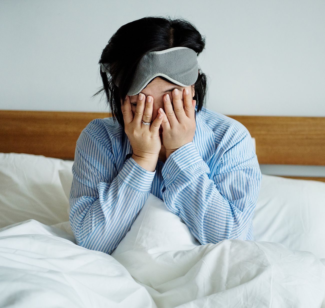
Definition and Purpose of a Sleep Study
A sleep study, formally known as a polysomnography (PSG) or other specialized sleep assessments, serves as a cornerstone diagnostic tool in sleep medicine. Its primary objective is to systematically record physiological parameters during sleep to identify disruptions, disorders, and underlying pathologies that impair sleep quality or daytime functioning. By leveraging advanced monitoring technologies, sleep studies enable clinicians to differentiate between primary sleep disorders, comorbid conditions, and secondary sleep disturbances, thereby guiding targeted therapeutic interventions.The clinical significance of sleep studies extends beyond mere symptom assessment, as they provide objective, quantifiable data that correlate with patient-reported symptoms. This distinction is critical, as many sleep disorders—such as obstructive sleep apnea (OSA) or periodic limb movement disorder (PLMD)—often present with subjective complaints (e.g., fatigue, poor concentration) that lack specificity without physiological validation. Sleep studies thus bridge the gap between clinical suspicion and definitive diagnosis, ensuring precision in treatment planning.
Core Objectives and Diagnostic Scope
The central purpose of a sleep study is to:Sleep studies are particularly indispensable in cases where:
Primary Conditions Identified Through Sleep Studies
The following table outlines the most common sleep disorders diagnosed via sleep studies, their key symptoms, the diagnostic role of PSG, and treatment implications. Data is derived from clinical guidelines by the American Academy of Sleep Medicine (AASM) and peer-reviewed literature.| Condition | Key Symptoms | Diagnostic Role of Sleep Study | Treatment Implications |
|---|---|---|---|
| Obstructive Sleep Apnea (OSA) |
|
|
|
| Insomnia Disorder |
|
|
|
| Restless Legs Syndrome (RLS) |
|
|
|
| Narcolepsy |
|
|
|
| Circadian Rhythm Sleep-Wake Disorders |
|
|
|
Procedural Distinctions: Sleep Study Modalities
While polysomnography (PSG) remains the gold standard for comprehensive sleep assessment, alternative modalities are employed based on clinical indication, resource availability, and patient needs. The following outlines key procedural distinctions:1. In-Laboratory Polysomnography (Attended PSG)
Types of Sleep Studies and Their Applications
Sleep studies, or polysomnographies, are essential diagnostic tools in sleep medicine, designed to assess physiological parameters during sleep to identify disorders such as sleep apnea, insomnia, periodic limb movement disorder, and parasomnias. The selection of an appropriate study type depends on clinical indications, patient demographics, symptom severity, and logistical considerations. Below is a structured comparison of the most common sleep study modalities, their procedural distinctions, and guidelines for patient-specific application.Comparison of Sleep Study Modalities
The following table outlines the key characteristics of polysomnography (PSG), home sleep tests (HST), multiple sleep latency tests (MSLT), and maintenance of wakefulness tests (MWT), including their purpose, duration, equipment, and ideal patient scenarios. This framework facilitates clinical decision-making based on diagnostic needs and patient profiles.| Study Type | Purpose | Duration | Equipment Used | Ideal Patient Scenarios |
|---|---|---|---|---|
| Polysomnography (PSG) | Comprehensive evaluation of sleep architecture, respiratory effort, oxygen saturation, limb movements, and brain wave activity. Diagnoses complex or severe sleep disorders (e.g., obstructive sleep apnea, central sleep apnea, REM sleep behavior disorder, narcolepsy). | Overnight (6–8 hours), occasionally split-night studies (diagnostic + therapeutic phase). | EEG, EOG, EMG, ECG, respiratory effort belts, nasal/oral airflow sensors, pulse oximetry, video/audio monitoring, and sometimes polysomnographic technologists for real-time observation. |
|
| Home Sleep Apnea Test (HSAT) | Screening and diagnosis of obstructive sleep apnea (OSA) in patients with high pre-test probability. Not intended for complex sleep disorders or those requiring detailed sleep staging. | Single night (4–8 hours), typically during habitual sleep. | Portable devices measuring airflow (nasal pressure transducer or thermistor), respiratory effort (chest/abdomen belts or inductance plethysmography), and oxygen saturation (pulse oximetry). Some include actigraphy or heart rate monitoring. |
|
| Multiple Sleep Latency Test (MSLT) | Assessment of daytime sleepiness and diagnosis of narcolepsy or idiopathic hypersomnia. Measures the time taken to fall asleep across multiple nap opportunities. | 4–5 naps scheduled at 2-hour intervals (total duration: ~8 hours). | EEG, EOG, EMG, and sometimes video monitoring. Conducted in a sleep lab under controlled conditions. |
|
| Maintenance of Wakefulness Test (MWT) | Evaluation of a patient’s ability to stay awake in a quiet, darkened environment. Used to assess fitness to drive or work in safety-sensitive professions. | 4 trials (20–40 minutes each), separated by 2-hour intervals (total duration: ~4–6 hours). | EEG, EOG, EMG, and sometimes video monitoring. Conducted in a sleep lab with standardized protocols. |
|
Selection Criteria for Sleep Study Modalities
The choice of sleep study modality is influenced by patient demographics, symptom severity, and clinical context. Below are guidelines for selecting the appropriate study based on specific profiles:Pediatric Patients: PSG is the gold standard due to the need for detailed sleep staging, safety monitoring, and the inability of children to tolerate home-based devices. HSATs are contraindicated unless approved by a pediatric sleep specialist for mild OSA screening.
Geriatric Patients: PSG remains preferred for complex cases (e.g., suspected REM sleep behavior disorder or periodic limb movement disorder). HSATs may be considered for high-risk patients (e.g., those with mobility issues or severe comorbidities) where transport to a lab is challenging. MSLT or MWT may be used to assess treatment response in conditions like narcolepsy or OSA.
Mild vs. Severe Symptoms:
- Mild symptoms (e.g., occasional snoring, mild daytime fatigue): HSAT may suffice for OSA screening, followed by PSG if results are inconclusive or complex.
- Severe symptoms (e.g., gasping awakenings, uncontrolled hypertension, suspected central apnea): PSG is mandatory to rule out life-threatening conditions and guide treatment.
- Excessive daytime sleepiness without clear OSA: MSLT is indicated to evaluate narcolepsy or idiopathic hypersomnia.
- Occupational or regulatory requirements (e.g., commercial drivers): MWT is used to assess wakefulness maintenance post-treatment.
Procedure for Conducting a Home Sleep Apnea Test (HSAT)
HSATs provide a convenient alternative to in-lab PSG for diagnosing OSA in select patients. Below is a step-by-step protocol for setup, data collection, and limitations.Prerequisites for HSAT: The test is only appropriate for patients with a high pre-test probability of OSA (e.g., STOP-Bang score ≥5 or clinical suspicion). Exclusion criteria include suspected central sleep apnea, complex sleep disorders, or unstable medical conditions.
-
Patient Preparation:
- Instruct the patient to avoid caffeine, alcohol, and sedatives for 24 hours prior to the test.
- Ensure the patient follows their usual sleep schedule and uses their typical sleep position.
- Provide written or verbal instructions on device application and troubleshooting (e.g., ensuring sensors are securely attached).
-
Equipment Setup:
- Position the portable device on a stable surface near the patient’s bed (e.g., nightstand).

Equipment and Technology Used in Sleep Studies
Sleep studies rely on advanced, multi-modal technology to capture physiological and behavioral data with high precision. The integration of specialized sensors, recording devices, and analytical software enables clinicians to diagnose sleep disorders, assess treatment efficacy, and refine therapeutic approaches. Below are the essential tools used in polysomnography (PSG) and ambulatory sleep monitoring, categorized by their functional roles in capturing neural, muscular, respiratory, and cardiovascular parameters.
Core Equipment in a Polysomnography Sleep Lab
The sleep laboratory employs a synchronized array of devices to record simultaneous physiological signals. These instruments are designed to minimize artifact interference while ensuring high-fidelity data acquisition. The primary components include:
-
Polysomnograph Machine (PSG System)
A centralized recording device that digitizes and synchronizes signals from multiple sensors. Modern systems feature:
- High-resolution analog-to-digital converters (ADC) for signal amplification and noise reduction.
- Multi-channel input capabilities (typically 16–32 channels) to accommodate EEG, EMG, EOG, respiratory, and cardiac sensors.
- Built-in event markers for annotating sleep stages, arousals, or patient-reported symptoms (e.g., leg movements).
- Software integration for real-time monitoring and automated scoring (e.g., hypnogram generation via algorithms like R&K or AASM criteria).
-
Electroencephalogram (EEG) Sensors
Surface electrodes placed on the scalp to measure brainwave activity across different frequencies (delta, theta, alpha, beta, gamma). Standard placements follow the 10-20 International System, with electrodes labeled (e.g., Fp1, C3, O2) to correspond to specific brain regions.
EEG frequencies and their clinical significance:
Frequency Band Range (Hz) Associated Sleep Stage Delta 0.5–4 Deep sleep (N3) Theta 4–8 Light sleep (N1, N2) Alpha 8–12 Awake/relaxed or drowsiness Beta 12–30 Active wakefulness or REM Gamma 30–100 Cognitive processing (less studied in sleep) -
Electromyogram (EMG) Electrodes
Surface or needle electrodes placed on muscles to detect electrical activity during sleep. Key applications include:
- Chin EMG: Assesses muscle tone to differentiate REM (atonia) from NREM sleep.
- Leg EMG: Monitors periodic limb movements (PLMs) associated with restless legs syndrome (RLS) or PLMD.
- Submental EMG: Helps identify arousals or sleep fragmentation.
-
Electrooculogram (EOG) Sensors
Electrodes placed near the outer canthi of the eyes to record eye movements (saccades and slow drifts). EOG distinguishes:
- REM sleep (characterized by rapid eye movements).
- NREM sleep (minimal eye movement).
- Eye movement disorders (e.g., periodic eye movement disorder).
-
Respiratory Monitoring Devices
A combination of sensors tracks breathing patterns, airflow, and thoracic/abdominal effort. Critical tools include:
-
Thermistors/Nasal Pressure Transducers: Measure airflow through the nose/mouth to detect apneas (cessation of airflow) or hypopneas (reduced airflow).
Apnea-Hypopnea Index (AHI) Calculation:
AHI = (Number of Apneas + 3 × Number of Hypopneas) / Total Sleep Time (hours) Thresholds for diagnosis:- 5–14: Mild OSA
- 15–29: Moderate OSA
- ≥30: Severe OSA
- Respiratory Effort Belts (REBs): Strain gauges placed around the chest/abdomen to detect paradoxical breathing (asynchronous chest/abdomen movement) in OSA or central sleep apnea (CSA).
- Capnography (End-Tidal CO₂ Monitoring): Tracks CO₂ levels to differentiate obstructive (elevated CO₂ during apnea) from central apneas (flat CO₂ trace).
-
Thermistors/Nasal Pressure Transducers: Measure airflow through the nose/mouth to detect apneas (cessation of airflow) or hypopneas (reduced airflow).
-
Pulse Oximeter
A non-invasive sensor (typically on the finger) that measures:
- Peripheral capillary oxygen saturation (SpO₂): Identifies desaturations (<90%) linked to apneas or hypopneas.
- Pulse rate: Detects tachycardia/bradycardia during arousals or sleep-disordered breathing.
- Oxygen desaturation index (ODI): Number of ≥3% SpO₂ drops per hour of sleep (correlates with OSA severity).
Calibration and Positioning of EEG Electrodes for Polysomnography
Accurate EEG placement ensures reliable brainwave measurement and minimizes artifacts. The process involves anatomical landmarks, impedance checks, and troubleshooting common issues.
-
Preparation and Landmark Identification
- Cleanse the scalp with alcohol or abrasive gel to reduce impedance (<5 kΩ).
- Identify key landmarks using a tape measure:
10-20 System Guidelines:
- Nasion (Nz) and inion (Iz): Midline reference points.
- Preauricular points: Align electrodes horizontally/vertically from these.
- Cz (vertex): 10% of nasion-inion distance from nasion.
- Fp1/Fp2: 10% from nasion along the midline to preauricular line.
- C3/C4: 20% lateral from midline at Cz level.
- Apply conductive gel to electrodes to ensure low-resistance contact.
-
Electrode Placement and Impedance Testing
- Follow the standard montage (e.g., F4-M1, C4-M1, O2-M1 for right hemisphere activity).
- Verify impedance for each electrode (<5 kΩ for EEG; <10 kΩ for EOG/EMG). High impedance may indicate:
- Insufficient gel or dried contact.
- Loose electrode or hair obstruction.
- Skin contamination (e.g., oils, lotions).
- Perform a light check (brief EEG recording) to confirm waveform presence and symmetry between homologous electrodes (e.g., C3 vs. C4).
-
Troubleshooting Common Artifacts
-
Muscle Artifact (EMG Contamination):
-
<
- Caffeine and Alcohol: Refrain from consuming caffeine (coffee, tea, soda, chocolate) or alcohol for 12 hours prior to the study, as these substances alter sleep stages and respiratory patterns.
- Heavy Meals and Spicy Foods: Eat a light dinner 2–3 hours before bedtime to prevent discomfort or reflux, which may interfere with sensor placement or breathing measurements.
- Hydration: Drink water moderately; excessive fluid intake may require frequent bathroom visits, while dehydration can affect sensor adhesion and physiological readings.
- Nicotine: Avoid smoking or vaping for 6–8 hours before the study, as nicotine reduces sleep quality and may influence respiratory metrics.
- Sleep Aids: Prescription sleep medications (e.g., zolpidem) may be permitted if approved by the sleep specialist but should be documented for analysis.
- Respiratory Medications: Bronchodilators or nasal decongestants (e.g., pseudoephedrine) should be used cautiously, as they can mask obstructive sleep apnea (OSA) or alter airflow signals.
- Over-the-Counter Drugs: Pain relievers (e.g., NSAIDs) or antihistamines (e.g., diphenhydramine) may be restricted, as they can suppress REM sleep or respiratory drive.
- Continuous Positive Airway Pressure (CPAP): If the patient uses CPAP, they should bring their device and mask for titration studies, but discontinue use the night before unless instructed otherwise.
- Napping: Avoid daytime naps on the study day to prevent sleep inertia, which can skew results.
- Bedtime Routine: Maintain a consistent sleep schedule for 3–4 days prior to align circadian rhythms with the study timeline.
- Clothing: Wear loose, comfortable clothing (e.g., a T-shirt and shorts) to facilitate sensor application and movement.
- Personal Items: Bring essentials such as earplugs, a pillow, a book, or a tablet for relaxation, but avoid electronic devices that emit light (e.g., smartphones).
- The sleep technician reviews the patient’s medical history, current medications, and any allergies to adhesives or sensors.
- Informed consent is obtained, explaining the procedure, potential discomfort, and data usage for diagnostics.
- A pre-sleep questionnaire may be administered to assess baseline symptoms (e.g., snoring, leg movements).
- Brain Activity: Electroencephalogram (EEG) electrodes placed at standard locations (e.g., C3/A2, O2/A1) to record sleep stages.
- Eye Movements: Electrooculogram (EOG) electrodes near the outer canthi to detect REM sleep.
- Muscle Activity: Electromyogram (EMG) sensors on the chin (submental) and legs (tibialis anterior) to monitor arousal and periodic limb movements.
- Heart Rate and Breathing: Electrocardiogram (ECG) leads and respiratory effort belts (thoracic/abdominal) or nasal/oral airflow sensors.
- Oxygen Saturation: Pulse oximetry on a finger to track blood oxygen levels (SpO₂).
- Body Position: A position sensor (e.g., mercury switch) to log sleep posture.
- The patient is directed to a private, dimly lit room with a comfortable bed, ambient temperature (65–68°F or 18–20°C), and white noise if requested.
- The technician calibrates all devices, checks sensor connectivity, and ensures the recording system (e.g., Embla, Compumedics) is operational.
- A video camera may be installed for behavioral observations (e.g., apnea events, parasomnias).
- The patient is left undisturbed, with a call button for emergencies or discomfort (e.g., itchy electrodes, bathroom needs).
- Technicians periodically check sensor integrity (e.g., electrode impedance <10 kΩ) but avoid unnecessary interruptions.
- Light sleep or awakenings may be noted if the patient reports discomfort, but efforts are made to minimize disruptions.
- Upon waking, the technician removes sensors gently, cleans the skin, and checks for any residual irritation.
- The patient completes a post-sleep questionnaire to assess sleep quality, symptoms (e.g., gasping, leg jerks), and comfort level.
- A sleep diary may be provided to log home sleep habits for comparison.
- Preliminary findings (e.g., suspected OSA severity) may be discussed, but formal results require physician interpretation.
- Electrode Dislodgment or Poor Signal Quality:
- Use hypoallergenic adhesive and skin prep solutions (e.g., Nuprep) to improve conductivity.
- Secure electrodes with double-sided tape or headbands for EEG/EOG sensors.
- Reapply sensors if impedance exceeds thresholds (e.g., >5 kΩ for EEG).
- Artifact from Movement or Sweating:
- Apply waterproof electrodes (e.g., for EMG) and use lightweight cables to reduce friction.
- Use dry electrodes or wireless sensors (e.g., for ECG) in active patients.
- Sensor Malfunction or Equipment Failure:
- Perform pre-study equipment checks, including battery levels and cable integrity.
- Have backup sensors and a technician on standby for rapid replacements.
- Anxiety or Insomnia from Novel Environment:
- Offer relaxation techniques (e.g., guided breathing, white noise) and allow the patient to bring familiar items (e.g., a pillow, pajamas).
- Schedule the study at the patient’s usual bedtime to align circadian rhythms.
- Provide clear instructions to reduce uncertainty (e.g., "You may fall asleep quickly; we’ll monitor you silently").
- Discomfort from Sensors or Restrictions:
- Use minimally invasive sensors (e.g., nasal pressure cannula instead of a mask for airflow).
- Apply topical numbing cream (e.g., lidocaine) for sensitive skin before electrode placement.
- Allow limited movement (e.g., turning over) if sensors permit, but advise against sitting up.
- Frequent Bathroom Needs:
- Restrict fluid intake 2 hours before bedtime unless medically necessary.
- Provide a portable commode or schedule bathroom breaks during light sleep phases.
- Use the call button for non-urgent needs (e.g., adjusting blankets, repositioning sensors).
- Gently tug on sensor cables if they feel loose, but avoid pulling electrodes.
- Notify technicians immediately for pain, itching, or respiratory distress (e.g., choking sensation from airflow sensors).
- Avoid touching sensors unless necessary, as this can introduce artifacts.
- Arrive at the sleep center 1–2 hours before your scheduled bedtime to complete paperwork and sensor application.
- You will be in a private room with a comfortable bed; technicians will monitor you silently via cameras and sensors.
- Sensors will be placed on your scalp, face, chest, legs, and finger to measure brain activity, breathing, and oxygen levels.
- Do not expect to sleep perfectly—it is
- Normal: <5 events/hour.
- Mild Obstructive Sleep Apnea (OSA): 5–14 events/hour.
- Moderate OSA: 15–29 events/hour.
- Severe OSA: ≥30 events/hour. Note: AHI ≥15 in symptomatic patients or ≥5 in asymptomatic patients with comorbidities (e.g., hypertension) may warrant treatment.
- Normal: ≥85%.
- Borderline: 75–84%.
- Poor: <75%. Low sleep efficiency may indicate insomnia, circadian rhythm disorders, or restless legs syndrome (RLS).
- Normal: 60–120 minutes (time from sleep onset to first REM period).
- Shortened (<60 min): Suggestive of narcolepsy type 1 or REM sleep behavior disorder (RBD).
- Absent/Fragmented REM: Associated with depression, alcohol use, or certain medications.
- Normal: <15 events/hour.
- Mild PLMD: 15–29 events/hour.
- Moderate-Severe PLMD: ≥30 events/hour. PLMD is often comorbid with OSA or RLS and may contribute to sleep fragmentation.
- Normal: <5 desaturation events/hour (drop ≥3% from baseline).
- Abnormal: ≥15 events/hour (indicative of OSA or other hypoxemic disorders).
- Normal: <10 arousals/hour.
- Abnormal: ≥15 arousals/hour (suggestive of sleep fragmentation due to PLMD, OSA, or central sleep apnea).
- AHI ≥5 with symptoms (e.g., snoring, daytime sleepiness) or ≥15 regardless of symptoms.
- Oxygen desaturation episodes (SpO₂ <90% for ≥10 seconds).
- Presence of obstructive apneas/hypopneas on PSG.
- Positive Airway Pressure (PAP) therapy (e.g., CPAP, APAP, BiPAP).
- Oral appliance therapy (for mild-moderate OSA).
- Lifestyle modifications (weight loss, avoidance of alcohol/sedatives, positional therapy).
- Surgical interventions (e.g., uvulopalatopharyngoplasty, maxillomandibular advancement) for refractory cases.
- Repeat PSG or HSAT after 3–6 months to assess adherence and efficacy (e.g., residual AHI <5).
- Monitor for treatment-emergent side effects (e.g., CPAP intolerance, dental issues).
- Annual follow-up for long-term management, especially in patients with comorbidities (e.g., cardiovascular disease).
- PLMI ≥15/hour with associated symptoms (e.g., insomnia, daytime fatigue).
- PLMD often coexists with RLS or OSA.
- Sleep fragmentation due to limb movements (e.g., >85% of movements cause arousals).
- Dopamine agonists (e.g., pramipexole, ropinirole) for PLMD/RLS overlap.
- Iron supplementation if ferritin <50 mcg/L (common in RLS/PLMD).
- Alpha-2-delta ligands (e.g., gabapentin, pregabalin) for refractory cases.
- Behavioral interventions (e.g., leg exercises, avoidance of caffeine/alcohol).
- Repeat PSG after 3–6 months to evaluate treatment response (reduced PLMI and improved sleep architecture).
- Monitor for augmentation (worsening of symptoms with long-term dopamine agonist use).
- Comanagement with a neurologist if RLS is suspected.
- Mean sleep latency ≤8 minutes on Multiple Sleep Latency Test (MSLT) with ≥2 sleep-onset REM periods (SOREMPs).
- Short REM latency (<15 minutes) on PSG.
- Cataplexy (sudden muscle weakness triggered by emotion) or hypnagogic hallucinations.
- Low hypocretin-1 levels in cerebrospinal fluid (CSF).
- Stimulant medications (e.g., modafinil, armodafinil) for daytime sleepiness.
- Sodium oxybate (GHB) for cataplexy and disrupted nighttime sleep.
- Antidepressants (e.g., venlafaxine, fluoxetine) for cataplexy/hallucinations.
- Scheduled naps (10–20 minutes) to mitigate sleepiness.
- Repeat MSLT after 3–6 months to assess treatment efficacy (improved sleep latency, reduced SOREMPs).
- Annual monitoring for medication side effects (e.g., dependence, mood changes).
- Patient education on safety precautions (e.g., avoidance of heavy machinery due to sudden sleep attacks).
Patient Preparation and Procedure for Sleep Studies
A sleep study, particularly a polysomnography (PSG), requires meticulous preparation to ensure accurate diagnostics and patient comfort. Proper patient preparation enhances data quality, minimizes disruptions, and reduces anxiety, while the procedural steps must be standardized to maintain consistency across clinical settings. This section outlines the preparatory checklist for patients, the structured workflow of an in-lab PSG, and strategies to address common procedural challenges.
Patient Preparation Checklist
Effective preparation before a sleep study significantly impacts the reliability of results. Patients must adhere to dietary, medication, and environmental guidelines to avoid confounding variables that could distort sleep architecture or physiological recordings.Dietary Restrictions and Timing
Avoiding stimulants and heavy meals ensures minimal disruption to sleep patterns and sensor accuracy. Patients should:
Medication Guidelines
Medications that influence sleep or respiration should be reviewed with a healthcare provider. Key considerations include:
Environmental Adjustments
Preparing the home environment before the study reduces anxiety and promotes natural sleep:
Step-by-Step Process of a Full-Night Polysomnography
A PSG involves monitoring physiological parameters throughout the night in a controlled sleep laboratory. The process is structured to ensure patient comfort, data integrity, and adherence to clinical protocols.1. Pre-Study Consultation and Consent
2. Sensor Application and Setup
The technician applies electrodes, sensors, and transducers to measure:
3. Sleep Environment Configuration
4. Patient Monitoring and Overnight Recording
5. Post-Study Debriefing and Sensor Removal
Common Challenges and Mitigation Strategies
Sleep studies are susceptible to technical and patient-related disruptions that can compromise data validity. Proactive strategies and troubleshooting protocols address these challenges efficiently.Technical Challenges and Solutions
Patient-Related Challenges and Solutions
Communication Protocols for Patient Discomfort
Patients should be instructed to:
Patient Instruction Sheet Template
What to Expect During Your Sleep Study

Data Interpretation and Clinical Implications of Sleep Studies
Sleep studies generate comprehensive physiological data that require systematic interpretation to diagnose sleep disorders, guide treatment decisions, and inform clinical management. Accurate analysis of metrics such as the Apnea-Hypopnea Index (AHI), sleep efficiency, and REM latency—among others—enables clinicians to correlate findings with specific pathologies. This section outlines a structured framework for interpreting sleep study results, their translation into evidence-based treatment plans, and their broader applications in legal and forensic contexts, including patient-centered reporting.
Framework for Interpreting Sleep Study Results
The interpretation of sleep study data relies on standardized metrics derived from polysomnography (PSG) or home sleep apnea testing (HSAT). Key parameters are evaluated against established clinical thresholds to diagnose sleep disorders and assess severity. Below are the primary metrics, their definitions, and diagnostic thresholds:- Apnea-Hypopnea Index (AHI):
AHI = (Number of apneas + hypopneas) / Total sleep time (events/hour).
- Sleep Efficiency:
Sleep Efficiency = (Total sleep time / Time in bed) × 100%.
- REM Latency:
- Periodic Limb Movement Index (PLMI):
PLMI = Number of periodic limb movements per hour of sleep.
- Oxygen Desaturation Index (ODI):
- Arousal Index:
Translation of Findings into Treatment Plans
Sleep study results directly inform therapeutic strategies tailored to the diagnosed condition. Below is a comparative table outlining diagnostic findings, recommended treatments, and follow-up protocols for three common sleep disorders:
Condition Diagnostic Findings Recommended Treatments Follow-Up Protocols Obstructive Sleep Apnea (OSA) Periodic Limb Movement Disorder (PLMD) Narcolepsy Type 1 Role of Sleep Studies in Legal and Forensic Contexts
Sleep studies play a critical role in legal and forensic evaluations, particularly in assessing an individual’s fitness to perform safety-sensitive tasks, such as driving or operating heavy machinery. Courts and regulatory bodies (e.g., Department of Transportation, occupational health programs) rely on sleep medicine expertise to determine liability, workplace accommodations, or disability claims. Key applications include:- Fitness to Drive Evaluations:
Sleep disorders such as OSA, narcolepsy, and severe insomnia can impair cognitive and motor function, increasing the risk of accidentsSleep studies transcend their role as mere diagnostic tools, serving as a gateway to improved health outcomes by illuminating the complexities of sleep-related disorders. From the meticulous calibration of EEG electrodes in a lab setting to the seamless integration of wearable actigraphy in home environments, each component of the process reflects a convergence of clinical expertise and technological innovation. As emerging technologies continue to refine accuracy and accessibility, sleep studies remain essential for tailoring treatments, mitigating risks, and empowering patients to reclaim restorative sleep. Their impact extends beyond individual well-being, influencing public health policies, occupational safety standards, and the broader understanding of sleep’s critical function in human physiology.
FAQ
what is a sleep study test?
Q: What is a sleep study test and what does it involve?
what is a sleep study like?
Q: What is a sleep study like when you go to a sleep lab?
what is a sleep study doctor called?
Q: What is a sleep study doctor called?
what is a sleep study called?
Q: What is a sleep study called in medical terms?
what is a sleep study for sleep apnea?
Q: What is a sleep study for sleep apnea specifically?
what is a sleep study and how is it done?
Q: What is a sleep study and how is it done step by step?
-
Muscle Artifact (EMG Contamination):
-
Polysomnograph Machine (PSG System)
A centralized recording device that digitizes and synchronizes signals from multiple sensors. Modern systems feature:
- Position the portable device on a stable surface near the patient’s bed (e.g., nightstand).
Leave a Comment
Comments are moderated before appearing. The data you submit is processed according to the Privacy Policy of Voltefac.