Tonsillitis What Causes Underlying Factors And Triggers
Table of Contents
- Medical Definition and Basic Anatomy of Tonsillitis
- Anatomical Location and Immune Function
- Pathophysiological Changes in Inflamed Tonsils
- Comparative Analysis: Acute vs. Chronic Tonsillitis
- Key Differentiating Factors in Clinical Practice
- Primary Causes and Pathogens in Tonsillitis
- Bacterial Pathogens and Transmission Routes
- Viral Triggers and Recurrent Tonsillitis Patterns
- Environmental Factors Exacerbating Tonsil Vulnerability
- Flowchart: Progression from Infection to Systemic Symptoms
- Risk Factors and Predisposing Conditions in Tonsillitis
- Immune System-Related Risk Factors and Their Impact on Disease Severity
- Comparison of Genetic and Acquired Predisposing Conditions
- Occupational and Lifestyle Habits Increasing Tonsillitis Risk
- Mechanisms Linking Chronic Sinusitis and Adenoid Hypertrophy to Recurrent Tonsillitis
- Diagnostic Methods and Clinical Workflow in Tonsillitis
- Patient History and Symptom Assessment
- Physical Examination Techniques
- Differential Diagnosis Table: Tonsillitis vs. Related Conditions
- Rapid Antigen Detection Tests for Group A Streptococcus
- Role of Imaging in Complex Tonsillitis Cases
- Complications and Systemic Impacts of Tonsillitis
- Local Complications and Anatomical Spread Pathways
- Systemic Complications and Autoimmune Sequelae
- Recurrent Tonsillitis and Obstructive Sleep Apnea
- FAQ
- What causes tonsillitis?
- What causes a sore throat?
- What causes the pain in a sore throat?
- What causes bacterial tonsillitis?
- What are the causes of tonsillitis in adults?
- What are the causes and treatment of tonsillitis?
Tonsillitis, an inflammatory condition affecting the tonsils, arises from a complex interplay of microbial pathogens, immune system vulnerabilities, and environmental exposures. Positioned as critical sentinels in the throat’s lymphatic defense, the tonsils frequently encounter infections that disrupt their protective function, leading to symptoms ranging from localized pain to systemic complications. Understanding the root causes—whether bacterial colonization, viral infiltration, or exacerbating lifestyle factors—is essential for accurate diagnosis and targeted intervention, particularly in distinguishing between acute and chronic presentations that demand differing clinical approaches.
The progression of tonsillitis often begins with subtle anatomical changes, such as crypt enlargement or follicular hypertrophy, which escalate into visible markers like exudate and erythema. While bacterial agents like Streptococcus pyogenes dominate acute cases, viral triggers and recurrent infections complicate management, especially in high-risk populations. Environmental stressors further compound susceptibility, underscoring the need for a multifaceted examination of both biological and external contributors to this prevalent yet often underestimated condition.
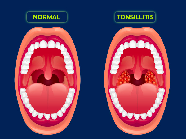
Medical Definition and Basic Anatomy of Tonsillitis
The tonsils are lymphoid tissues located in the oropharynx, serving as the first line of defense in the immune system. Positioned symmetrically on either side of the throat—palatine tonsils at the back of the oral cavity, lingual tonsils at the base of the tongue, and pharyngeal tonsils (adenoids) in the nasopharynx—they trap and neutralize pathogens entering the respiratory or digestive tracts. Tonsillitis refers to the inflammation of these tissues, primarily the palatine tonsils, due to infection or irritation, impairing their function and causing localized discomfort.
The tonsillar tissue comprises crypts—deep invaginations lined with epithelial cells—and follicles containing immune cells (lymphocytes, macrophages). During inflammation, these structures exhibit hallmark signs: erythema (redness) from increased blood flow, swelling due to edema and cellular infiltration, and exudate (purulent discharge) from bacterial infection or viral cytolysis. Severe cases may involve tonsillar hypertrophy (enlargement) or peritonsillar abscess (quinsy), where pus accumulates in surrounding tissues.
Anatomical Location and Immune Function
The palatine tonsils, the most clinically relevant in tonsillitis, are situated between the anterior and posterior tonsillar pillars, forming part of Waldeyer’s ring—a lymphatic barrier in the oropharynx. Their reticulum network of lymphocytes (B and T cells) detects antigens via M cells in the crypt epithelium, initiating an immune response. Key functions include:Disruption of these processes—whether by viral replication (e.g., adenovirus) or bacterial colonization (e.g., Streptococcus pyogenes)—leads to inflammation. Chronic irritation (e.g., allergens, smoking) may cause lymphoid hyperplasia, where tonsillar tissue proliferates to compensate for dysfunction.
Pathophysiological Changes in Inflamed Tonsils
Tonsillitis manifests through morphological and histological alterations in the tonsillar tissue, detectable via physical examination or biopsy. Below is a text-based cross-sectional diagram description of inflamed tonsils (acute bacterial tonsillitis):```
| ORAL CAVITY |
|---|
| [Anterior Tonsillar Pillars] |
| [Erythematous Mucosa] |
| [Enlarged Crypts with Purulent Exudate] |
| [Hyperemic Follicles (White-Yellow Nodules)] |
| [Edematous Tonsillar Tissue] |
| [Fibrinous Pseudomembrane (if diphtheria)] |
| [Posterior Tonsillar Pillars] |
Key Features:
Comparative Analysis: Acute vs. Chronic Tonsillitis
The clinical presentation and progression of tonsillitis vary significantly between acute and chronic forms. Below is a comparative table summarizing distinguishing features:| Feature | Acute Tonsillitis | Chronic Tonsillitis |
|---|---|---|
| Primary Cause |
|
|
| Symptoms |
|
|
| Duration | 3–7 days (self-limiting; bacterial cases may require antibiotics). | Weeks to months; recurrent episodes (≥5/year in children). |
| Typical Age Groups |
|
|
| Complications |
|
|
| Diagnostic Markers | Rapid antigen detection test (RADT) for GAS; throat culture for confirmation. |
Clinical history of recurrent episodes; tonsillar biopsy if malignancy suspected. |
Key Differentiating Factors in Clinical Practice
The distinction between acute and chronic tonsillitis influences treatment strategies. Acute cases often resolve with supportive care or antibiotics (e.g., penicillin for GAS), while chronic cases may require tonsillectomy if complications arise. Red flags for chronic progression include:Note: Chronic tonsillitis in adults with HPV-16 positivity warrants oncological evaluation due to increased squamous cell carcinoma risk.
Primary Causes and Pathogens in Tonsillitis
Tonsillitis arises predominantly from infectious agents, with bacterial and viral pathogens accounting for the majority of cases. While bacterial infections often necessitate targeted antibiotic therapy, viral triggers typically resolve spontaneously but may contribute to recurrent or chronic tonsillitis. Environmental factors further modulate susceptibility by compromising mucosal defenses, thereby amplifying infection risk. This section categorizes the primary pathogens, outlines their transmission dynamics, and examines how seasonal and environmental variables influence disease presentation.Bacterial Pathogens and Transmission Routes
Bacterial tonsillitis is most commonly associated with Streptococcus pyogenes (Group A Streptococcus, GAS), which is responsible for 30–40% of bacterial cases in children and adolescents. Other notable pathogens include Streptococcus pneumoniae, Haemophilus influenzae, Moraxella catarrhalis, and, less frequently, Streptococcus agalactiae (Group B Streptococcus) in immunocompromised individuals.Transmission mechanisms for bacterial tonsillitis primarily involve:
Streptococcus pyogenes produces pyogenic exotoxins (SpeA, SpeB, SpeC), which trigger systemic symptoms such as scarlet fever rash, fever, and pharyngotonsillar exudates, distinguishing it from viral tonsillitis.Risk multipliers for bacterial spread include:
Viral Triggers and Recurrent Tonsillitis Patterns
Viral pathogens account for 50–70% of tonsillitis cases, often presenting with milder symptoms but higher recurrence rates. Common viral agents include:Seasonal and epidemiological patterns of viral tonsillitis:
Recurrent viral tonsillitis (defined as ≥3 episodes/year) is frequently associated with EBV or adenovirus persistence, particularly in individuals with genetic predispositions (e.g., HLA-DQB1*0301 alleles) or tonsillar lymphoid hyperplasia.Atypical presentations requiring differential diagnosis:
Environmental Factors Exacerbating Tonsil Vulnerability
Environmental exposures weaken mucosal barriers, increasing susceptibility to both bacterial and viral tonsillitis. Key risk multipliers include:Airborne irritants and pollutants:
Crowded or poorly ventilated spaces:
Climatic and occupational hazards:
Flowchart: Progression from Infection to Systemic Symptoms
Bacterial Tonsillitis (e.g., Streptococcus pyogenes)```
[Initial Exposure] → Respiratory droplets/fomites →
[Colonization] → Adherence to tonsillar crypts via M protein →
[Invasion] → Epithelial breach → Pyogenic exotoxins (SpeA/SpeB) trigger inflammation →
[Local Symptoms] → Erythematous tonsils, exudates, cervical lymphadenopathy →
[Systemic Response] → Fever (>38.3°C), headache, ASO titer elevation →
[Complications] → Peritonsillar abscess, rheumatic fever (if untreated).
```
Viral Tonsillitis (e.g., Adenovirus/EBV)
```
[Initial Exposure] → Aerosolized droplets →
[Colonization] → Viral replication in tonsillar epithelium →
[Immune Response] → Cytokine storm (IFN-γ, IL-6) →
[Local Symptoms] → Punctate exudates, petechiae, pharyngeal edema →
[Systemic Response] → Low-grade fever, malaise, lymphadenopathy (posterior cervical) →
[Recurrence Risk] → Persistent viral reservoirs in tonsillar tissue → Chronic fatigue (EBV) or reactivation (adenovirus).
```
Key Differentiators in Symptom Progression
| Feature | Bacterial (S. pyogenes) | Viral (Adenovirus/EBV) |
|---|---|---|
| Onset | Abrupt (1–3 days) | Gradual (3–7 days) |
| Exudate | Thick, purulent | Scant, punctate or none |
| Fever Pattern | High (>38.5°C), spikes | Low-grade or intermittent |
| Lymphadenopathy | Anterior cervical, tender | Posterior cervical, non-tender |
| Associated Symptoms | Headache, abdominal pain | Fatigue, splenomegaly (EBV) |
| Complications | Scarlet fever, glomerulonephritis | Hemorrhagic cystitis (adenovirus) |

Risk Factors and Predisposing Conditions in Tonsillitis
Tonsillitis, an inflammatory condition primarily affecting the palatine tonsils, exhibits significant variability in severity and recurrence among individuals. While pathogen exposure and immune response play critical roles, certain risk factors and predisposing conditions—ranging from immune system deficiencies to chronic anatomical abnormalities—substantially influence susceptibility. These factors often exacerbate the disease by impairing immune clearance, prolonging inflammation, or facilitating recurrent infections. Understanding these elements is essential for targeted prevention, early intervention, and personalized management strategies in clinical practice.The interplay between genetic predispositions and acquired conditions further complicates the etiology of tonsillitis. While some individuals inherit a heightened susceptibility due to familial patterns, others develop vulnerabilities through environmental exposures or systemic diseases. Below, the key risk factors are categorized and analyzed to elucidate their mechanisms and clinical implications.
Immune System-Related Risk Factors and Their Impact on Disease Severity
Impaired immune function significantly alters the course of tonsillitis, often leading to prolonged infections, treatment resistance, and increased complications. The following conditions disrupt the body’s ability to mount an effective response against pathogens, thereby heightening susceptibility to recurrent or severe tonsillitis:- Primary Immunodeficiencies (PIDs):
Conditions such as common variable immunodeficiency (CVID), selective IgA deficiency, or DiGeorge syndrome impair humoral or cellular immunity, reducing the tonsils’ ability to trap and eliminate pathogens. Patients with CVID, for instance, experience recurrent bacterial infections (e.g., Streptococcus pyogenes) due to defective antibody production, while those with DiGeorge syndrome lack T-cell-mediated immunity, increasing viral susceptibility.
- Secondary Immunodeficiencies:
Acquired immunosuppression from HIV/AIDS (CD4+ T-cell depletion), chemotherapy/radiotherapy (lymphocyte depletion), or chronic steroid use (immunosuppression) creates an environment where opportunistic pathogens thrive. In HIV/AIDS, oral candidiasis and EBV-driven lymphoproliferative disorders often coexist with tonsillitis, complicating diagnosis and management.
- Chronic Granulomatous Disease (CGD):
A rare inherited disorder characterized by defective phagocyte function leads to recurrent abscess formation in the tonsils due to impaired oxidative killing of bacteria (e.g., Staphylococcus aureus).
- Complement Deficiencies:
Defects in the alternative or classical complement pathways (e.g., C3 or C5-C9 deficiencies) impair opsonization and membrane attack complex formation, increasing susceptibility to neisserial infections and post-streptococcal sequelae like glomerulonephritis.
Clinical Note: Patients with HIV/AIDS and CD4 counts <200 cells/µL exhibit a 10-fold higher risk of recurrent Candida albicans tonsillitis compared to immunocompetent individuals (CDC, 2019).
Comparison of Genetic and Acquired Predisposing Conditions
The following table contrasts inherited genetic factors with acquired conditions that predispose individuals to recurrent tonsillitis, highlighting their mechanisms and clinical relevance.| Genetic Predispositions | Acquired Conditions |
|---|---|
|
|
Occupational and Lifestyle Habits Increasing Tonsillitis Risk
Certain occupational exposures and lifestyle behaviors elevate the risk of tonsillitis by directly damaging mucosal barriers or facilitating pathogen transmission. The following habits create environments conducive to recurrent infections:Key Mechanism: Chronic irritation or mechanical stress disrupts tonsillar crypt integrity, allowing pathogens to colonize deeper tissues and evade immune clearance.
- Tobacco and Vaping:
Smoking (cigarette, cigar) and vaping induce oxidative stress and mucociliary dysfunction, impairing tonsillar drainage. Smokers have a 2.5-fold higher risk of S. aureus tonsillitis due to altered microbiome composition (e.g., Prevotella dominance) (Am J Respir Crit Care Med, 2019). E-cigarettes, while less carcinogenic, still reduce tonsillar IgA levels by 30% (Nicotine Tob Res, 2021).
- Alcohol Consumption:
Chronic alcohol abuse suppresses immune surveillance (e.g., neutrophil dysfunction) and irritates the oropharynx, increasing susceptibility to mixed bacterial-viral infections. Heavy drinkers exhibit delayed tonsillar healing post-infection (Alcohol Clin Exp Res, 2018).
- Poor Oral Hygiene:
Accumulation of dental plaque and periodontal pathogens (e.g., Fusobacterium nucleatum) creates a reservoir for tonsillar colonization. Patients with untreated caries have a 60% higher risk of recurrent Streptococcus tonsillitis (J Clin Periodontol, 2020).
- Environmental Pollutants:
Exposure to airborne particulates (PM2.5) or chemical irritants (e.g., ammonia in cleaning products) compromises tonsillar epithelial barriers, as demonstrated in industrial workers with chronic tonsillar inflammation (Occup Environ Med, 2019).
Mechanisms Linking Chronic Sinusitis and Adenoid Hypertrophy to Recurrent Tonsillitis
Chronic sinusitis and adenoid hypertrophy indirectly contribute to recurrent tonsillitis through anatomical, immunological, and microbiological pathways. The following step-by-step breakdown outlines these connections:1. Anatomical Continuity and Pathogen Drainage:
The nasopharynx serves as a shared conduit for the sinuses, adenoids, and tonsils. In chronic sinusitis, mucosal edema and polyp formation obstruct normal drainage, causing postnasal drip that
Diagnostic Methods and Clinical Workflow in Tonsillitis
The accurate diagnosis of tonsillitis requires a structured clinical approach that integrates patient history, physical examination, and targeted diagnostic tests. Early and precise identification distinguishes tonsillitis from other pharyngotonsillar infections, guiding appropriate antimicrobial therapy, surgical interventions, or supportive care. Misdiagnosis can lead to complications such as bacterial superinfections, abscess formation, or unnecessary antibiotic use. This section outlines the systematic diagnostic workflow, including history-taking, physical assessment techniques, and the role of laboratory and imaging modalities in complex presentations.
Patient History and Symptom Assessment
A detailed patient history serves as the foundation for differentiating tonsillitis from other pharyngotonsillar pathologies. Key historical features include:
Red flag symptoms prompting urgent evaluation include:
Physical Examination Techniques
The physical examination focuses on tonsillar grading, pharyngeal erythema, and associated signs of complications. Standardized grading scales, such as the Centor Criteria (modified for adults) or McIsaac Score, assist in assessing bacterial likelihood but require clinical correlation.Tonsillar grading scales evaluate erythema, exudate, and hypertrophy:
Key examination findings:
Instrumental aids:
Differential Diagnosis Table: Tonsillitis vs. Related Conditions
The following table compares key distinguishing features of tonsillitis with mononucleosis, diphtheria, and peritonsillar abscess to facilitate clinical differentiation.| Feature | Tonsillitis (Bacterial) | Mononucleosis (EBV) | Diphtheria | Peritonsillar Abscess |
|---|---|---|---|---|
| Onset | Acute (<7 days) | Gradual (1–2 weeks) | Indolent (1–6 weeks) | Acute, often follows tonsillitis |
| Fever | High (≥38.3°C), sudden | Moderate (37.8–38.8°C), prolonged | Low-grade or absent | High, spiking |
| Pharyngeal Appearance | Erythematous, exudative tonsils | Patchy exudate, petechiae, palatal petechiae | Grayish membrane (pseudomembrane) | Asymmetric swelling, uvular deviation |
| Lymphadenopathy | Cervical, tender | Posterior cervical, nontender | Minimal or absent | Unilateral, severe |
| Systemic Symptoms | Malaise, headache | Fatigue, pharyngitis, splenomegaly | Toxicity, bull neck (cervical lymphadenopathy) | Odynophagia, trismus, muffled "hot potato" voice |
| Diagnostic Tests | Rapid antigen test (GAS), throat culture | Monospot (heterophile antibodies), EBV serology | Throat culture (Corynebacterium diphtheriae), PCR | CT/ultrasound (fluid collection), needle aspiration |
| Complications | Peritonsillar abscess, rheumatic fever | Splenic rupture, airway obstruction | Myocarditis, neuropathy | Airway compromise, sepsis |
Rapid Antigen Detection Tests for Group A Streptococcus
Rapid antigen detection tests (RADTs) for GAS provide point-of-care results within 5–15 minutes, facilitating timely antibiotic initiation. These tests detect group A carbohydrate antigens on the bacterial cell wall using lateral flow immunoassays or enzyme-linked immunosorbent assays (ELISA).Mechanism:
Limitations and Performance Characteristics:
Clinical guidelines (e.g., Infectious Diseases Society of America) recommend:
Role of Imaging in Complex Tonsillitis Cases
Imaging is reserved for complicated tonsillitis, where physical examination findings suggest abscess formation, deep-space infections, or non-resolving symptoms. Modalities include ultrasound (US), computed tomography (CT), and magnetic resonance imaging (MRI), each with distinct advantages.Ultrasound (First-Line for Abscess Suspicion):

Complications and Systemic Impacts of Tonsillitis
Tonsillitis, when left untreated or recurrent, can progress beyond localized inflammation to cause severe local and systemic complications. Untreated acute tonsillitis or chronic infections may lead to abscess formation, autoimmune sequelae, or obstructive respiratory disorders, particularly in high-risk populations such as children. The anatomical proximity of the tonsils to critical structures—including the pharynx, retropharyngeal space, and vascular supply—facilitates the spread of infection, while systemic immune responses may trigger autoimmune conditions through molecular mimicry. This section examines the progression pathways of local complications, systemic sequelae, and their long-term impacts, including obstructive sleep apnea in recurrent cases.Local Complications and Anatomical Spread Pathways
Local complications arise from the extension of infection beyond the tonsillar tissue, often due to delayed or inadequate treatment. The tonsils lack a fibrous capsule, allowing pathogens to disseminate into adjacent spaces via lymphatic or direct tissue invasion. Key complications include peritonsillar abscess (quinsy), retropharyngeal abscess, and deep neck space infections, each with distinct anatomical spread mechanisms.Peritonsillar Abscess (Quinsy)
A peritonsillar abscess develops when an acute tonsillitis infection extends into the peritonsillar space, typically between the tonsillar capsule and the pharyngeal constrictor muscles. The infection spreads through:
Clinical Progression and Risk Factors
Retropharyngeal and Deep Neck Space Infections
Retropharyngeal abscesses occur when infection spreads posteriorly into the retropharyngeal space, a potential space between the buccopharyngeal fascia and prevertebral fascia. This pathway is more common in children due to:
Deep neck space infections (e.g., parapharyngeal or lateral pharyngeal abscesses) may result from:
Diagnostic Considerations
Systemic Complications and Autoimmune Sequelae
Systemic complications of tonsillitis primarily stem from streptococcal infections, particularly Streptococcus pyogenes (Group A Streptococcus, GAS). These complications arise from:High-Risk Populations for Systemic Complications
The following groups exhibit increased susceptibility to autoimmune sequelae, particularly if untreated or recurrent infections occur:
- Children aged 5–15 years:
- Adults with recurrent tonsillitis:
- Immunocompromised individuals:
Pathophysiology of Autoimmune Triggers
Molecular mimicry plays a central role in GAS-induced autoimmune diseases. Key mechanisms include:
Long-Term Sequelae
Prevention Strategies
Recurrent Tonsillitis and Obstructive Sleep Apnea
Chronic or recurrent tonsillitis leads to tonsillar hypertrophy, a compensatory enlargement of lymphoid tissue in response to persistent antigenic stimulation. This hypertrophy can obstruct the upper airway, contributing to obstructive sleep apnea (OSA)—a condition characterized by repeated airway collapse during sleep, leading to hypoxia, fragmented sleep, and systemic complications.Pathophysiology of Tonsillar Hypertrophy and OSA
Clinical Manifestations and Sleep Study Findings
The following table summarizes key features of tonsillar hypertrophy-related OSA:
| Feature | Tonsillar Hypertrophy | Obstructive Sleep Apnea (OSA) Findings |
|---|---|---|
| Anatomical Changes | Enlarged tonsils (>3+ on Friedman scale), narrowed oropharyngeal airway. | Reduced minimal cross-sectional area (MCA) of the airway (<100 mm²). |
| Sleep Study Metrics (Polysomnography) | Chronic inflammation may contribute to sleep fragmentation. |
|
Leave a Comment
Comments are moderated before appearing. The data you submit is processed according to the Privacy Policy of Voltefac.