What Is Plaque On Teeth And Its Critical Impact On Oral Health
Table of Contents
- Definition and Basic Composition of Dental Plaque
- Primary Components of Dental Plaque
- Bacterial Taxonomy and Functional Roles in Plaque
- Mechanism of Plaque Formation: From Initial Attachment to Mature Biofilm
- Chemical Composition of Plaque at Different Stages
- Plaque’s Role in Oral Health and Disease Progression
- Mechanisms of Plaque-Induced Dental Caries and Enamel Demineralization
- Plaque’s Contribution to Gingivitis and Periodontitis
- Surface Topography and Plaque Retention
- Flowchart: Progression from Plaque Accumulation to Advanced Periodontal Disease
- Visual and Physical Characteristics of Dental Plaque
- Color Variations and Texture of Plaque
- Differentiating Soft Plaque from Calcified Tartar
- Anatomical Accumulation Patterns of Plaque
- Tactile Sensation During Plaque Removal
- Prevention and Removal Techniques for Dental Plaque
- Manual Plaque Removal Techniques
- Optimal Brushing Techniques
- Interdental Cleaning Tools
- Electric Toothbrush Usage for Maximum Plaque Disruption
- Professional-Grade Plaque Removal Methods
- Scaling and Root Planing
- Plaque and Systemic Health: Biological Pathways and Clinical Implications
- Biological Pathways Linking Oral Plaque to Systemic Diseases
- Key Bacterial Strains and Mechanisms in Systemic Health Risks
- Plaque Control and Chronic Inflammatory Conditions: The Gut-oral Axis
- Innovations in Plaque Detection and Monitoring
- Emerging Technologies for Plaque Detection
- Real-Time Plaque Monitoring Devices
- Experimental Approaches to Inhibit Plaque Formation
- Future Trends in Plaque Management
- FAQ
- What is plaque on teeth made of?
- What causes plaque to form on teeth?
- What is plaque on teeth called in dental terms?
- How can you get rid of plaque on teeth?
- What is plaque on teeth for kids?
- What is tartar on teeth?
Dental plaque, a ubiquitous yet often overlooked biological film, plays a pivotal role in oral health by harboring harmful bacteria that erode tooth structure and trigger inflammatory diseases. Comprising a complex matrix of microbial colonies, salivary proteins, and food remnants, plaque evolves from a soft, adhesive biofilm into a hardened calculus that exacerbates dental decay and periodontal destruction. Understanding its composition—ranging from acid-producing streptococci to proteolytic Porphyromonas—reveals how even microscopic accumulations can precipitate systemic complications, from cardiovascular risks to metabolic disorders. This exploration dissects plaque’s formation, pathogenic mechanisms, and preventive strategies, bridging scientific rigor with actionable insights for maintaining optimal oral and systemic well-being.
The progression from initial bacterial adhesion to mature biofilm formation underscores plaque’s dual nature: a transient yet relentless threat when unchecked. While early-stage plaque remains reversible through meticulous hygiene, its unchecked maturation leads to irreversible damage, including enamel demineralization and gingival tissue degradation. Comparative analyses of plaque’s chemical evolution—from high-protein, low-mineral early stages to mineralized calculus—highlight critical windows for intervention. Equally critical is recognizing how plaque’s interaction with tooth morphology, particularly rough surfaces and interdental spaces, accelerates disease progression, demanding tailored removal techniques. Beyond its local effects, emerging evidence links oral biofilms to broader systemic inflammation, positioning plaque as a sentinel marker for chronic diseases.
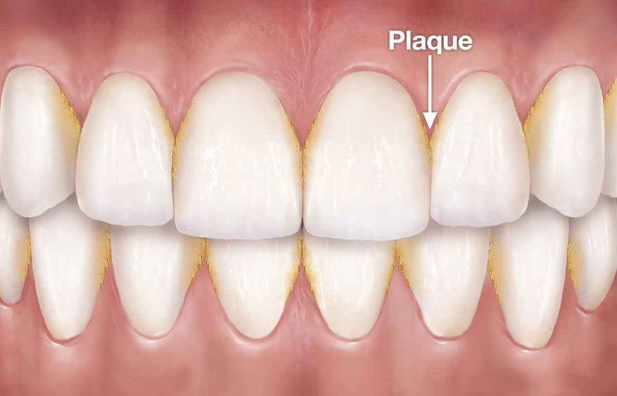
Definition and Basic Composition of Dental Plaque
Dental plaque represents a complex, organized microbial community adhering to tooth surfaces, constituting the primary etiological agent in caries, gingivitis, and periodontal diseases. Its formation initiates within minutes of tooth brushing and progresses through structured stages, transitioning from a loose accumulation of bacteria to a dense, structured biofilm. The composition of plaque evolves dynamically, incorporating salivary proteins, bacterial metabolites, and extracellular polymeric substances (EPS) that stabilize its structure. Understanding its biological nature—including bacterial taxonomy, metabolic functions, and physicochemical interactions—is essential for developing targeted preventive and therapeutic strategies.
The biological foundation of dental plaque lies in its heterogeneous composition, where microorganisms coexist in a symbiotic matrix of organic and inorganic components. Salivary glycoproteins, such as mucins and proline-rich proteins, serve as initial adhesion mediators, while food debris and bacterial byproducts contribute to the nutrient-rich microenvironment. The extracellular matrix, primarily composed of polysaccharides (e.g., glucans and fructans) produced by bacterial enzymes, provides structural integrity and protection against host defenses and antimicrobial agents.
Primary Components of Dental Plaque
Dental plaque comprises four fundamental elements: bacteria, salivary proteins, food debris, and the extracellular matrix. Bacteria constitute the core of plaque, accounting for 70–80% of its dry mass, while salivary proteins and food remnants provide substrates for microbial metabolism. The extracellular matrix, derived from bacterial secretions and host-derived components, binds the community together and facilitates nutrient exchange.Bacterial Taxonomy and Functional Roles in Plaque
The bacterial population in dental plaque is diverse, with over 700 species identified, though fewer than 50 dominate in healthy individuals. These bacteria are categorized based on their metabolic and structural contributions to biofilm formation and pathogenicity. Key functional groups include:- Acidogenic bacteria: Primarily Streptococcus mutans and Streptococcus sobrinus, these organisms ferment carbohydrates into organic acids (e.g., lactic, acetic, formic acids), lowering pH and demineralizing tooth enamel.
Mechanism of Plaque Formation: From Initial Attachment to Mature Biofilm
Plaque development follows a sequential process governed by physicochemical and microbial interactions. The stages are as follows:1. Acquired Pellicle Formation (0–2 hours post-cleaning)
Salivary proteins adsorb onto hydroxyapatite crystals of tooth enamel, forming a thin (0.1–0.5 µm) conditioning film. This layer, rich in proline-rich proteins and statherin, mediates bacterial adhesion through electrostatic and hydrophobic interactions.
2. Initial Bacterial Attachment (2–4 hours)
Early colonizers, primarily Streptococcus spp. and Actinomyces spp., bind to pellicle components via specific adhesins. These bacteria exhibit reversible attachment, allowing detachment if mechanical forces (e.g., saliva flow) exceed adhesive strength.
3. Irreversible Attachment and Microcolony Formation (4–12 hours)
Bacteria produce extracellular polysaccharides (e.g., dextrans from sucrose metabolism) and synthesize adhesins that anchor them permanently to the tooth surface. S. mutans dominates in sucrose-rich environments due to its high glucosyltransferase activity.
4. Coaggregation and Biofilm Maturation (12–24 hours to days)
Secondary colonizers bind to early colonizers via coaggregation, a species-specific interaction mediated by lectin-like proteins. The biofilm expands vertically, with anaerobic species occupying deeper layers. The extracellular matrix, composed of EPS and host-derived components (e.g., fibrinogen), encases the community, enhancing resistance to antimicrobials and host immune responses.
5. Mature Biofilm (Days to weeks)
The biofilm reaches a steady state, with a stratified structure: aerobic/ facultative bacteria near the tooth surface and anaerobes in deeper layers. Metabolic byproducts (e.g., short-chain fatty acids, ammonia) create a microenvironment conducive to pathogen survival. Quorum sensing mechanisms regulate bacterial gene expression, coordinating functions such as virulence factor production and biofilm dispersal.
Chemical Composition of Plaque at Different Stages
The physicochemical properties of plaque evolve as it matures, influencing its pathogenicity and susceptibility to disruption. The following table compares key compositional parameters between early and mature plaque:| Stage | Protein Content (%) | Mineral Content (%) | Bacteria Density (CFU/mm²) | Extracellular Polysaccharides (%) | pH Range |
|---|---|---|---|---|---|
| Early Plaque (0–24 hours) | 30–40 | 5–10 (primarily salivary minerals) | 10³–10⁴ | 5–10 (glucans from sucrose metabolism) | 6.5–7.5 (neutral) |
| Mature Plaque (48+ hours) | 20–30 (decreased due to bacterial metabolism) | 15–25 (calcium phosphate deposition) | 10⁶–10⁸ | 20–30 (diverse EPS, including fructans) | 5.5–6.5 (acidic, especially post-sugar exposure) |
The transition from early to mature plaque reflects a shift from a reversible microbial community to a structured, highly organized biofilm. This progression underscores the importance of mechanical disruption (e.g., brushing) and chemical inhibition (e.g., fluoride) in preventing pathological outcomes.
Plaque’s Role in Oral Health and Disease Progression
Dental plaque is a primary etiological factor in oral diseases, acting as a biofilm that facilitates microbial colonization and metabolic activity. Its role extends beyond mere accumulation; it actively contributes to the biochemical and inflammatory processes underlying dental caries, gingivitis, and periodontitis. Understanding these mechanisms clarifies why plaque control is essential for maintaining oral health and preventing progressive tissue destruction.The progression of plaque-related diseases involves complex interactions between microbial communities, host immune responses, and structural changes in oral tissues. While dental caries primarily results from acid-mediated demineralization, periodontal diseases stem from chronic inflammation and collagen degradation. Surface topography further influences plaque retention, with rough surfaces exacerbating microbial accumulation and disease risk.
Mechanisms of Plaque-Induced Dental Caries and Enamel Demineralization
Dental caries develops when plaque bacteria metabolize fermentable carbohydrates, producing organic acids (primarily lactic, acetic, and formic acids) that lower the pH at the tooth surface. This acidogenic process disrupts the equilibrium between demineralization and remineralization, leading to enamel dissolution. The critical pH threshold for enamel demineralization is pH 5.5, below which hydroxyapatite crystals begin to dissolve, initiating cavity formation.Key stages in caries progression:
Critical pH for enamel demineralization: <5.5 (varies with fluoride exposure and salivary flow).
Plaque’s Contribution to Gingivitis and Periodontitis
Plaque-associated gingivitis arises from the host’s inflammatory response to bacterial toxins, particularly lipopolysaccharides (LPS) and proteases from Gram-negative anaerobes like Porphyromonas gingivalis. This response triggers a cascade of immune-mediated tissue damage, characterized by:If untreated, gingivitis progresses to periodontitis, where chronic inflammation destroys periodontal ligaments and alveolar bone. Key pathogenic mechanisms include:
Periodontitis progression stages:
1. Initial lesion: Reversible inflammation (2–4 days).
2. Early lesion: Plasma cell infiltration (4–7 days).
3. Established lesion: Collagen destruction begins (2–3 weeks).
4. Advanced lesion: Bone loss and pocket formation (>3 weeks).
Surface Topography and Plaque Retention
Tooth surface characteristics significantly influence plaque accumulation and disease risk. Smooth surfaces (e.g., facial/lingual enamel) allow for easier mechanical removal during mastication and oral hygiene, whereas rough surfaces (e.g., proximal contacts, restored teeth, or exposed root surfaces) enhance biofilm adherence. Studies show:Comparison of plaque impact by surface type:
| Surface Type | Plaque Retention Risk | Disease Association | Removal Difficulty |
|---|---|---|---|
| Smooth enamel | Low (self-cleansing) | Minimal caries/gingivitis | Easy (brushing) |
| Rough enamel (e.g., proximal) | Moderate (food impaction) | Interproximal caries | Moderate (flossing required) |
| Restored surfaces (composite/amalgam) | High (microgaps, roughness) | Recurrent caries, periodontitis | Difficult (specialized tools) |
| Root surfaces (cementum) | Very high (porous, exposed) | Root caries, attachment loss | Challenging (scaling) |
Flowchart: Progression from Plaque Accumulation to Advanced Periodontal Disease
The following structured pathway illustrates the sequential and interdependent stages of plaque-induced periodontal breakdown, emphasizing critical decision points where intervention can alter disease trajectories.-
Plaque Formation
- Initial colonization (0–24 hours): Streptococcus sanguinis, Actinomyces.
- Maturation (2–7 days): Gram-negative anaerobes emerge (Fusobacterium, Prevotella).
-
Gingival Inflammation (Gingivitis)
- Host response: Cytokine release (IL-1, TNF-α), neutrophil recruitment.
- Clinical signs: Redness, swelling, bleeding on probing (BOP).
- Reversibility: Removable with professional cleaning and improved oral hygiene.
-
Transition to Periodontitis
- Trigger: Persistent plaque, systemic factors (diabetes, smoking).
- Pathogenic shift: P. gingivalis, T. denticola dominate.
- Tissue damage: Collagenase/MMP activity increases.
-
Established Periodontitis
- Bone loss: Radiographic evidence of alveolar ridge resorption.
- Pocket formation: >4 mm depth, anaerobic niches.
- Tooth mobility: Ligament fiber destruction.
-
Advanced Periodontitis
- Severe attachment loss: >5 mm pocketing.
- Systemic risks: Linked to cardiovascular disease, preterm birth.
- Therapeutic challenge: Requires surgical intervention (flap debridement, bone grafts).
Critical intervention points:
Gingivitis stage: 100% reversible with plaque control. Early periodontitis: Reversible with scaling/root planing (SRP) and maintenance. Advanced stages: Irreversible; focuses on halting progression and restoring function.
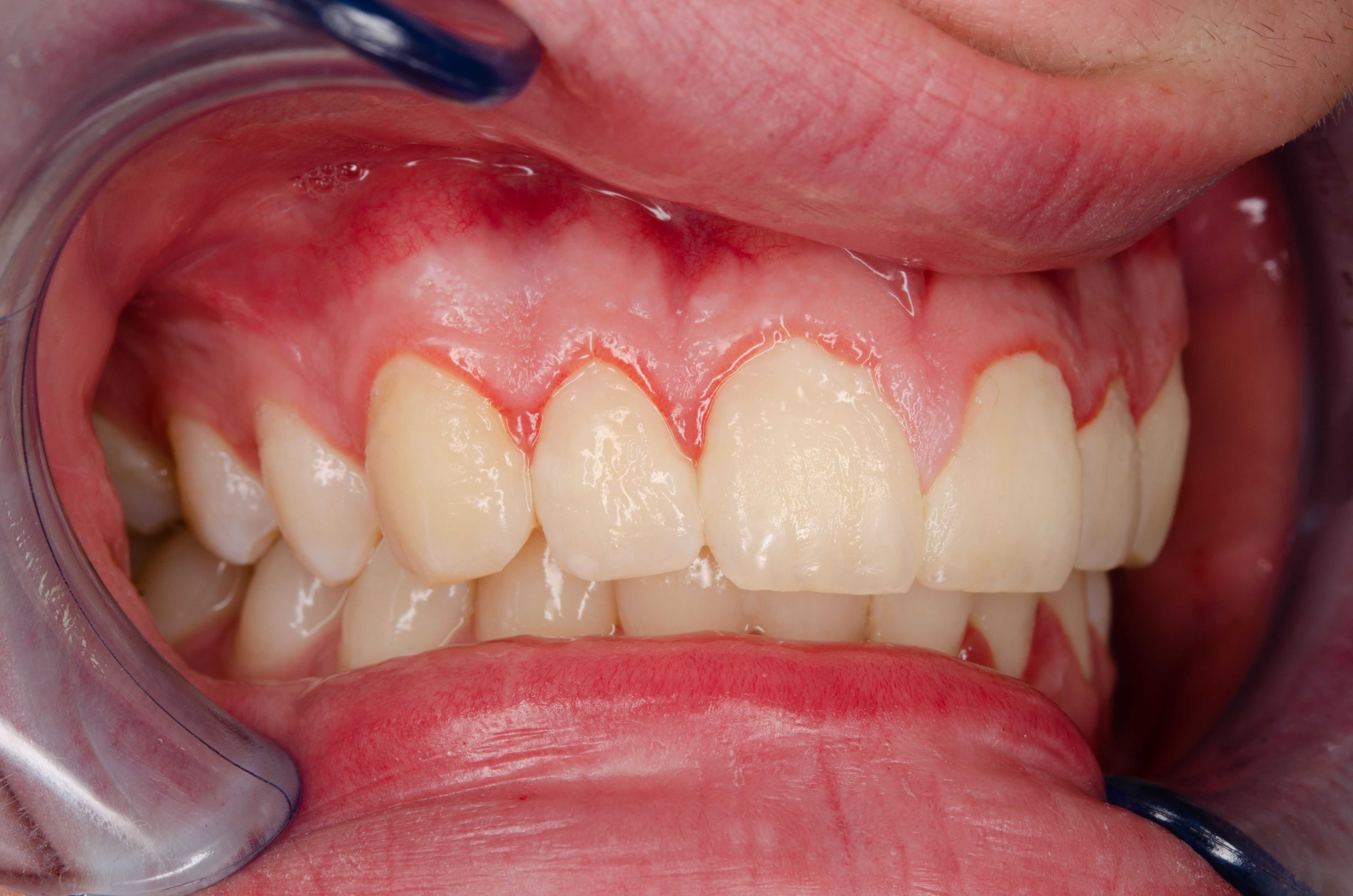
Visual and Physical Characteristics of Dental Plaque
Dental plaque exhibits distinct visual and tactile properties that vary depending on its age, composition, and location within the oral cavity. These characteristics serve as critical indicators for oral health assessment, distinguishing between early-stage soft plaque and advanced calcified deposits. Understanding these features enables clinicians and patients to identify high-risk areas for caries, gingivitis, and periodontal disease progression.Color Variations and Texture of Plaque
Plaque appears in multiple colors due to dietary stains, bacterial metabolism, and mineralization processes. Fresh dental plaque typically presents as a translucent or whitish film when newly formed, often indistinguishable against tooth enamel under standard lighting. As it matures, it may develop a yellowish or brownish hue due to:The texture of plaque transitions from slimy and adhesive in its soft form to rough and granular upon calcification. Early plaque adheres loosely to tooth surfaces, while older deposits may exhibit a chalky or crystalline consistency when scraped.
Differentiating Soft Plaque from Calcified Tartar
Visual and tactile assessments are essential for distinguishing between soft plaque and hardened calculus, as treatment approaches differ significantly.Visual Cues:
Tactile Cues:
A dental explorer can further differentiate the two:
Anatomical Accumulation Patterns of Plaque
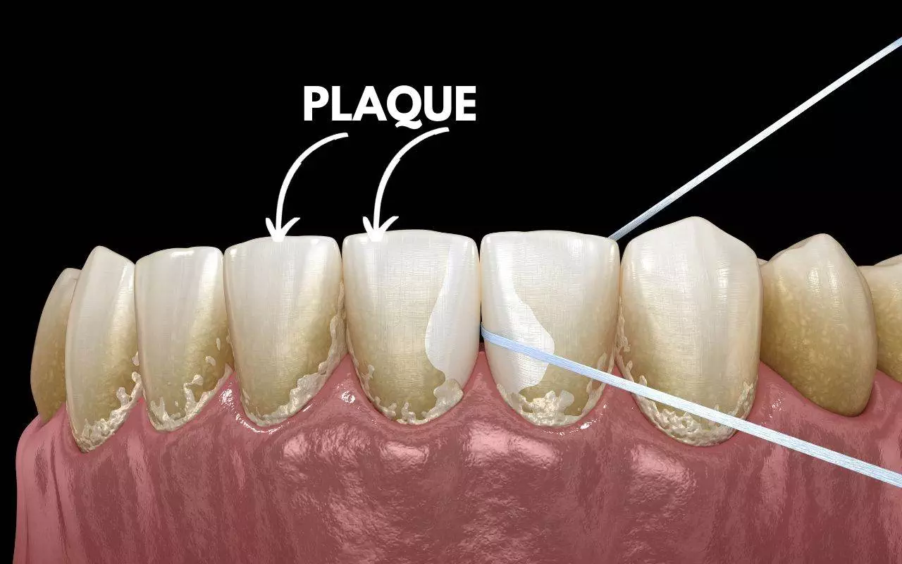
Plaque preferentially accumulates in high-risk zones where mechanical cleaning is challenging, creating ideal conditions for microbial colonization. Key anatomical landmarks include:Interdental Spaces (Embrasures):
Plaque thrives in the gingival embrasures (spaces between teeth) due to:
Gingival Margins and Sulci:
The gingival crevice (sulcus) is a primary site for plaque accumulation, especially:
Occlusal and Lingual Surfaces:
Orthodontic and Prosthetic Appliances:
Tactile Sensation During Plaque Removal
The resistance encountered during plaque removal correlates with its maturity and mineralization state. A dental probe or explorer provides immediate feedback on plaque consistency:When manually removed, soft plaque feels yielding and stringy, similar to wet cotton candy or thin mucus. It adheres lightly to the probe and can be wiped away with minimal pressure, often leaving a slightly sticky residue on the instrument. In contrast, calcified tartar offers firm resistance, requiring sustained scraping motion to dislodge. The tactile sensation resembles grating against a rough surface, with particles detaching in chunks rather than dissolving. Subgingival calculus may feel denser and more tenacious, sometimes lodging in the probe’s tip due to its adherence to root surfaces.Stages of Resistance:
| Plaque Stage | Removal Resistance | Instrument Feedback | Outcome |
|---|---|---|---|
| Newly formed plaque | Minimal resistance | Probe glides smoothly; plaque lifts easily | Tooth surface appears clean |
| Mature soft plaque | Moderate adherence | Probe catches briefly; plaque stretches | Partial removal; residual biofilm |
| Early calculus | Increased grit | Probe scrapes with audible scratching | Chalky deposits remain |
| Mature tartar | High resistance | Probe binds briefly; chunks detach | Rough surface; scaling required |
Prevention and Removal Techniques for Dental Plaque
Effective plaque removal is fundamental to maintaining oral health, preventing caries, gingivitis, and periodontal disease. While plaque forms continuously on tooth surfaces, its accumulation can be minimized through systematic mechanical disruption and professional interventions. Manual techniques, such as brushing and interdental cleaning, form the first line of defense, while professional-grade methods—like scaling and polishing—address deeper deposits and subgingival plaque. The choice of method depends on individual oral anatomy, plaque retention risk, and adherence to a consistent regimen.Optimal plaque control requires a combination of daily self-care and periodic professional maintenance. Research indicates that improper brushing techniques or infrequent cleaning can leave up to 60% of plaque undisturbed, particularly in interproximal and subgingival areas (American Dental Association, 2020). Below, structured protocols for manual and professional plaque removal are outlined, along with comparative analysis to guide evidence-based oral hygiene practices.
Manual Plaque Removal Techniques
Manual plaque removal relies on mechanical disruption through brushing and interdental cleaning. The efficacy of these methods depends on technique precision, tool selection, and frequency. Studies demonstrate that proper brushing reduces plaque by 40–60%, while interdental cleaning can eliminate an additional 20–30% of plaque in hard-to-reach areas (Journal of Clinical Periodontology, 2018).Optimal Brushing Techniques
The Bass technique and Stillman technique are gold-standard methods for plaque removal, each targeting specific plaque retention sites.Bass Technique (Sulcular Brushing):
Purpose: Disrupts plaque at the gingival margin and within sulci. Steps: 1. Position the bristle tips at a 45-degree angle to the gumline.
2. Apply gentle pressure to seat the bristles into the sulcus.
3. Use vibrating or circular motions (20–30 strokes per tooth).
4. Move systematically around the arch, ensuring full coverage of all surfaces.
Effectiveness: Reduces gingival inflammation by 30–50% when performed correctly (American Academy of Periodontology, 2019).
Stillman Technique (Gentle Circular Motion):
Purpose: Ideal for recession-prone areas or exposed roots. Steps: 1. Angle the brush 45 degrees toward the gumline.
2. Apply light pressure and roll the brush away from the gumline in small circles.
3. Focus on buccal and lingual surfaces of anterior teeth.
Effectiveness: Particularly beneficial for root sensitivity reduction and plaque control in gingival pockets (≤3 mm).
Interdental Cleaning Tools
Interdental plaque accounts for 30–40% of total plaque accumulation (Journal of Periodontal Research, 2017). Tools such as dental floss, interdental brushes, and water flossers target these areas where toothbrush bristles cannot reach.-
Dental Floss:
- Types: Waxed (reduces fraying), unwaxed (thinner for tight contacts), or flavored varieties.
- Technique: 1. Wind 18–24 inches of floss around middle fingers, leaving 1–2 inches working length.
- Effectiveness: Removes 90% of interproximal plaque when used daily (NIH, 2021).
-
Interdental Brushes:
- Selection: Choose brush diameters matching the interdental space (e.g., #000 for tight contacts, #3 for wider gaps).
- Technique: 1. Insert brush gently into the space until resistance is felt.
- Effectiveness: Superior for orthodontic patients or implant sites, reducing plaque by 45% in hard-to-reach areas (Journal of Clinical Dentistry, 2020).
-
Water Flossers:
- Mechanism: Uses pressurized water streams to disrupt plaque and massage gums.
- Technique: 1. Fill the reservoir with warm water (optional: add antimicrobial mouthwash).
- Effectiveness: Reduces plaque by 20–30% and improves gingival bleeding scores by 50% in patients with periodontal disease (Cochrane Review, 2019).
2. Gently slide between teeth using a sawing motion (avoid snapping to prevent gum trauma).
3. Curve floss around the tooth in a C-shape and move up/down 3–4 times per contact.
2. Use light scrubbing motions (5–10 strokes per tooth).
3. Clean buccal and lingual surfaces of adjacent teeth.
2. Direct the tip 1–2 mm into the sulcus at a 90-degree angle.
3. Use low-to-medium pressure and sweep across teeth in short bursts.
4. Clean one quadrant at a time, focusing on gingival margins.
Electric Toothbrush Usage for Maximum Plaque Disruption
Electric toothbrushes enhance plaque removal through oscillating, rotating, or sonic vibrations, which increase brushing efficiency by 20–30% compared to manual brushes (American Dental Association, 2022). Proper use requires adherence to specific protocols to avoid abrasion or ineffective cleaning.Step-by-Step Guide for Electric Toothbrush Optimization:
1. Brush Head Selection:
Use small-headed brushes for posterior teeth and multi-tufted heads for orthodontic appliances. Replace heads every 3–4 months or when bristles fray. 2. Pressure Application:
Apply light pressure (electric toothbrushes often have pressure sensors to indicate excessive force). Incorrect pressure (>200 g) can lead to enamel abrasion and gingival recession. 3. Brushing Duration and Technique:
Duration: 2 minutes total, divided into 30-second quadrants. Motion: For sonic/oscillating brushes: Hold at a 45-degree angle and let the brush do the work. For rotating brushes: Use small circular motions (2–3 per tooth). Timing: Spend 10–15 seconds per tooth, ensuring gingival margins and occlusal surfaces are covered. 4. Frequency:
Brush twice daily, with a third session post-lunch if plaque accumulation is high. Timing: Brush before breakfast (to remove overnight plaque) and before bed (to prevent nocturnal plaque buildup).
Professional-Grade Plaque Removal Methods
While manual techniques address supragingival plaque, professional interventions are essential for subgingival plaque, calculus, and biofilm embedded in periodontal pockets. These methods are performed by dental hygienists or periodontists and are critical for preventing periodontitis progression and tooth loss.Scaling and Root Planing
Scaling:
Definition: Mechanical removal of supragingival and subgingival plaque, calculus, and stains using hand instruments (scalers/curetttes) or ultrasonic devices. Types: Hand Scaling: Precise for root surfaces and furcations (e.g., Gracey curettes). Ultrasonic Scaling: Uses vibrations (25–50 kHz) to disrupt calculus and irrigate pockets with antimicrobial solutions. Effectiveness: Reduces periodontal pocket depth by 1–3 mm in 50–70% of sites (Journal of Periodontology, 2021). Antimicrobial irrigation (e.g., chlorhexidine) during scaling enhances plaque reduction by 25–40%.
Root Planing:
Definition: Smoothing root surfaces to remove embedded plaque and calculus, eliminating bacterial niches. Purpose: Reduces toxin-induced inflammation by removing endotoxin-laden biofilm. Facilitates reattachment of gingival tissues to root surfaces. Technique: Performed under local anesthesia for deep pockets (>5 mm). Combines scaling + root debridement to achieve a glass-smooth surface. Outcome: 60–80% reduction in bleeding on probing post
Plaque and Systemic Health: Biological Pathways and Clinical Implications
Emerging evidence establishes a bidirectional relationship between oral plaque and systemic health, where dysbiotic microbial communities in dental biofilm contribute to chronic inflammatory diseases beyond the oral cavity. Research increasingly supports the concept of "oral systemic health"—a framework recognizing oral pathogens as modifiable risk factors for conditions such as cardiovascular disease, metabolic disorders, and respiratory infections. The biological mechanisms underlying these associations involve bacterial translocation, immune dysregulation, and systemic inflammation, with specific bacterial strains acting as key mediators.The gut-oral axis further complicates this interplay, as oral pathogens may alter gut microbiota composition, exacerbate metabolic dysfunction, and promote low-grade inflammation. Poor plaque control disrupts this balance, amplifying systemic risks through persistent activation of pro-inflammatory pathways. Below, the discussion explores the hypothesized pathways, bacterial contributors, and clinical implications of this emerging paradigm.
Biological Pathways Linking Oral Plaque to Systemic Diseases
The connection between dental plaque and systemic health operates through multiple interconnected mechanisms, primarily involving bacterial translocation, immune-mediated inflammation, and metabolic dysregulation. Key pathways include:- Bacterial Translocation and Endotoxemia
Oral pathogens, particularly gram-negative bacteria (e.g., Porphyromonas gingivalis, Aggregatibacter actinomycetemcomitans), release lipopolysaccharides (LPS)—endotoxins that trigger systemic immune responses. These endotoxins enter the bloodstream via ulcerated gingival tissues or through microaspirations, inducing endotoxemia, a state associated with elevated levels of pro-inflammatory cytokines (e.g., TNF-α, IL-6). Chronic endotoxemia is linked to endothelial dysfunction, atherosclerosis progression, and insulin resistance.- Immune System Activation and Chronic Inflammation
Oral pathogens stimulate innate and adaptive immune responses, including the activation of T-helper 17 (Th17) cells and neutrophil extracellular traps (NETs), which release reactive oxygen species (ROS). Persistent immune activation contributes to low-grade inflammation, a hallmark of systemic conditions such as type 2 diabetes and Alzheimer’s disease. The "inflammatory theory of atherosclerosis" posits that periodontal pathogens accelerate plaque formation in arteries by promoting monocyte adhesion and foam cell development.- Metabolic Dysregulation via Gut-oral Axis
Oral bacteria and their metabolites (e.g., trimethylamine N-oxide (TMAO) produced by Fusobacterium nucleatum) may influence gut microbiota composition, disrupting metabolic homeostasis. Dysbiosis in the gut-oral axis is associated with increased intestinal permeability ("leaky gut"), allowing bacterial products to enter circulation and exacerbate metabolic syndrome. Studies suggest that oral P. gingivalis infection in mice induces gut dysbiosis, reducing beneficial Lactobacillus species and increasing pro-inflammatory Desulfovibrio.- Direct Invasion and Tissue Damage
Certain oral pathogens (e.g., Streptococcus sanguinis, Staphylococcus aureus) possess adhesins and invasins that facilitate colonization of distant tissues, including the heart valves (infective endocarditis) and lungs (pneumonia). Microaspiration of oral bacteria during sleep or poor oral hygiene is a recognized risk factor for community-acquired pneumonia, particularly in elderly or immunocompromised individuals.
Key Bacterial Strains and Mechanisms in Systemic Health Risks
Specific bacterial species within plaque exhibit unique virulence factors that directly contribute to systemic disease progression. Below are the most studied pathogens, their mechanisms, and associated health risks:
Critical Note: The presence of these bacteria in plaque does not guarantee systemic disease, but their abundance and virulence factor expression correlate with increased risk in susceptible individuals.Porphyromonas gingivalis Mechanisms: Produces gingipains (cysteine proteases) that degrade host proteins (e.g., IgG, fibrinogen) and LPS that suppresses adaptive immunity via T-cell anergy. Its fimbriae promote biofilm formation and tissue invasion. Associated Diseases: Cardiovascular Disease (CVD): Linked to atherosclerosis via induction of matrix metalloproteinases (MMPs), which destabilize arterial plaques. Alzheimer’s Disease (AD): Detected in amyloid plaques of AD patients; hypothesized to contribute to neuroinflammation via TLR4-mediated pathways. Rheumatoid Arthritis (RA): Cross-reactivity between P. gingivalis peptides and citrullinated proteins may trigger autoimmune responses. - Aggregatibacter actinomycetemcomitans
Mechanisms: Secretes leukotoxin (LtxA), which lyses neutrophils and monocytes, impairing immune clearance. Its collagenase facilitates tissue invasion. Associated Diseases: Myocardial Infarction (MI): Higher serum antibodies against A. actinomycetemcomitans correlate with coronary artery disease (CAD) progression. Preterm Low Birth Weight (PLBW): Maternal periodontal infection increases risk via fetal inflammatory response syndrome (FIRS). - Fusobacterium nucleatum
Mechanisms: Acts as a "bridge organism" in polymicrobial biofilms, recruiting pathogens like P. gingivalis and Prevotella intermedia. Produces butyrate and TMAO, which alter gut microbiota and promote endothelial dysfunction. Associated Diseases: Colorectal Cancer (CRC): Enriched in CRC tumors; its FadA adhesin interacts with host E-cadherin, promoting tumor cell invasion. Pregnancy Complications: Linked to pre-eclampsia via systemic inflammation and placental dysfunction. - Streptococcus mutans
Mechanisms: Produces dextransucrase, forming sticky biofilms and acidogenic metabolites that demineralize teeth. Its surface protein C (SpaC) may contribute to thrombosis by activating platelets. Associated Diseases: Diabetes Mellitus: Poor glycemic control increases S. mutans colonization, creating a bidirectional cycle of hyperglycemia and periodontal inflammation. Osteoporosis: Chronic inflammation from periodontal disease may accelerate bone resorption via RANKL/OPG pathway dysregulation. - Prevotella intermedia
Mechanisms: Produces prostaglandin E2 (PGE2) and collagenase, which degrade extracellular matrices and induce vasodilation. Associated Diseases: Adverse Pregnancy Outcomes: Associated with preterm birth via amniotic fluid infection and chorioamnionitis. Chronic Obstructive Pulmonary Disease (COPD): Colonization of lower airways exacerbates chronic bronchitis through neutrophil recruitment. Plaque Control and Chronic Inflammatory Conditions: The Gut-oral Axis
Poor plaque control perpetuates a vicious cycle of systemic inflammation, particularly in individuals with pre-existing chronic conditions. The gut-oral axis serves as a critical mediator, where oral dysbiosis influences gut microbiota composition, intestinal permeability, and metabolic function. Key interactions include:- Disruption of Gut Microbiota Balance
Oral pathogens (e.g., P. gingivalis, F. nucleatum) may translocate to the gut via microaspiration or bloodstream, altering microbial ecology. For example:
Reduction in Lactobacillus and Bifidobacterium species, which are protective against inflammation. Increase in Proteobacteria (e.g., Escherichia coli, Klebsiella), associated with metabolic endotoxemia. - Increased Intestinal Permeability ("Leaky Gut")
Chronic periodontal inflammation elevates zonulin levels—a protein that disrupts tight junctions in the gut epithelium. This allows LPS and bacterial metabolites (e.g., lipoteichoic acid) to enter circulation, triggering:
Systemic inflammation via TLR4/NF-κB pathway activation. Insulin resistance by impairing insulin receptor signaling in adipocytes. - Exacerbation of Metabolic Syndrome
The gut-oral axis contributes to visceral obesity and non-alcoholic fatty liver disease (NAFLD) through:
Increased TMAO production, which promotes atherosclerosis and glucose intolerance. Altered bile acid metabolism, reducing FXR (farnesoid X receptor) activation and worsening lipid dysregulation. - Autoimmune and Neurodegenerative Disease Progression
Oral pathogens may mimic host antigens, triggering autoimmune responses (e.g., P. gingivalis in RA) or cross-seeding amyloid plaques in Alzheimer’s disease. Additionally, periodontal pathogens induce microglial activation, accelerating neurodegenerativeInnovations in Plaque Detection and Monitoring
Advancements in dental technology have transformed plaque detection from subjective visual assessments to precise, real-time monitoring systems. Emerging innovations leverage digital diagnostics, biosensors, and AI-driven analytics to enhance early intervention, personalize oral hygiene protocols, and inhibit biofilm formation. These developments address limitations in traditional plaque detection methods, such as disclosing tablets, by integrating quantitative data, predictive modeling, and targeted antimicrobial strategies. The integration of these technologies holds promise for reducing caries, periodontal disease, and systemic health risks associated with chronic oral inflammation.The evolution of plaque detection technologies reflects a shift toward preventive, data-driven dentistry. While conventional tools like disclosing tablets remain cost-effective for visualizing plaque deposits, modern approaches—such as intraoral cameras, salivary biomarkers, and AI-assisted imaging—offer deeper insights into biofilm composition, bacterial load, and individual risk profiles. Experimental therapies, including biofilm-disrupting enzymes and probiotics, further complement these diagnostic innovations by targeting plaque formation at a molecular level. Below, the key innovations in detection, monitoring, and inhibition are explored, alongside projected trends in plaque management.
Emerging Technologies for Plaque Detection
Digital imaging and AI-driven tools have redefined plaque visualization by providing high-resolution, quantifiable assessments. Intraoral cameras equipped with fluorescence or polarized light enhance contrast between plaque and tooth surfaces, enabling dentists to identify early-stage biofilm accumulation with greater accuracy. For instance, the DIAGNOcam system (KaVo) uses blue light to detect demineralized areas and plaque deposits, while 3Shape TRIOS integrates 3D scanning with plaque-mapping software to generate patient-specific risk assessments.AI-assisted diagnostic platforms analyze intraoral images to classify plaque distribution, thickness, and bacterial dominance. Machine learning algorithms, trained on datasets from disclosing tablet studies, can predict caries risk by correlating plaque patterns with microbial profiles. OralID (Israeli startup) employs AI to process smartphone-captured images, providing real-time feedback on plaque levels and brushing efficacy. These tools reduce human error and standardize plaque evaluation, particularly in large-scale screenings or tele-dentistry applications.
"AI-driven plaque analysis bridges the gap between qualitative visual assessments and quantitative microbial risk stratification, enabling proactive interventions." — Journal of Dental Research (2023)Real-Time Plaque Monitoring Devices
Biosensors and salivary diagnostics represent a paradigm shift from periodic plaque checks to continuous monitoring of oral microbial activity. Saliva-based tests, such as the OralDNA Labs or SalivaCheck, detect DNA from pathogenic bacteria (e.g., Porphyromonas gingivalis, Streptococcus mutans) through polymerase chain reaction (PCR) or next-generation sequencing. These tests quantify bacterial load and identify dysbiotic shifts linked to periodontal disease or caries progression, allowing for personalized antimicrobial therapies.Wearable or portable biosensors, such as those developed by BioSerenity or Sensilab, incorporate electrochemical or optical sensors to monitor metabolic byproducts of plaque bacteria (e.g., volatile sulfur compounds, lactate). For example, a smart toothbrush prototype by Colgate-P&G integrates a biosensor that measures biofilm disruption in real time, adjusting brushing pressure or duration based on feedback. These devices enable patients to track plaque accumulation between dental visits, fostering adherence to preventive protocols.
"Salivary microbiome profiling and biosensors enable dynamic risk assessment, shifting oral health management from reactive to predictive." — Nature Reviews Microbiology (2022)Experimental Approaches to Inhibit Plaque Formation
Innovations in antimicrobial agents and biofilm-disrupting therapies aim to prevent plaque accumulation at its biological foundation. Enzymatic plaque inhibitors, such as dextranases and mutanases, degrade the extracellular polysaccharides (e.g., glucans) that stabilize biofilms. Clinical trials of mutanase-based mouthwashes (e.g., GB1000, developed by Genecopoeia) have shown up to 40% reduction in S. mutans adhesion compared to placebo, with potential for integration into toothpaste formulations.Probiotics targeting oral dysbiosis have gained traction as adjunctive therapies. Lactobacillus reuteri and Streptococcus salivarius strains, when delivered via lozenges or chewing gum (e.g., BLIS K12 by BLIS Technologies), compete with pathogenic bacteria for adhesion sites and modulate immune responses. Studies indicate these probiotics reduce plaque formation by 25–35% over 3–6 months, particularly in high-risk populations like orthodontic patients or those with dry mouth.
Nanotechnology-based approaches, such as nanohydroxyapatite or quaternary ammonium nanoparticle mouthwashes, disrupt biofilm integrity by penetrating microbial clusters. NanoSilver-coated toothbrushes (e.g., SilverCare) release silver ions to inhibit bacterial growth, while dendrimer-based antimicrobials (e.g., SPL7013) target biofilm matrix components without resistance development. These methods are under investigation for sustained-release applications in dental materials.
Future Trends in Plaque Management
The next decade of plaque management will likely converge nanotechnology, microbiome engineering, and smart diagnostics into integrated systems. Below are key trends poised to redefine oral health care:
- Nanotechnology Integration
Nanoparticles embedded in toothpaste, mouthwashes, or dental sealants will deliver targeted antimicrobials (e.g., gold nanoparticles for P. gingivalis inhibition) or remineralizing agents (e.g., calcium phosphate nanoparticles) with controlled release kinetics. Nano-robotic systems, though experimental, could theoretically "clean" plaque at the microscopic level via magnetic or enzymatic guidance.- AI and IoT-Enabled Smart Toothbrushes
Toothbrushes with pressure sensors, Bluetooth connectivity, and AI feedback (e.g., Fairywill’s AI-powered brush) will provide real-time brushing analytics, plaque removal efficiency scores, and personalized instructions. Future iterations may incorporate saliva sensors to adjust brushing protocols based on microbial activity detected post-use.- Microbiome-Targeted Therapies
CRISPR-based gene editing could disable virulence factors in plaque-forming bacteria, while phage therapy (bacteriophage cocktails) may selectively lyse pathogenic strains without disrupting commensal flora. Fecal microbiota transplants (FMT) for oral dysbiosis are under preliminary study, though ethical and safety concerns remain.- Wearable Oral Health Monitors
Smart mouthguards or dental retainers with embedded biosensors will track pH, bacterial metabolites, and plaque thickness in real time, syncing data to mobile apps for continuous monitoring. Projects like Oral Health AI’s "SmartGuard" prototype aim to predict caries or gingivitis flare-ups via machine learning.- Personalized Antimicrobial Delivery
3D-printed dental appliances (e.g., night guards, aligners) infused with antimicrobial peptides (AMPs) or enzymes will provide localized, prolonged inhibition of plaque. Saliva-on-chip devices could enable point-of-care testing for bacterial resistance, tailoring therapies to individual microbiomes.- Blockchain for Oral Health Records
Decentralized oral health platforms may use blockchain to securely store plaque-related data (e.g., microbial profiles, treatment responses), enabling longitudinal tracking and cross-referencing with systemic health records. This could improve coordination between dentists, physicians, and researchers in managing oral-systemic diseases."The future of plaque management lies in closed-loop systems—where diagnostics, therapeutics, and patient behavior are dynamically integrated via real-time data, reducing reliance on invasive procedures." — Frontiers in Bioengineering and Biotechnology (2023)Dental plaque emerges not merely as a nuisance but as a multifaceted biological entity whose management directly influences both oral and systemic health outcomes. From its inception as a soft, reversible biofilm to its progression into calcified tartar, plaque’s trajectory underscores the necessity of proactive prevention—through evidence-based brushing techniques, interdental cleaning, and professional interventions—to disrupt its pathogenic potential. The interplay between plaque’s microbial composition, host immune responses, and anatomical vulnerabilities reveals a dynamic ecosystem where early intervention can avert severe complications, including cavities, periodontitis, and extrapolated systemic risks. As innovations in plaque detection—from AI-driven diagnostics to microbiome-targeted therapies—reshape preventive care, the foundation remains unchanged: vigilance in oral hygiene and a nuanced understanding of plaque’s biology. By addressing plaque with both scientific precision and practical diligence, individuals can safeguard not only their teeth but also their long-term well-being.
FAQ
What is plaque on teeth made of?
Plaque is a sticky, colorless film made mostly of bacteria (over 300 types), their byproducts, saliva, food debris, and dead cells. The bacteria metabolize sugars from food, producing acids that erode tooth enamel and irritate gums.
What causes plaque to form on teeth?
Plaque forms when bacteria in your mouth combine with starches and sugars from food, creating a biofilm that adheres to tooth surfaces. Poor oral hygiene, frequent snacking, dry mouth, or improper brushing allows plaque to build up quickly.
What is plaque on teeth called in dental terms?
In dental terminology, plaque is simply called "dental plaque" or "biofilm." When it hardens and mineralizes, it becomes "calculus" (commonly known as tartar).
How can you get rid of plaque on teeth?
Plaque can be removed by brushing your teeth at least twice daily with fluoride toothpaste, flossing daily, and using an antiseptic mouthwash. Regular dental cleanings (every 6 months) are essential, as plaque hardens into tartar, which only professionals can remove.
What is plaque on teeth for kids?
Plaque in kids is the same bacterial biofilm as in adults but forms faster due to softer enamel, more frequent sugar exposure, or less thorough brushing. It can cause cavities, gum disease, and bad breath, making early oral hygiene critical.
What is tartar on teeth?
Tartar (or calculus) is hardened plaque that has been mineralized by calcium and phosphate in saliva, forming a rough, yellowish deposit on teeth. It cannot be removed by brushing alone and requires professional scraping (scaling) to prevent gum disease and decay.
Leave a Comment
Comments are moderated before appearing. The data you submit is processed according to the Privacy Policy of Voltefac.