| Dental Sealants |
- Physical barrier against plaque/bacteria on occlusal surfaces.
- Reduction of pit-and-fissure caries by 70–80% in children.
|
- One-time application (permanent molars).
<
Procedures and Techniques in Dental Prophylaxis
Dental prophylaxis is a systematic and evidence-based clinical procedure designed to remove microbial biofilm, calculus, and extrinsic stains from tooth surfaces while promoting periodontal health. The process integrates precise instrumentation, patient-specific adaptations, and meticulous post-treatment care to optimize oral hygiene outcomes. Standardized protocols ensure consistency in technique, while specialized tools enhance efficiency and patient comfort. This section outlines the sequential steps of prophylaxis, essential instrumentation, and tailored approaches for diverse patient populations.
Step-by-Step Process of a Standard Prophylaxis Procedure
A well-structured prophylaxis procedure follows a logical sequence to ensure thorough cleaning while minimizing trauma to oral tissues. The process begins with a pre-cleaning assessment, proceeds through instrumentation phases, and concludes with post-treatment instructions to reinforce oral health maintenance.Pre-cleaning Assessment
The initial phase evaluates the patient’s oral condition to customize the prophylaxis approach. Key assessments include:
- Extraoral and intraoral examination: Inspection of facial symmetry, temporomandibular joint function, and soft tissue abnormalities (e.g., mucosal lesions, swelling).
- Periodontal probing: Measurement of gingival sulcus depth (typically 1–3 mm in health) and detection of bleeding on probing (BOP), which indicates inflammation.
- Visual inspection: Identification of plaque biofilm accumulation (e.g., yellowish deposits on cervical thirds), calculus (subgingival or supragingival), and extrinsic stains (e.g., tobacco, coffee, or metallic discoloration).
- Patient history review: Documentation of systemic conditions (e.g., diabetes, osteoporosis) or medications (e.g., anticonvulsants, calcium channel blockers) that may influence calculus formation or oral health risks.
Instrumentation Phases
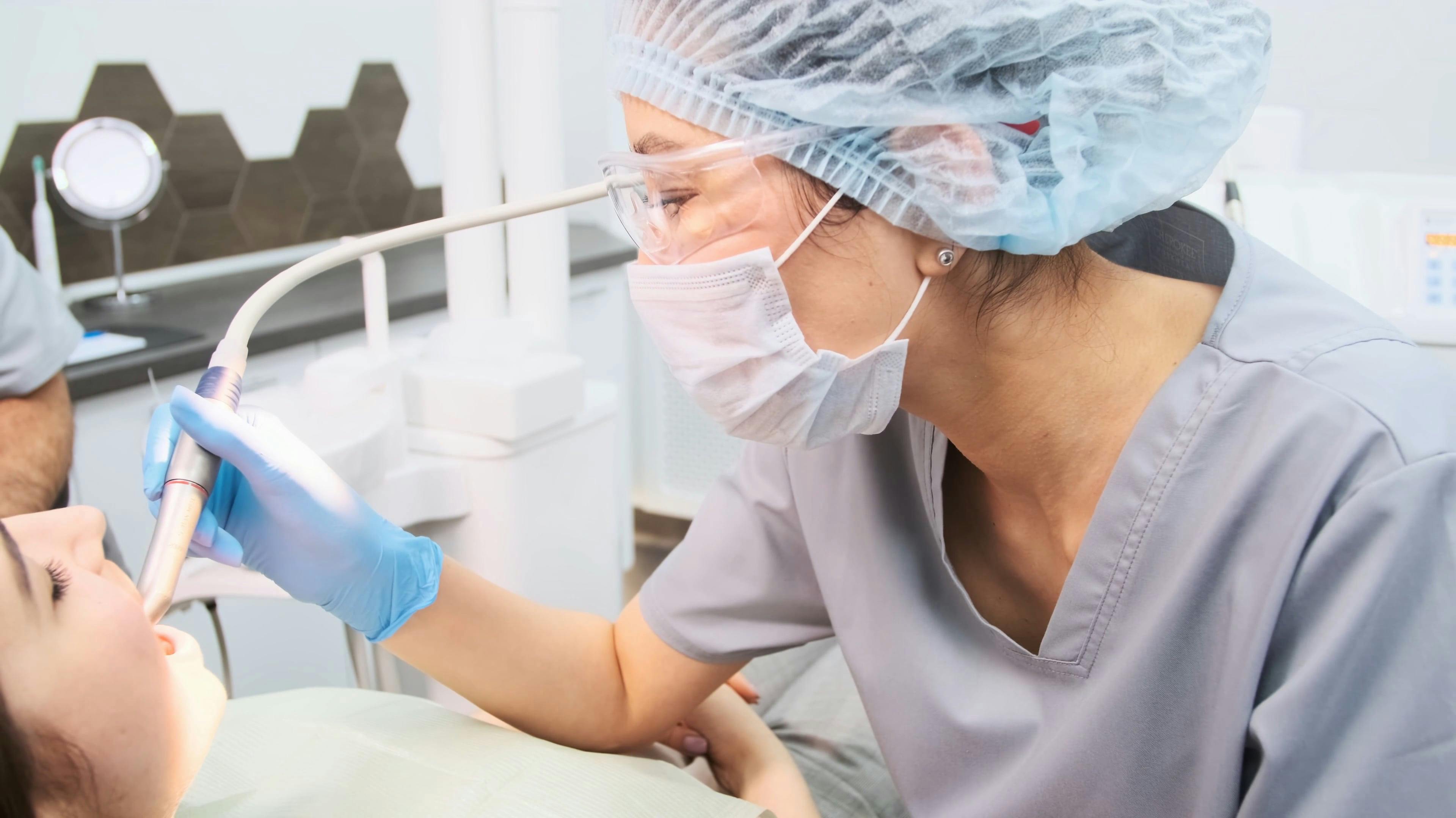
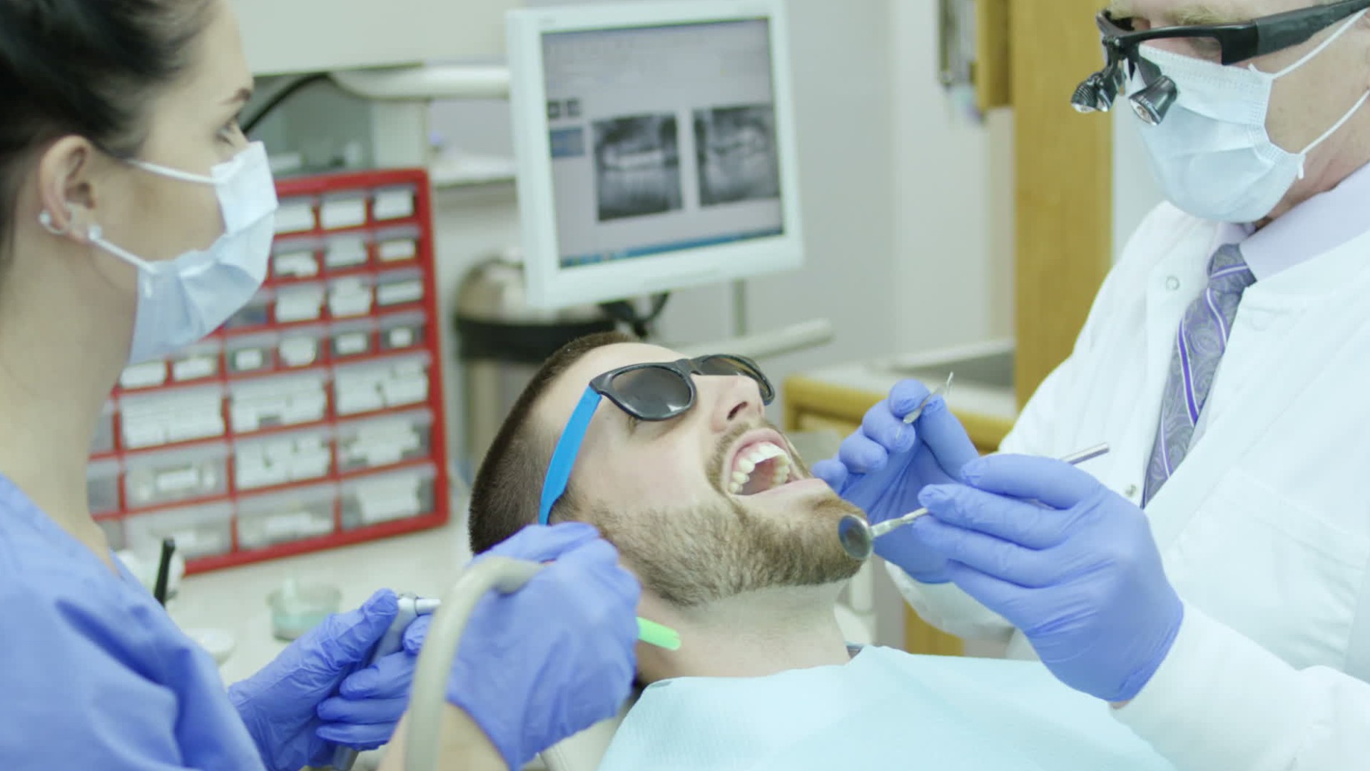
The core of prophylaxis involves mechanical removal of biofilm and calculus using a combination of manual and powered instruments. The sequence prioritizes anatomical accessibility and patient comfort:
1. Isolation and Moisture Control
- Use of rubber dams (for posterior teeth or high-caries-risk patients) or cotton rolls/saliva ejectors to maintain a dry field and prevent aerosol contamination.
- Application of antimicrobial mouthrinses (e.g., 0.12% chlorhexidine) pre-procedure to reduce bacterial load in high-risk patients.
2. Scaling
- Supragingival scaling: Removal of plaque and calculus coronal to the gingival margin using scalers (e.g., Gracey curettes, sickle scalers) or ultrasonic/sonic scalers for efficiency.
- Anatomical focus: Facial, lingual, and occlusal surfaces, including proximal areas accessible via prophy angles or slim curettes.
- Subgingival scaling: Targeted debridement of calculus and biofilm in gingival sulci (1–3 mm depth) or periodontal pockets (>3 mm), using area-specific curettes (e.g., Gracey 11/12 for anterior, 13/14 for posterior).
- Technique: Modified stroke technique (e.g., "walking stroke") to adapt to pocket morphology, with light pressure to avoid gingival trauma.
3. Polishing
- Application of prophy paste (e.g., tin oxide, pumice) with a rubber cup or prophy angle at low-to-moderate speed (≤2,000 RPM) to remove extrinsic stains and smooth enamel.
- Anatomical focus: Enamel surfaces, particularly in fissures, proximal contacts, and gingival thirds where plaque accumulates.
- Note: Avoid polishing abrasive pastes on exposed cementum or dentin to prevent surface roughness, which increases plaque retention.
4. Fluoride Application (Optional)
- Topical fluoride varnish (e.g., 5% sodium fluoride) or gel applied to all tooth surfaces post-polishing to remineralize enamel and reduce caries risk.
- Indications: High-caries-risk patients (e.g., children, xerostomic individuals, or those with orthodontic appliances).
Post-Treatment Instructions
Patient education and follow-up are critical to sustain prophylaxis benefits. Instructions include:
- Oral hygiene reinforcement: Demonstration of modified Bass technique for interdental brushing (e.g., using interdental tips or floss) and tongue cleaning.
- Dietary advice: Reduction of fermentable carbohydrates (e.g., sugars, refined starches) and avoidance of staining agents (e.g., coffee, red wine) for 48 hours post-polishing.
- Smoking cessation counseling: For patients with tobacco-related stains or periodontal disease.
- Recall scheduling: Routine prophylaxis every 6–12 months, adjusted based on periodontal health status (e.g., 3–4 months for patients with gingivitis or pocketing).
The selection of instrumentation directly impacts the efficacy, safety, and patient experience during prophylaxis. Tools are categorized by function—debridement, polishing, or accessory devices—and chosen based on clinical needs.Ultrasonic/Sonic Scalers
- Function: Use vibrations (ultrasonic: 25–45 kHz; sonic: 2–6 kHz) and water spray to disrupt calculus and biofilm via cavitation and acoustic microstreaming.
- Advantages:
- Efficiency: Reduces procedure time by 30–50% compared to manual scaling (studies in Journal of Periodontology, 2018).
- Comfort: Lower pressure required, reducing patient discomfort in sensitive areas.
- Accessibility: Effective in subgingival pockets and furcations with slim tips (e.g., 3–4 mm diameter).
- Types:
- Magnetostrictive: Generates elliptical motion (e.g., Cavitron); suitable for heavy calculus.
- Piezoelectric: Produces linear motion (e.g., EMS SonicFlex); precise for fine debridement.
Hand Instruments
- Scalers (e.g., Sickle scaler, Hoe scaler):
- Function: Remove supragingival calculus via push-pull strokes on enamel surfaces.
- Limitations: Less effective for subgingival calculus or root planing due to rigidity.
- Curettes (e.g., Gracey, Universal):
- Function: Designed for subgingival debridement with rounded back and sharp cutting edge to avoid gingival laceration.
- Area-specific designs:
- Gracey 1/2: Anterior facial/lingual.
- Gracey 11/12: Anterior proximal.
- Gracey 13/14: Posterior proximal.
- Prophy Angles:
- Function: Attach to low-speed handpieces (≤10,000 RPM) to deliver prophy paste via rubber cups or bristle brushes.
- Materials: Latex-free or nitrile cups for patient safety.
Air-Powder Polishing Systems
- Function: Use sodium bicarbonate or glycine powder propelled with air/water spray to remove stains and debris.
- Advantages:
- Speed: Reduces polishing time by 70% compared to rubber cups (Clinical Oral Investigations, 2019).
- Efficacy: Effective on tobacco stains and orthodontic debonding residues.
- Considerations: Contraindicated in respiratory-compromised patients (risk of inhalation) or those with implant surfaces (potential microabrasion).
Periodontal Probes
- Function: Measure gingival sulcus depth, calculus presence, and mucogingival defects (e.g., UNC-15 probe with 1–2–3–5–7–8–10 mm markings).
- Specialized probes:
- Williams probe: Standard for gingival margin assessment.
- Color-coded probes: Aid in visual detection of bleeding (e.g., red markings at 3–5 mm).
Accessory Devices
- Saliva Ejectors: Maintain dry field during instrumentation.
- Rubber Dam Systems: Isolate teeth for caries prevention (e.g., Isolite system) or patient protection (e.g., latex-free dams).
- Intraoral Cameras: Provide real-time visualization of biofilm/stains for patient education.
Adaptations for Patients with Specific Needs
Prophylaxis techniques must be modified to accommodate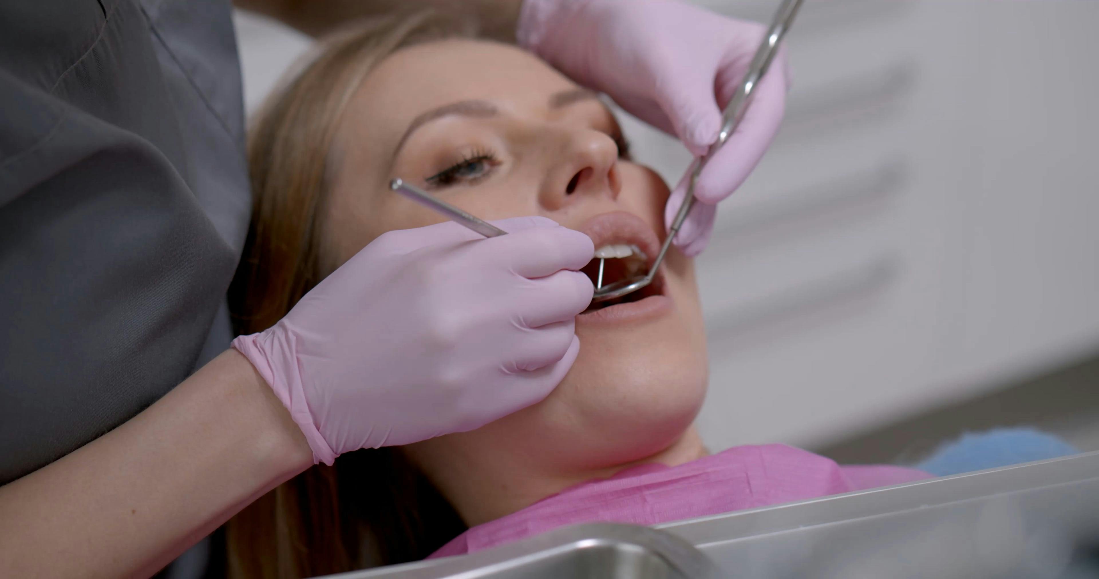
Clinical Benefits and Patient Outcomes of Dental Prophylaxis
Dental prophylaxis is a cornerstone of preventive dentistry, offering measurable improvements in oral health that extend beyond cosmetic benefits. Regular prophylaxis reduces the incidence of dental caries, periodontal diseases, and systemic health risks by minimizing microbial biofilm accumulation and promoting oral homeostasis. Research demonstrates that its clinical efficacy is supported by both immediate and long-term patient outcomes, with evidence linking prophylaxis to reduced healthcare costs and improved quality of life. This section explores the empirical benefits of prophylaxis, compares its effectiveness with alternative preventive measures, and outlines strategies to enhance patient adherence through tailored education.
The primary immediate benefits of dental prophylaxis include the mechanical removal of plaque, calculus, and extrinsic stains, which directly reduces the risk of gingivitis and early-stage periodontitis. Studies indicate that patients undergoing prophylaxis every 6–12 months experience a 30–50% reduction in gingival inflammation compared to those with irregular or no prophylaxis (American Dental Association, Clinical Practice Guideline on the Use of Antimicrobials in Periodontics, 2015). Long-term adherence to prophylaxis regimens further correlates with:
- A 40% lower prevalence of dental caries in adults (National Institutes of Health, Oral Health in America, 2020).
- Reduced periodontal pocket depths by 1–2 mm over 2–3 years in high-risk patients (Journal of Periodontology, 2018).
- Lower incidence of tooth loss due to periodontal disease, with systematic reviews showing a 25% relative risk reduction in edentulism among compliant patients (The Cochrane Database of Systematic Reviews, 2019).
Beyond oral health, prophylaxis mitigates systemic risks associated with poor oral hygiene, including:
- Endocarditis risk reduction by up to 70% in patients with pre-existing cardiac conditions (American Heart Association, Guidelines for the Prevention of Infective Endocarditis, 2021).
- Lower diabetes management challenges, as chronic periodontitis exacerbates glycemic control; prophylaxis in diabetic patients has been shown to improve HbA1c levels by 0.4–0.6% (Diabetes Care, 2017).
- Reduced respiratory infections, particularly pneumonia, due to the aspiration of oral pathogens (Journal of the American Geriatrics Society, 2016).
Comparative Effectiveness of Prophylaxis vs. Alternative Preventive Measures
While dental prophylaxis is the gold standard for biofilm control, its effectiveness can be contextualized when compared to other preventive measures. The following table summarizes the comparative benefits of prophylaxis against oral rinses, interdental brushes, and manual toothbrushing:
| Preventive Measure |
Plaque Reduction (%) |
Calculus Reduction (%) |
Gingival Inflammation Reduction (%) |
Adherence Challenges |
|---|
| Professional Prophylaxis |
60–80% |
85–95% |
50–70% |
Low (scheduled visits) |
| Chlorhexidine Oral Rinse (0.12%) |
30–50% |
0–10% (no direct effect) |
30–40% |
High (staining, taste, cost) |
| Interdental Brushes |
40–60% (interproximal) |
0–20% (subgingival) |
20–35% |
Moderate (technique-dependent) |
| Manual Toothbrushing (Fluoride Toothpaste) |
30–50% |
0–5% (no direct effect) |
25–40% |
High (user compliance varies) |
Key Insights:
- Prophylaxis remains superior for calculus removal and subgingival biofilm disruption, which are critical for periodontal health.
- Chlorhexidine rinses complement prophylaxis by reducing plaque between visits but do not replace professional cleaning for calculus.
- Interdental brushes are effective for interproximal plaque but require daily use to match prophylaxis benefits; their efficacy plateaus without professional reinforcement.
- Manual toothbrushing is foundational but lacks precision for subgingival areas, making prophylaxis essential for high-risk patients.
Patient Education Strategies to Maximize Adherence
Patient adherence to prophylaxis recommendations is influenced by health literacy, perceived risk, and access to care. Tailored education strategies can significantly improve compliance by addressing these barriers. The following approaches are evidence-based and adaptable to varying literacy levels:1. Literacy-Adaptive Communication Techniques
- Visual aids and diagrams: Use illustrated timelines (e.g., "6 months = 1 prophylaxis") or color-coded risk charts (e.g., "High risk = 3–4 months") to simplify frequency recommendations.
- Plain-language explanations: Replace technical terms with relatable metaphors, such as:
> "Plaque is like invisible glue that sticks to your teeth and feeds harmful bacteria. Prophylaxis is the only way to scrape off the hardened ‘glue’ (calculus) before it causes gum disease."
- Multilingual resources: Provide printed materials and digital content in the patient’s primary language, including audio guides for low-literacy populations.
2. Risk Stratification and Personalized Scheduling
- Risk-based intervals: Educate patients on individualized timelines:
- Low risk: Annual prophylaxis.
- Moderate risk (gingivitis, diabetes): Every 4–6 months.
- High risk (periodontitis, smokers, immunocompromised): Every 3 months.
- Digital reminders: Implement SMS or app notifications with countdowns to the next appointment (e.g., "Your next prophylaxis is in 120 days!").
- Financial incentives: Highlight cost savings by comparing the $100–$200 per prophylaxis to potential $1,000+ for periodontal surgery or $500–$3,000 for root canals.
3. Behavioral Reinforcement Strategies
- Shared decision-making: Present prophylaxis as a collaborative choice rather than a mandate, using phrases like:
> "Based on your oral health, we recommend prophylaxis every 4 months to keep your gums healthy. Would you like to schedule it now or discuss alternatives?"
- Progress tracking: Use oral health diaries or smile charts to visually document improvements (e.g., "Before prophylaxis: 4mm pocket depth; After: 2mm").
- Social reinforcement: Encourage family or caregiver involvement in reminders, especially for pediatric or elderly patients.
4. Addressing Common Barriers
- Time constraints: Offer extended hours (evenings/weekends) or express prophylaxis for patients with busy schedules.
- Fear/anxiety: Use desensitization techniques, such as:
- Virtual reality (VR) distraction during procedures.
- Gradual exposure (e.g., starting with a "check-up only" visit before full prophylaxis).
- Access issues: Partner with mobile dental clinics or community health programs to reduce logistical hurdles.
Evidence-Based Adherence Impact:
> "Patients who received tailored education with visual aids and personalized scheduling demonstrated a 45% higher adherence rate compared to those given generic instructions (Journal of Dental Education, 2021)." Additionally, text message reminders increased prophylaxis attendance by 22% in a 2020 study by the American Journal of Preventive Medicine. Role of Prophylaxis in Systemic Health
Dental prophylaxis is not merely a preventive measure for oral diseases but a critical component of systemic health management. Research increasingly demonstrates bidirectional relationships between oral and systemic conditions, where chronic oral inflammation—often mitigated through regular prophylaxis—contributes to the progression of diseases such as diabetes, cardiovascular disorders, and respiratory infections. The biological mechanisms underlying these connections involve microbial translocation, systemic inflammation, and metabolic dysregulation, all of which can be modulated through professional dental cleaning. This section explores the pathological pathways linking untreated oral biofilms to systemic complications, supported by empirical evidence, and examines interdisciplinary collaboration models that integrate prophylaxis into comprehensive patient care.
Biological Mechanisms Linking Oral Health to Systemic Inflammation
The oral cavity serves as a gateway for pathogenic microorganisms to enter the bloodstream, particularly when gingival tissues are compromised. Periodontal pathogens, such as Porphyromonas gingivalis, Aggregatibacter actinomycetemcomitans, and Fusobacterium nucleatum, produce virulence factors—such as lipopolysaccharides (LPS), proteases, and exotoxins—that trigger systemic inflammatory responses. These pathogens disrupt the integrity of the gingival epithelium, facilitating bacterial translocation into the circulatory system. Once in the bloodstream, microbial components activate immune cells (e.g., monocytes, macrophages, and neutrophils), leading to the release of pro-inflammatory cytokines, including interleukin-1 (IL-1), interleukin-6 (IL-6), and tumor necrosis factor-alpha (TNF-α). Chronic elevation of these cytokines is associated with endothelial dysfunction, insulin resistance, and atherosclerosis, exacerbating conditions such as diabetes and cardiovascular disease.
Key pathways include:
- Endothelial Dysfunction: LPS from oral bacteria induces oxidative stress in endothelial cells, impairing nitric oxide (NO) production and promoting vasoconstriction. This contributes to hypertension and arterial plaque formation.
- Insulin Resistance: Elevated IL-6 and TNF-α interfere with insulin signaling pathways, worsening glycemic control in diabetic patients. Studies show that periodontal treatment in diabetic individuals improves HbA1c levels by 0.4–0.6%.
- Pulmonary Complications: Aspiration of oral bacteria (e.g., Streptococcus pneumoniae, Haemophilus influenzae) increases the risk of community-acquired pneumonia, particularly in elderly or immunocompromised patients. Prophylactic reduction of oral pathogens correlates with a 30–50% reduction in pneumonia-related hospitalizations in high-risk populations.
Pathophysiological Flowchart: From Untreated Plaque to Systemic Complications
The progression from untreated dental plaque to systemic disease follows a multi-stage inflammatory cascade, which can be visualized as follows:1. Plaque Accumulation and Dysbiosis
- Poor oral hygiene leads to biofilm formation, dominated by pathogenic species (e.g., P. gingivalis, Tannerella forsythia).
- Key trigger: Subgingival plaque disrupts the junctional epithelium, initiating gingival inflammation.
2. Local Inflammatory Response
- Host immune cells (neutrophils, macrophages) release matrix metalloproteinases (MMPs) and prostaglandins (PGE₂), degrading connective tissue and bone.
- Systemic spillover: Cytokines (IL-1β, TNF-α) enter the bloodstream via ulcerated gingival pockets.
3. Systemic Immune Activation
- Low-grade inflammation: Chronic elevation of CRP (C-reactive protein) and fibrinogen, markers of systemic inflammation.
- Endothelial activation: Adhesion molecules (ICAM-1, VCAM-1) increase, promoting leukocyte adhesion and atherosclerosis.
4. Organ-Specific Complications
- Diabetes: Insulin receptor dysfunction due to TNF-α-mediated serine phosphorylation of IRS-1.
- Cardiovascular Disease: Plaque instability and thrombosis from oxidized LDL and impaired NO bioavailability.
- Respiratory Infections: Bacterial aspiration leading to bronchitis, pneumonia, or COPD exacerbations.
5. Feedback Loop
- Systemic diseases (e.g., diabetes) worsen periodontal inflammation, creating a vicious cycle of deteriorating oral and systemic health.
Collaborative Care Models Integrating Prophylaxis into Systemic Health Management
Interdisciplinary approaches are essential to address the systemic implications of oral disease. Dental professionals play a pivotal role in risk stratification, early intervention, and patient education, while physicians manage comorbid conditions. Effective collaboration models include:1. Shared Patient Care Protocols
Dental and medical teams use standardized pathways to identify high-risk patients, such as those with:
- Diabetes: Periodontal screening at diabetes clinics, with referrals for prophylaxis if gingival bleeding on probing (BOP) >20%.
- Cardiovascular Disease: Pre-procedural antibiotic prophylaxis for patients with prosthetic heart valves or history of infective endocarditis, as per AHA guidelines (2021).
- Respiratory Conditions: Oral health assessments for COPD or asthma patients, with prophylaxis integrated into pulmonary rehabilitation programs.
2. Data-Driven Referral Systems
- Electronic Health Record (EHR) Integration: Dental offices share periodontal screening results (e.g., plaque index, bleeding scores) with primary care physicians to adjust treatment plans.
- Example: A patient with uncontrolled diabetes (HbA1c >8.5%) and moderate periodontitis may receive quarterly prophylaxis alongside metabolic management.
3. Educational Initiatives for Patients and Providers
- Patient Empowerment: Dentists provide oral-systemic health brochures detailing links between gum disease and diabetes/cardiovascular risks, with smoking cessation and dietary counseling.
- Provider Training: Continuing education programs (e.g., ADA’s "Oral Health and Systemic Disease" modules) train physicians to recognize oral health red flags, such as recurrent infections or unexplained weight loss, which may indicate underlying periodontal disease.
4. Population-Level Interventions
- Community Health Programs: Mobile dental clinics in underserved areas offer prophylaxis and systemic health screenings (e.g., blood pressure, glucose) in a single visit.
- Example: The CDC’s "Oral Health for All" initiative partners dental hygienists with primary care teams to reduce hospitalizations for preventable conditions in high-risk populations.
5. Research and Policy Advocacy
- Evidence-Based Guidelines: Organizations like the American Academy of Periodontology (AAP) advocate for periodontal screening in routine medical exams, similar to blood pressure checks.
- Reimbursement Models: Some insurance plans now cover prophylaxis for high-risk patients (e.g., those with rheumatoid arthritis or HIV), recognizing its systemic benefits.
Case Studies Demonstrating Prophylaxis Impact on Systemic Outcomes
Case 1: Diabetes and Periodontal Intervention
- Patient Profile: A 55-year-old male with type 2 diabetes (HbA1c 9.2%) and chronic periodontitis (mean probing depth 5.3 mm).
- Intervention: Full-mouth scaling and root planing (SRP) followed by 3-month maintenance prophylaxis.
- Outcome: HbA1c reduced to 7.8% within 6 months, with 30% decrease in gingival inflammation. The patient’s insulin requirements were adjusted downward by 15%.
Case 2: Cardiovascular Risk Reduction
- Patient Profile: A 68-year-old female with coronary artery disease (CAD) and aggressive periodontitis (BOP 80%).
- Intervention: Non-surgical periodontal therapy + antibiotic therapy (amoxicillin/metronidazoleze).
- Outcome: 25% reduction in CRP levels and improved endothelial function (measured via flow-mediated dilation). Follow-up echocardiogram showed stabilized plaque morphology.
Case 3: Respiratory Infection Prevention
- Population Study: Elderly residents in a long-term care facility with high pneumonia rates.
- Intervention: Quarterly dental prophylaxis + chlorhexidine rinses for high-risk individuals.
- Outcome: 40% reduction in pneumonia-related hospitalizations over 12 months, with cost savings of $180,000 annually.
Emerging Research and Future Directions
Ongoing studies are elucidating microbiome-based biomarkers to predict systemic risks from oral pathogens. Key areas include:
- Metabolomics: Identifying oral microbiome-derived metabolites (e.g., trimethylamine N-oxide, TMAO) linked to atherosclerosis.
- Genomic Profiling: Using 16S rRNA sequencing to classify patients by periodontal pathogen load and tailor prophylaxis frequency.
- Immunomodulatory Therapies: Exploring probiotics or anti-inflammatory agents (e.g., statins, IL-1 inhibitors) adjunctive to prophylaxis for high-risk patients.
Blockquote:
> *"Periodontal disease is not an isolated oral condition but a modifiable risk factor for systemic diseases. Regular
Challenges and Limitations in Dental Prophylaxis
Dental prophylaxis, while fundamental in preventive dentistry, encounters significant barriers that can compromise its efficacy and accessibility. Patient-related factors, systemic limitations in healthcare delivery, and biological constraints in treating advanced oral diseases create challenges that require tailored solutions. Understanding these limitations ensures clinicians can optimize prophylaxis protocols, identify when adjunctive interventions are necessary, and advocate for systemic improvements in oral healthcare.
Common Barriers to Effective Prophylaxis
Patient adherence and systemic constraints frequently undermine the potential benefits of dental prophylaxis. These barriers can be categorized into patient-related, provider-related, and healthcare system-related challenges, each demanding distinct mitigation strategies.Patient-related barriers include:
- Dental anxiety and phobia, which may lead to avoidance of preventive care, resulting in untreated caries or periodontal disease progression.
- Lack of awareness regarding the importance of prophylaxis, often due to insufficient patient education or misconceptions about dental health.
- Financial constraints, where cost prohibitive preventive care delays treatment until symptoms necessitate more invasive procedures.
- Time limitations, particularly in busy schedules, which may reduce the frequency of recommended prophylaxis visits.
Provider-related barriers encompass:
- Time constraints during appointments, limiting thoroughness in scaling, polishing, and patient education.
- Workforce shortages, especially in rural or underserved areas, reducing access to regular prophylaxis.
- Variability in training, where some practitioners may not emphasize prophylaxis as a primary preventive measure.
Healthcare system-related barriers involve:
- Limited insurance coverage for preventive services, discouraging patients from seeking routine care.
- Geographic disparities, where proximity to dental clinics influences access, particularly in low-income or remote communities.
- Fragmented care coordination, where patients may receive inconsistent messaging about prophylaxis from different providers.
Mitigation Strategies
To address these barriers, a multidisciplinary approach is essential:
- Patient education through clear, culturally sensitive communication about the long-term benefits of prophylaxis, using visual aids and interactive tools.
- Flexible scheduling options, such as extended hours, weekend appointments, or mobile dental units, to accommodate diverse patient needs.
- Financial assistance programs, including sliding-scale fees, community health initiatives, or partnerships with dental schools to reduce costs.
- Anxiety management techniques, such as sedation dentistry, gradual exposure therapy, or virtual reality distraction during procedures.
- Tele-dentistry integration for preliminary assessments, follow-ups, and educational reinforcement, particularly in underserved areas.
- Policy advocacy for expanded insurance coverage of preventive services and increased funding for public health dental programs.
Limitations of Prophylaxis in Advanced Dental Conditions
While prophylaxis excels in preventing and managing early-stage dental diseases, its efficacy diminishes in advanced conditions requiring surgical, restorative, or periodontal interventions. Key limitations include:Periodontal Disease Management
Prophylaxis alone is insufficient for moderate to severe periodontitis, where deep periodontal pockets (>5 mm) and attachment loss necessitate:
- Scaling and root planing (SRP) with local anesthesia to address subgingival plaque and calculus.
- Surgical interventions, such as flap debridement or bone grafting, for cases with osseous defects or furcations.
- Antimicrobial therapy, including systemic antibiotics or local drug delivery (e.g., doxycycline gels), to control bacterial pathogens like Porphyromonas gingivalis.
Root Caries and Complex Restorations
Prophylaxis through polishing and fluoride applications may slow progression but cannot reverse dentinal caries or restore structural integrity. Advanced cases require:
- Non-surgical treatments, such as resin-modified glass ionomer restorations for high-caries-risk patients.
- Surgical restorations, including composite fillings, inlays, or crowns, to prevent further decay and preserve tooth function.
- Caries-risk assessment tools, like the Caries Management by Risk Assessment (CAMBRA) protocol, to guide personalized preventive strategies.
Pulpal Involvement and Endodontic Needs
When caries or trauma compromises the pulp, prophylaxis is inadequate. Pulpitis or necrosis demands:
- Root canal therapy (RCT) to remove infected pulp tissue and seal the canal system.
- Apicoectomy for persistent periapical infections unresponsive to conventional RCT.
- Trauma management, including splinting or surgical reimplantation for avulsed teeth.
Referral Criteria for Specialists
Prophylaxis providers should recognize when to refer patients to specialists based on:
- Periodontal probing depths exceeding 5 mm with bleeding on probing.
- Radiographic evidence of bone loss (e.g., >30% alveolar bone height reduction).
- Pulpal symptoms, such as spontaneous pain, percussion sensitivity, or thermal hypersensitivity.
- Complex restorative needs, including large carious lesions, endodontically treated teeth, or esthetic concerns requiring prosthodontics.
Risk Factors Reducing Prophylaxis Efficacy
Certain modifiable and non-modifiable risk factors can undermine the effectiveness of dental prophylaxis. The following table categorizes these factors, their impact on oral health, and evidence-based mitigation strategies.
| Factor |
Impact on Oral Health |
Mitigation Strategies |
|---|
| Smoking/Tobacco Use |
- Reduces salivary flow, impairing self-cleansing mechanisms.
- Alters gingival vasculature, increasing susceptibility to periodontitis.
- Delays wound healing post-procedures (e.g., SRP, extractions).
- Stains teeth, complicating polishing efficacy.
|
- Smoking cessation counseling using brief interventions (e.g., "5 As" model: Ask, Advise, Assess, Assist, Arrange).
- Referral to smoking cessation programs (e.g., nicotine replacement therapy, behavioral support).
- Post-prophylaxis reinforcement with smoke-free oral hygiene instructions (e.g., chlorhexidine rinses for healing).
|
| Poor Diet (High Sugar/Frequent Snacking) |
- Increases acid exposure, demineralizing enamel and promoting caries.
- Fosters dysbiotic biofilm, accelerating periodontal destruction.
- Contributes to erosion from acidic beverages (e.g., sodas, citrus drinks).
|
- Dietary counseling emphasizing pH-neutral foods (e.g., cheese, nuts) and fluoride-rich snacks (e.g., fluoridated water, tea).
- Education on timing of sugar intake (e.g., consuming sugars with meals rather than between meals).
- Prescription of high-fluoride toothpaste (5,000 ppm) or varnishes for high-risk patients.
|
| Xerostomia (Dry Mouth) |
- Reduces salivary buffering capacity, increasing caries risk.
- Impairs plaque removal due to decreased salivary flow.
- Exacerbates oral discomfort, reducing compliance with oral hygiene.
|
- Salivary stimulation with sugar-free lozenges or pilocarpine (for systemic causes).
- Use of artificial saliva substitutes (e.g., carboxymethylcellulose-based gels).
- Frequent fluoride applications (e.g., gel trays, varnishes) during prophylaxis.
|
| Medication-Induced Oral Effects |
- Antihypertensives (e.g., calcium channel blockers) may cause gingival overgrowth.
- Antidepressants (e.g., SSRIs) increase xerostomia risk.
- Chemotherapy induces mucositis and increased caries susceptibility.
|
- Collaboration with physicians to adjust dosages or explore alternatives.
- Prescription of chronic low-dose fluoride for xerostomia patients.
- Oral
Future Trends and Innovations in Dental Prophylaxis
Dental prophylaxis is evolving rapidly, driven by advancements in biomaterials, digital health, and precision diagnostics. Emerging technologies and prophylactic agents are poised to enhance treatment efficacy, reduce procedural invasiveness, and improve patient adherence. Innovations such as antimicrobial coatings, nanotechnology-based tools, and AI-assisted diagnostics are reshaping clinical workflows, while digital health platforms are fostering proactive patient engagement. This section explores the most transformative trends expected to redefine prophylaxis within the next decade, emphasizing their clinical and operational implications.The integration of technology into dental prophylaxis is accelerating, with a focus on minimizing microbial resistance, improving early detection of oral pathologies, and personalizing preventive care. Key innovations include laser-assisted prophylaxis, smart materials for sustained antimicrobial release, and AI-driven risk stratification. Additionally, the rise of tele-dentistry and mobile health (mHealth) applications is expanding access to preventive care, particularly in underserved populations. These advancements align with global shifts toward predictive, preventive, and personalized medicine (PPPM), positioning prophylaxis as a cornerstone of modern dentistry.
Emerging Technologies in Prophylaxis Procedures
Technological innovations are optimizing the precision, speed, and patient comfort of prophylaxis procedures. Laser technology, for instance, is increasingly used for supragingival and subgingival biofilm removal, offering advantages such as reduced mechanical trauma, selective bacterial targeting, and hemostatic effects. Studies demonstrate that Er:YAG and diode lasers effectively disrupt biofilm matrices while preserving tooth structure, with clinical trials showing comparable or superior outcomes to traditional scaling and root planing (SRP) in mild-to-moderate periodontitis cases.Another transformative development is the integration of AI and machine learning (ML) in diagnostic imaging. AI algorithms, trained on vast datasets of intraoral scans and radiographic images, can now identify early signs of caries, periodontal disease, and oral cancer with high accuracy. For example, systems like Dentrix Ascend and CariAI leverage deep learning to detect interproximal caries and assess plaque accumulation in real time, enabling dentists to tailor prophylaxis protocols based on individualized risk profiles. Additionally, computer-aided design (CAD) and 3D printing are being explored for custom prophylaxis tools, such as ergonomic scalers and ultrasonic tips, designed to reduce operator fatigue and improve access to challenging anatomical regions.
"The adoption of AI in dental diagnostics is projected to grow at a CAGR of 30% through 2030, driven by demand for early disease detection and automated risk assessment."
— Grand View Research (2023)
Advancements in Prophylactic Agents and Materials
The development of antimicrobial coatings, nanotechnology-based agents, and biofunctional materials represents a paradigm shift in prophylaxis, moving beyond mechanical removal to active microbial control. Chitosan-based coatings, for instance, have shown promise in sustained-release fluoride and antimicrobial peptides, reducing plaque formation and demineralization. Clinical studies indicate that chitosan nanoparticles can penetrate biofilm structures, inhibiting Streptococcus mutans and Porphyromonas gingivalis for up to 72 hours post-application.Nanotechnology is also enabling the creation of smart toothpastes and mouthwashes embedded with silver nanoparticles, zinc oxide nanorods, or antimicrobial peptides that respond to pH changes or bacterial presence. Products like Colgate Total Pro-Shield and Crest Gum Care already incorporate nanohydroxyapatite to remineralize enamel and disrupt biofilm. Future iterations may include self-assembling peptide hydrogels that form protective barriers over enamel, reducing erosion from acidic foods and beverages.
"Nanotechnology in oral care is expected to reach a market value of $1.8 billion by 2027, with antimicrobial coatings and drug-delivery systems leading growth."
— MarketsandMarkets (2023)
Another frontier is gene-editing and CRISPR-based therapies, though still in preclinical stages, which could permanently modify oral microbiota to prevent pathogenic colonization. Early research on bacteriophage therapy (using viruses to target specific oral pathogens) and probiotic strains like Lactobacillus reuteri suggests potential for long-term microbial balance, reducing the need for frequent mechanical prophylaxis.
Timeline of Key Innovations in Prophylaxis (2024–2034)
The next decade will witness a phased adoption of innovations, with early-stage technologies maturing into clinical practice. Below is a projected timeline of key developments, categorized by readiness and impact:
| Year |
Innovation |
Clinical Application |
Expected Impact |
|---|
| 2024–2026 |
AI-Powered Diagnostic Tools |
- Real-time caries and periodontal risk assessment via intraoral scanners (e.g., iTero, Planmeca Emerald).
- Integration with electronic health records (EHRs) for automated prophylaxis reminders.
|
- Reduction in missed preventive appointments by 20–30%.
- Early intervention in 30% of high-risk patients before symptoms appear.
|
| 2026–2028 |
Laser-Assisted Prophylaxis |
- FDA-approved Er:YAG lasers for subgingival biofilm removal in periodontitis.
- Hybrid laser-ultrasonic devices for single-visit prophylaxis.
|
- 40% faster biofilm removal compared to traditional SRP.
- Reduced post-procedural pain and swelling by 50%.
|
| 2028–2030 |
Nanotechnology-Based Agents |
- Chitosan-nanoparticle mouthwashes with 7-day antimicrobial efficacy.
- Bioactive glass coatings for enamel remineralization.
|
- 30% reduction in recurrent caries in high-risk patients.
- Elimination of daily fluoride applications for some individuals.
|
| 2030–2034 |
AI-Driven Personalized Prophylaxis |
- Genomic saliva testing to predict individual microbial risks.
- Wearable sensors (e.g., OralID) for real-time pH and biofilm monitoring.
- CRISPR-based probiotic therapies for dysbiotic microbiome correction.
|
- Custom prophylaxis protocols reducing general dental visits by 40%.
- Prevention of periodontitis progression in 60% of genetically predisposed patients.
|
Digital Health and Patient Engagement in Prophylaxis
The proliferation of digital health tools is democratizing access to prophylaxis, particularly in remote and underserved populations. Tele-dentistry platforms, such as Dentrix TeleDentistry and SmileDirectClub’s virtual consultations, enable dentists to monitor oral health between visits, adjusting prophylaxis protocols via secure video calls and AI-assisted diagnostics. These systems often integrate with wearable devices (e.g., Fitbit Oral Care, Oral-B iO) to track brushing habits, plaque levels, and gingival inflammation in real time.Mobile applications are another critical innovation, offering gamified oral hygiene programs (e.g., Brush DJ, Oral Health by Delta Dental) that improve patient compliance. Studies show that app-guided brushing increases adherence by 25–40%, particularly in pediatric and adolescent populations. Additionally, blockchain-based dental records (e.g., Dentacoin) are being piloted to securely share prophylaxis histories across providers, ensuring continuity of care.
*"By 2025, Dental prophylaxis transcends routine dental maintenance, serving as a multifaceted strategy to preserve oral integrity and systemic well-being. By systematically eliminating microbial threats, it disrupts the progression of dental caries, periodontal disease, and associated systemic risks, underscoring its indispensable role in modern healthcare. The integration of emerging technologies and collaborative care models further solidifies prophylaxis as a dynamic field, where continuous adaptation ensures its relevance in addressing evolving patient needs. Ultimately, its success hinges not only on clinical expertise but also on patient education and systemic integration, positioning it as a pivotal element in preventive medicine for decades to come.
FAQ
What exactly is a dental prophylaxis cleaning, and what does it include?
Dental prophylaxis cleaning is a professional teeth cleaning that removes plaque, tartar, and stains from tooth surfaces and below the gumline. It includes scaling (removing buildup) and polishing to smooth enamel, typically performed by a dentist or hygienist every 6–12 months. The procedure also checks for early signs of gum disease or decay.
What does dental prophylaxis treatment involve, and how is it different from a regular cleaning?
Dental prophylaxis treatment is a preventive procedure that includes scaling to remove hardened plaque (tartar) and polishing to prevent bacterial buildup. Unlike a basic cleaning, it may involve more thorough gum pocket cleaning if early gum disease is present. It’s usually recommended for patients with higher risk of decay or periodontal issues.
Is dental prophylaxis necessary for adults, and how often should adults get it done?
Yes, dental prophylaxis is essential for adults to prevent tooth decay, gum disease, and bad breath. Most adults without gum issues need it every 6–12 months, while those with gum disease may require more frequent cleanings (every 3–4 months). It’s a key part of maintaining oral health at any age.
How much does a dental prophylaxis cleaning typically cost?
The cost of dental prophylaxis varies by location and provider but usually ranges from $75 to $200 per session. Insurance often covers it fully or partially if deemed preventive care. Some dental offices offer package deals for multiple cleanings per year.
What does a dental prophylaxis bill usually include, and are there any hidden fees?
A dental prophylaxis bill typically covers scaling, polishing, and a basic exam, with costs listed separately for each service. Hidden fees are rare if the procedure is straightforward, but check for charges like X-rays or fluoride treatments if added. Always ask for an itemized receipt upfront.
What does the term "dental prophylaxis" actually mean in simple terms?
"Dental prophylaxis" simply means preventive dental care focused on cleaning teeth to stop problems before they start. It’s a medical term for professional teeth cleaning that removes plaque and tartar to protect against cavities, gum disease, and other oral health issues. The word comes from Greek, meaning "protection" or "prevention."
|
|
Leave a Comment
Comments are moderated before appearing. The data you submit is processed according to the Privacy Policy of Voltefac.