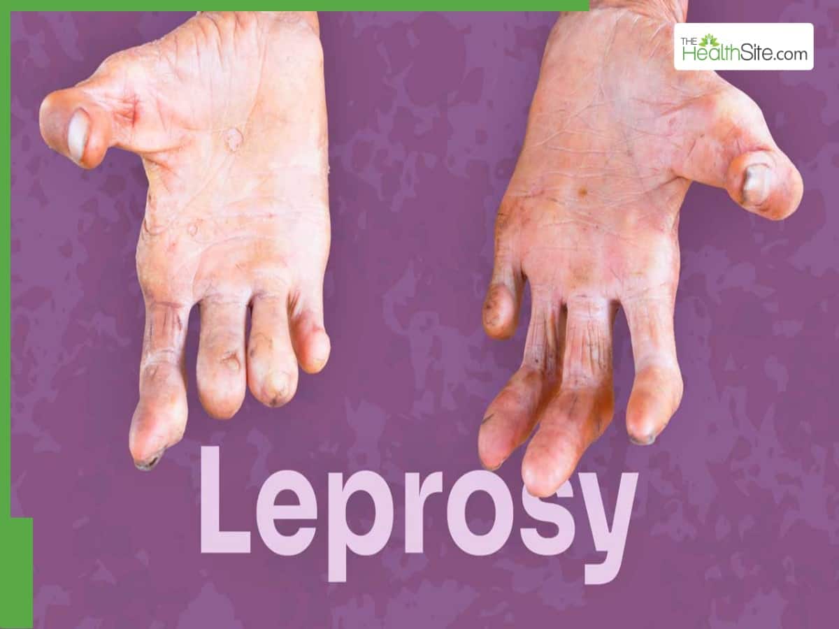
What Is Leprosy Its History Medical Impact And Modern Solutions

Published

Table of Contents

Leprosy, an ancient disease rooted in human history, persists as a misunderstood yet curable condition affecting millions globally. Despite its eradication in many regions, stigma and misconceptions surrounding Mycobacterium leprae continue to fuel discrimination, overshadowing the medical advancements that now render it treatable. From biblical plagues to modern public health campaigns, leprosy’s journey reflects humanity’s evolving relationship with disease—balancing scientific progress against enduring social prejudices. This exploration examines its origins, transmission, clinical progression, and the transformative therapies reshaping its legacy.

The disease’s historical stigma, particularly in medieval Europe, where sufferers faced exile and religious condemnation, contrasts sharply with today’s evidence-based treatments. Modern medicine has identified leprosy’s bacterial cause, developed multidrug therapies, and emphasized early intervention to prevent disability. Yet challenges remain, from drug resistance in endemic regions to the psychological toll of a condition still associated with shame. Understanding leprosy demands a synthesis of medical rigor and historical context to dismantle myths and highlight its preventable nature.

what is leprosy

Historical and Medical Background of Leprosy

Leprosy, a chronic infectious disease caused by Mycobacterium leprae, has left an indelible mark on human history, shaping religious beliefs, medical practices, and societal structures for millennia. Documented in ancient civilizations across Egypt, India, China, and the Middle East, leprosy was often misunderstood, feared, and systematically marginalized. Its progression from a medical condition to a symbol of divine punishment or moral corruption reflects broader historical trends in disease perception, while key scientific breakthroughs—such as the identification of its bacterial agent in the 19th century—marked turning points in its treatment and societal reintegration.

The study of leprosy’s historical and medical evolution reveals how civilizations grappled with its physical and psychological toll, from ritualistic isolation in ancient India to the establishment of leper colonies in medieval Europe. Below, the origins, ancient documentation, and medical milestones are examined, followed by an analysis of its stigmatization and contrasting portrayals in religious texts versus modern medicine.

Origins and Earliest Recorded Cases of Leprosy

The precise origins of leprosy remain debated, but skeletal and textual evidence suggests its presence in human populations as early as 600 BCE. The disease likely emerged in tropical regions, where Mycobacterium leprae thrives in cooler climates and armadillos (a natural reservoir). Archaeological findings, such as mummified remains with characteristic bone deformities in ancient Egypt and Peru, indicate leprosy’s global spread via trade and migration routes.

Ancient civilizations documented leprosy through varied lenses:

  • Egypt (c. 1550 BCE): The Ebers Papyrus, a medical text from the New Kingdom, describes skin lesions resembling leprosy, though treatments included herbal remedies and magical incantations.
  • India (c. 800–200 BCE): The Sushruta Samhita, an Ayurvedic treatise, categorizes leprosy (Kushtha) into types based on symptoms, advocating dietary restrictions and oil-based therapies.
  • China (c. 3rd century BCE): The Huangdi Neijing (Yellow Emperor’s Inner Canon) references "leprous" skin conditions, linking them to imbalances in yin and yang, while later dynasties recorded quarantine practices.
  • "The leprosy of the skin, the scabs, the itch, the eczema, the scurf, the burn, the frostbite, the yellow crust, the white leprosy, the black leprosy, the leprosy of the nails, the leprosy of the hair, the leprosy of the beard."Sushruta Samhita (4th century BCE), describing leprosy’s diverse manifestations.

    Chronological Timeline of Medical Discoveries

    The understanding of leprosy evolved through empirical observations, religious interpretations, and scientific advancements. Below is a chronological overview of pivotal discoveries:
    1. Ancient Texts (c. 1500 BCE–500 CE):
      Leprosy appears in the Bible (Leviticus 13–14), where priests diagnose and isolate afflicted individuals, associating the disease with ritual impurity. Similarly, Hindu scriptures (e.g., Manusmriti) prescribe social exclusion for those with Kushtha, reflecting moral and spiritual dimensions of illness.
    2. Medieval Europe (500–1500 CE):
      The Black Death (1347–1351) exacerbated fears of leprosy, leading to the establishment of leper houses in Europe. Medical texts like those of Avicenna (11th century) distinguished leprosy from other skin diseases but reinforced its link to sin.
    3. 1873: Identification of Mycobacterium leprae:
      Armadillo Hansen, a Norwegian physician, discovered the bacillus under a microscope, proving leprosy’s infectious nature. This breakthrough dismantled supernatural explanations and paved the way for bacteriological research.
    4. 1940s: Sulphone Drugs:
      Promin (sulfone therapy) became the first effective treatment, reducing bacterial load and preventing disability. Later, dapsone and rifampicin (1960s–1980s) improved cure rates.
    5. 1991: WHO’s Multidrug Therapy (MDT):
      The World Health Organization introduced MDT, combining dapsone, rifampicin, and clofazimine, achieving over 99% cure rates and eliminating leprosy as a public health threat in many regions.
    6. 2010s–Present: Genomic and Armadillo Research:
      Advances in genomics revealed Mycobacterium leprae’s reduced genome, while studies on nine-banded armadillos (natural hosts) in the U.S. (e.g., Louisiana) provided insights into transmission dynamics.

    Stigmatization and Societal Consequences

    Leprosy’s association with shame, contagion fears, and divine punishment led to systemic discrimination. In medieval Europe, legal codes such as the Leprosy Statutes of England (1258) mandated:
  • Isolation: Lepers were expelled from communities, required to wear bell-clappers to announce their approach, and barred from public spaces.
  • Property Confiscation: Families of lepers could lose inheritance rights, and affected individuals were denied burial in consecrated ground.
  • Religious Expiation: The Catholic Church classified leprosy as a "living death," with afflicted individuals encouraged to seek absolution through pilgrimage or monastic life.
  • In India and Southeast Asia, caste systems reinforced exclusion, with lepers relegated to untouchable status. Colonial powers exacerbated stigma by segregating patients in colonies (e.g., Karigiri Leprosy Colony, India), perpetuating cycles of poverty and marginalization. Even after medical advancements, discrimination persisted in some regions until the late 20th century, with survivors facing employment bans and social ostracization.

    "The leper is not merely a victim of disease; he is a victim of the fear and ignorance of his fellow men." —WHO Leprosy Elimination Programme (1991), acknowledging systemic barriers to recovery.

    Comparison Table: Ancient vs. Modern Portrayals of Leprosy

    The following table contrasts how leprosy was depicted in religious and historical texts against its modern medical classification:
    <

    Causal Agent and Transmission Mechanics of Leprosy

    Mycobacterium leprae remains the sole etiological agent of leprosy, a slow-progressing bacterial infection with unique biological and epidemiological traits. Its pathogenicity is intricately linked to its metabolic adaptations, host tissue tropism, and inefficient transmission dynamics, which collectively shape its endemic persistence in specific populations. Understanding these mechanisms is critical for designing targeted interventions, particularly in regions where household transmission remains a primary concern.

    Biological Characteristics of Mycobacterium leprae

    Mycobacterium leprae exhibits distinct physiological and biochemical properties that influence its survival, infectivity, and disease manifestation. The bacterium is an obligate intracellular pathogen, meaning it relies on host cells for replication, which restricts its growth to cooler peripheral tissues. Key biological features include:

    - Growth Rate and Replication Cycle:
    The organism demonstrates an exceptionally slow doubling time of approximately 12–14 days under optimal laboratory conditions, with in vivo replication rates estimated at 11–20 days. This prolonged generation time contributes to the insidious progression of leprosy, often delaying clinical diagnosis for years. The slow growth also complicates cultivation in vitro, as standard mycobacterial media fail to support its replication, necessitating specialized techniques such as the mouse footpad model or armadillo inoculation for research.

    - Optimal Temperature and Tissue Tropism:
    M. leprae thrives at temperatures between 30–33°C, aligning with the cooler peripheral regions of the human body, such as the skin, nasal mucosa, and peripheral nerves. This thermal preference explains its predilection for dermatoneural lesions, where it disrupts Schwann cell function and axon integrity, leading to nerve damage and sensory loss. The bacterium’s inability to survive at core body temperatures (~37°C) further limits its systemic dissemination, confining infection to accessible peripheral sites.

    - Genomic and Metabolic Adaptations:
    The genome of M. leprae (approximately 3.3 million base pairs) encodes ~1,600 genes, many of which are pseudogenes or non-functional due to extensive degeneration. This genomic reduction reflects its parasitic lifestyle, with critical pathways for fatty acid synthesis and cell wall biosynthesis being absent, necessitating reliance on host-derived nutrients. The bacterium’s mycolic acid-rich cell wall enhances resistance to environmental stressors but also complicates antibiotic penetration, contributing to treatment challenges.

    Transmission Mechanisms and Epidemiological Patterns

    Leprosy transmission occurs primarily through respiratory droplets and prolonged close contact, with household transmission accounting for ~90% of cases in endemic regions. The inefficiency of transmission, combined with host susceptibility factors, underpins its focal distribution rather than widespread epidemics.

    - Respiratory Route and Infectious Dose:
    M. leprae is shed in nasal secretions and respiratory droplets from untreated multibacillary (MB) patients, particularly those with active bacterial replication (high bacillary load). However, transmission requires frequent and sustained exposure, as the infectious dose is estimated to be high (likely 10⁵–10⁷ bacilli). This explains why children under 15 years old—who lack prior immunity—are 90% more likely to develop leprosy when exposed, while adults often remain asymptomatic despite exposure. Studies in Brazil and India demonstrate that household contacts of MB patients have a 10–20% lifetime risk of infection, underscoring the role of intimate, prolonged cohabitation.

    - Environmental and Host Factors Influencing Transmission:
    Several variables modulate transmission risk:

  • Climate and Humidity: Higher transmission rates correlate with warm, humid environments, where M. leprae remains viable in aerosols for extended periods.
  • Crowding and Poor Sanitation: Overcrowded households with limited ventilation facilitate droplet persistence, as seen in urban slums and refugee camps.
  • Immunological Susceptibility: Genetic factors, such as polymorphisms in PARK2 and NRAMP1 genes, increase vulnerability, while BCG vaccination (used in some endemic regions) offers ~50% protection against leprosy.
  • - Limited Animal Reservoirs and Research Constraints:
    M. leprae infects nine-banded armadillos (Dasypus novemcinctus) as the only known natural non-human host, with ~5% of wild populations in the southern U.S. testing positive. Armadillos develop spontaneous leprosy-like lesions, making them invaluable for bacterial isolation and drug testing. However, their low prevalence in captivity and long incubation period (1–2 years) hinder large-scale research. Other animals, including mice, guinea pigs, and primates, require artificial inoculation to sustain infection, further limiting transmission studies. This scarcity of animal models has impeded understanding of zoonotic transmission risks, though no evidence supports armadillos as a significant source of human infection.

    WHO Stance on Transmission Risks and Preventive Measures

    The World Health Organization (WHO) emphasizes that leprosy is not highly infectious and does not spread casually. Transmission requires close, repeated contact over months or years, primarily within households. Key preventive strategies for high-risk populations include:
    "Leprosy is preventable through early diagnosis, treatment, and targeted interventions in high-burden communities. Household contacts of leprosy patients are at highest risk and should undergo annual clinical examinations. BCG vaccination reduces infection risk, particularly in children, while multidrug therapy (MDT) eliminates infectiousness within weeks of treatment initiation. Public health efforts must focus on stigma reduction, health education, and improving access to care in endemic regions."
    — WHO Global Leprosy Strategy (2021–2030)
    The WHO further advises that leprosy is not an occupational hazard for healthcare workers, provided standard precautions are followed. However, healthcare-associated transmission remains a theoretical risk in settings with delayed diagnosis or poor infection control. Data from Madagascar and Mozambique show that ~50% of new cases are household contacts, reinforcing the need for contact tracing and preventive therapy in endemic areas.

    what is leprosy - Ilustrasi 2

    Clinical Manifestations and Staging of Leprosy

    Leprosy presents with a spectrum of clinical features determined by the host’s immune response and the virulence of Mycobacterium leprae. The disease primarily affects the skin, peripheral nerves, and mucous membranes, with manifestations ranging from localized, self-limiting lesions to disseminated, progressive disease. Accurate classification into tuberculoid (TT) and lepromatous (LL) forms, along with intermediate borderline types, is critical for tailored management. This section explores the distinct clinical presentations, diagnostic differentiation techniques, and the role of immune-mediated reactional states in disease progression.

    Primary Forms of Leprosy: Tuberculoid and Lepromatous Variants

    The two polar forms of leprosy—tuberculoid (TT) and lepromatous (LL)—represent opposing ends of the disease spectrum, characterized by differing immune responses, lesion morphology, and bacterial load.

    Tuberculoid Leprosy (TT)

  • Skin Lesions: Well-defined, hypopigmented or erythematous plaques with raised, anesthetic borders. Lesions are typically asymmetrical, solitary, or few in number, and exhibit sharp margins due to localized granulomatous inflammation.
  • Nerve Damage: Single or few nerves are affected, often corresponding to the distribution of skin lesions (e.g., ulnar, great auricular, or posterior tibial nerves). Nerve enlargement (neuritis) is common, leading to motor weakness (e.g., claw hand, footdrop) and sensory loss (e.g., loss of pain/temperature sensation).
  • Bacterial Load: Low; acid-fast bacilli (AFB) are rarely detectable on slit-skin smears.
  • Progression: Slow; spontaneous healing may occur if treated early, but untreated cases can progress to disability.
  • Lepromatous Leprosy (LL)

  • Skin Lesions: Diffuse, symmetrical involvement with hypopigmented macules, plaques, or nodular lesions. Nodules are firm, skin-colored, or erythematous, often distributed symmetrically on the face (leonine facies), ears, and cooler extremities. Hypopigmented patches lack tactile sensation and may exhibit follicular involvement (e.g., madarosis—loss of eyebrows/eyelashes).
  • Nerve Damage: Multiple nerves are affected early, often painlessly, leading to chronic, irreversible damage. Common sites include the great auricular, ulnar, and peroneal nerves, resulting in muscle atrophy, contractures, and deformities (e.g., "claw hand" or "dropped foot").
  • Bacterial Load: High; AFB are abundant in skin smears, nasal secretions, and cooler body regions.
  • Progression: Rapid and progressive if untreated, with systemic involvement (e.g., laryngeal edema, orchitis, kidney damage).
  • Differentiation of Skin Lesions in Lepromatous Leprosy
    To distinguish between hypopigmented patches, plaques, and nodules in LL:

  • Hypopigmented Patches: Flat, ill-defined areas of depigmentation with loss of sensation (tested via monofilament or pinprick). May appear scaly or dry due to autonomic dysfunction.
  • Plaques: Elevated, irregularly shaped lesions with raised borders, often erythematous or violaceous. Sensory loss is patchy but more pronounced than in patches.
  • Nodules: Firm, discrete lumps (1–3 cm) arising from the dermis or subcutaneous tissue. May ulcerate or coalesce. Sensory testing reveals complete anesthesia within the lesion.
  • Sensory Testing Protocol:
    1. Pinprick: Use a sterile needle to assess pain sensation (absent in anesthetic areas).
    2. Temperature: Apply warm/cold objects to test thermoreception (loss indicates autonomic neuropathy).
    3. Vibration: Use a 128-Hz tuning fork on bony prominences (e.g., malleolus) to evaluate deep nerve function.
    4. Light Touch: Cotton wool or monofilament (10g) to map tactile deficits.

    Borderline Leprosy and the Immune Response Spectrum

    Leprosy exists as a continuum between TT and LL, with borderline forms (BT, BB, BL) reflecting intermediate immune responses. The Th1/Th2 paradigm explains this spectrum:
  • Th1-Dominant (TT): Strong cell-mediated immunity (CMI) with macrophage activation and granuloma formation, limiting bacterial spread.
  • Th2-Dominant (LL): Weak CMI with humoral immunity predominance, allowing uncontrolled bacterial replication.
  • Borderline Types and Clinical Features

  • Borderline Tuberculoid (BT): Resembles TT but with multiple lesions, less well-defined borders, and higher bacterial load than TT.
  • Borderline Borderline (BB): Unstable immune state; lesions may fluctuate between TT and LL. Prone to reactional states (Type 1 reactions).
  • Borderline Lepromatous (BL): High bacterial load with less nerve damage than LL but more systemic involvement (e.g., lymphadenopathy, iridocyclitis). Prone to Type 2 reactions (erythema nodosum leprosum, ENL).
  • Reactional States in Leprosy
    Acute immune-mediated episodes triggered by antibiotic therapy, spontaneous immune shifts, or bacterial antigens. Two primary types:
    1. Type 1 Reactions (Reversal Reactions)

  • Mechanism: Th1 shift in borderline cases, leading to granulomatous inflammation.
  • Features: Erythema, edema, tenderness in skin lesions/nerves; new nerve damage (e.g., footdrop, blindness).
  • Management: Corticosteroids (prednisolone) + multidrug therapy (MDT) adjustment.
  • 2. Type 2 Reactions (Erythema Nodosum Leprosum, ENL)
  • Mechanism: Type III hypersensitivity (immune complex deposition) in LL/BL.
  • Features: Recurrent, painful nodules (shins, face), fever, arthritis, orchitis, iridocyclitis.
  • Management: Thalidomide (or clofazimine) + NSAIDs/corticosteroids; avoid steroids in ENL (may worsen).
  • Staging of Leprosy: Symptoms, Diagnostic Tools, and Treatment Adjustments

    Leprosy progression correlates with immune status, bacterial load, and reactional activity. Below is a 4-column table mapping clinical features by stage (early vs. advanced), affected systems, diagnostic tools, and treatment modifications.
    Aspect Ancient Texts (Biblical/Hindu/Chinese) Modern Medical Literature
    Cause
    • Divine punishment (Bible: Leviticus 13–14).
    • Karmic retribution (Hindu: Manusmriti).
    • Imbalance of yin-yang or "wind-evil" (Chinese medicine).

    Infectious bacterial disease (Mycobacterium leprae), transmitted via respiratory droplets or broken skin.

    Treatment
    • Ritual purification (e.g., 7-day quarantine in Leviticus).
    • Herbal oils, diet restrictions (Ayurveda).
    • Acupuncture or mercury-based remedies (China).

    Multidrug therapy (MDT): Rifampicin, dapsone, clofazimine (WHO-recommended since 1991).

    Social Impact
    • Exile from communities (Europe: leper houses).
    • Caste-based segregation (India: achut, untouchable).
    • Forced labor or monastic isolation (Asia).

    Human rights frameworks (e.g., WHO’s 1998 "Stop Leprosy Stigma" campaign), legal protections against discrimination, and community reintegration programs.

    Stage Affected Body Systems & Symptoms Diagnostic Tools Treatment Adjustments
    Early Leprosy Skin- 1–5 hypopigmented/erythematous plaques or nodules
    - Asymmetry (TT) or symmetry (BL/LL)
    - Loss of sensation in lesions
    Slit-Skin Smear (SSS): AFB count (TT: 0–1+, LL: 3–6+)
    Skin Biopsy: Granulomas (TT: well-formed, LL: foamy macrophages)
    MDT (WHO-recommended)- Pauci-bacillary (PB) regimen (rifampicin + dapsone, 6 months) for TT/BT
    - Multi-bacillary (MB) regimen (rifampicin + dapsone + clofazimine, 12+ months) for BL/LL
    Nerves- Single nerve thickening (TT) or multiple nerve involvement (BL/LL)
    - Early sensory loss (numbness, burning)
    Nerve Function Tests- Monofilament (10g) for tactile sensation
    - Nerve conduction studies (NCS) for motor/sensory deficits
    Steroids for Type 1 reactions- Prednis

    Diagnosis and Modern Treatment Protocols for Leprosy

    The accurate diagnosis of leprosy remains critical for timely intervention, as delayed treatment can lead to irreversible nerve damage and disability. Modern diagnostic approaches integrate clinical evaluation, bacteriological assessments, and molecular techniques to ensure precise identification of Mycobacterium leprae infection. Concurrently, the World Health Organization (WHO) has standardized multidrug therapy (MDT) as the cornerstone of leprosy management, emphasizing early treatment to prevent progression and complications. Regional variations in treatment efficacy, drug resistance, and adherence challenges underscore the need for adaptive public health strategies in endemic areas.

    Diagnostic Methods for Leprosy Confirmation

    Leprosy diagnosis relies on a combination of clinical suspicion, microbiological evidence, and histopathological confirmation. Skin slit smear examination remains the primary bacteriological tool, where acid-fast bacilli (AFB) are detected under a microscope from skin lesions. The Bacteriological Index (BI) quantifies bacillary load, with higher scores correlating with multibacillary (MB) disease. However, AFB detection lacks sensitivity in paucibacillary (PB) cases, necessitating supplementary tests.

    Polymerase Chain Reaction (PCR) has revolutionized leprosy diagnostics by amplifying M. leprae DNA from clinical samples, including skin biopsies, nasal swabs, and slit smears. Target genes such as RLEP (repetitive element) and 16S rRNA enable detection even in AFB-negative cases, improving diagnostic accuracy in early or borderline leprosy. Histopathology via skin biopsies reveals granulomatous inflammation, nerve thickening, and AFB presence, aiding differentiation from other dermatological conditions like tuberculosis or deep fungal infections.

    Diagnostic Algorithm for Leprosy Suspicion:
  • Step 1: Clinical evaluation (hypopigmented/erythematous lesions, nerve thickening, anesthesia).
  • Step 2: Skin slit smear (BI assessment; BI ≥2 indicates MB classification).
  • Step 3: PCR confirmation (if AFB-negative or ambiguous clinical features).
  • Step 4: Histopathology (for atypical presentations or diagnostic uncertainty).
  • Step 5: Referral to dermatology/leprosy specialists for advanced staging.
  • WHO Multidrug Therapy (MDT) Regimen and Treatment Durations

    The WHO’s MDT regimen combines rifampicin, dapsone, and clofazimine to target M. leprae’s slow-growing, intracellular nature. Treatment duration varies based on disease classification:
  • Paucibacillary (PB) leprosy (≤5 lesions): Rifampicin (600 mg monthly) + Dapsone (100 mg daily) for 6 months.
  • Multibacillary (MB) leprosy (>5 lesions): Rifampicin (600 mg monthly) + Dapsone (100 mg daily) + Clofazimine (300 mg monthly, 50 mg daily) for 12 months.
  • Rifampicin inhibits RNA polymerase, dapsone disrupts folate synthesis, and clofazimine interferes with DNA replication, creating a synergistic effect. Early intervention prevents nerve damage by reducing bacillary load and inflammation, while prolonged therapy ensures sterilization of latent bacteria.

    Key MDT Principles:
  • Monthly supervised dosing (to improve adherence, especially in remote areas).
  • Fixed-dose combinations (FDCs) reduce pill burden and risk of resistance.
  • Post-treatment follow-up (to monitor for relapse, defined as ≥2 new lesions after completion).
  • Regional Efficacy of MDT and Challenges in Treatment Delivery

    MDT efficacy varies by region due to differences in endemic intensity, healthcare infrastructure, and patient compliance. In Brazil, high coverage (>90%) and integrated leprosy programs have reduced new cases by 75% since 2000, with MB cure rates exceeding 95% due to strong primary healthcare networks. Conversely, India, despite being the highest-burden country, faces challenges in rural adherence, with some states reporting <70% treatment completion due to stigma and long travel distances to clinics.

    Drug resistance remains a concern, particularly in East Africa and Southeast Asia, where dapsone-resistant M. leprae strains have emerged. Clofazimine resistance is rare but monitored in high-endemic areas. Patient adherence is further hindered by:

  • Stigma and social isolation, leading to late presentation.
  • Limited access to FDCs in conflict zones (e.g., parts of the Democratic Republic of Congo).
  • Lack of awareness among healthcare workers in low-resource settings, delaying diagnosis.
  • Case Study: MDT Challenges in Rural India
  • Village X (Bihar): 30% of MB patients defaulted treatment due to 30 km round-trip to the nearest clinic; introduction of mobile MDT vans improved completion rates to 85%.
  • State Y (Madhya Pradesh): 12% relapse rate linked to dapsone monotherapy in informal sectors; WHO-recommended FDCs reduced relapses by 40% within 2 years.
  • Diagnostic Confirmation Flowchart for Leprosy

    The following structured approach ensures systematic leprosy diagnosis, balancing clinical judgment with laboratory evidence:
    • Initial Clinical Assessment
      • Evaluate for hypopigmented/erythematous skin patches, nerve thickening, or loss of sensation (using a monofilament test).
      • Assess disability grade (WHO’s 2-4-0 system: eyes, hands, feet) to guide urgency.
    • Bacteriological Screening
      • Perform skin slit smear from earlobe, lesion edge, or thickened nerves (avoid ulcerated areas).
      • Stain smears with Ziehl-Neelsen and calculate BI score (0–6+ scale).
      • Interpretation:
        • BI 0–1+ → Likely PB leprosy (proceed to PCR if clinical suspicion persists).
        • BI ≥2 → MB leprosy (initiate MDT without delay).
    • Molecular and Histopathological Confirmation
      • For AFB-negative but clinically suspicious cases, extract DNA from skin biopsy or smear for RLEP-PCR or 16S rRNA-PCR.
      • Send biopsy samples to reference labs for acid-fast staining and granuloma analysis (e.g., foamy macrophages in lepromatous leprosy).
      • Rule out differential diagnoses (e.g., sarcoidosis, deep mycosis) via chest X-ray or fungal cultures.
    • Specialist Referral and Classification
      • Consult a dermatologist or leprologist for:
        • Ridley-Jopling classification (TT, BT, BB, BL, LL spectra).
        • Nerve function tests (NFTs) to assess disability risk.
      • Initiate MDT based on classification and monitor for type 1 (reversal) or type 2 (ENL) reactions.
    • Post-Diagnostic Management
      • Educate patients on MDT adherence, foot care, and stigma reduction via support groups.
      • Report cases to national leprosy control programs for surveillance and contact tracing.
    what is leprosy - Ilustrasi 3

    Complications and Long-Term Management of Leprosy

    Untreated leprosy progresses beyond skin lesions to cause irreversible physical disabilities and profound psychological distress, particularly in endemic regions where stigma persists. Complications arise from chronic nerve damage, immune-mediated reactions, and secondary infections, often leading to lifelong functional impairments. Effective long-term management integrates medical, rehabilitative, and psychosocial interventions to mitigate disabilities and restore dignity. This section examines the systemic complications of leprosy, their prevalence, and evidence-based strategies for rehabilitation and mental health support.

    Physical Complications and Their Systemic Impact

    Leprosy-induced complications primarily stem from peripheral neuropathy, autoimmune reactions (reactional states), and secondary infections, affecting multiple body systems. The most severe consequences include deformities of the hands and feet, blindness, and chronic pain, which significantly reduce quality of life. A 2020 WHO report highlighted that 20–40% of untreated leprosy patients develop grade 2 disabilities (G2D), defined as visible deformities or functional impairments in hands, feet, or eyes.

    The progression of complications can be categorized by affected systems:

    Grade 2 Disabilities (G2D) Criteria (WHO, 2021):
  • Hands: Loss of sensation in all fingers, claw hand deformity, or fixed flexion contractures.
  • Feet: Loss of sensation in all toes, rocker-bottom deformity, or ulceration.
  • Eyes: Lagophthalmos (incomplete eyelid closure) or corneal opacity.
  • Prevalence and Rehabilitation Options for Systemic Complications

    The following table summarizes key complications by body system, their estimated prevalence in untreated cases, and corresponding rehabilitation strategies. Data is derived from longitudinal studies in endemic regions (e.g., Brazil, India, and Southeast Asia).
    Body System Complication Prevalence (%) Rehabilitation Options
    Peripheral Nervous System Claw Hand (Ulnar Nerve Palsy) 15–30%
    • Surgical nerve decompression (e.g., ulnar nerve release).
    • Dynamic splinting to prevent contractures.
    • Occupational therapy for fine motor retraining.
    Foot Drop (Peroneal Nerve Damage) 20–40%
    • Ankle-foot orthotics (AFOs) to stabilize gait.
    • Tendon transfers (e.g., posterior tibial tendon to dorsiflexors).
    • Pressure-relieving footwear to prevent ulcers.
    Chronic Pain (Neuropathic) 30–50%
    • Pharmacological: Gabapentin, pregabalin, or amitriptyline.
    • Physical therapy: Transcutaneous electrical nerve stimulation (TENS).
    • Psychosocial interventions for pain coping strategies.
    Ocular System Lagophthalmos and Corneal Ulcers 5–15%
    • Tarsorrhaphy (partial eyelid suturing).
    • Artificial tears and lubricating ointments.
    • Regular ophthalmology follow-ups.
    Glaucoma (Secondary to Leprosy) 2–8%
    • Topical or oral antiglaucoma medications.
    • Laser trabeculoplasty or surgery if refractory.
    Musculoskeletal System Joint Contractures (e.g., Wrist Drop) 10–25%
    • Static progressive splints.
    • Physiotherapy for joint mobilization.
    • Surgical release if severe.
    Osteomyelitis (Secondary to Ulcers) 5–12%
    • Antibiotic therapy (e.g., fluoroquinolones, rifampicin).
    • Debridement and wound care.
    • Offloading devices (e.g., total contact casts).
    Integumentary System Chronic Ulcers (Lower Extremities) 25–50%
    • Pressure-relieving footwear (e.g., custom orthotics).
    • Hyperbaric oxygen therapy for non-healing wounds.
    • Skin grafts for large defects.

    Role of Physical Therapy and Assistive Devices

    Physical therapy is foundational in preventing and correcting deformities, with early intervention significantly improving outcomes. Nerve decompression surgery, performed during the reactional state (Type 1 or Type 2), can halt progression of nerve damage. For example, ulnar nerve decompression in early-stage claw hand deformities has shown 60–70% success in restoring grip strength (Indian Journal of Leprosy, 2019).

    Assistive devices are critical for functional independence:

  • Orthotics: Custom-made braces (e.g., wrist-hand orthoses) stabilize joints and reduce pain.
  • Prosthetics: Silicone or carbon-fiber prosthetics replace lost digits or limbs in severe cases.
  • Mobility Aids: Canes or walkers compensate for foot drop or balance issues.
  • A multidisciplinary approach, combining physiotherapy, occupational therapy, and orthotic fitting, reduces disability progression by 40–60% (WHO Leprosy News, 2022).

    Psychosocial Impact and Mental Health Support

    Stigma and discrimination remain pervasive in leprosy-affected communities, exacerbating mental health challenges. Studies in Brazil and India report that 30–50% of leprosy patients experience depression or anxiety, linked to social isolation, unemployment, and fear of rejection. The WHO’s Mental Health Gap Action Programme (mhGAP) emphasizes integrated care models for leprosy patients, including:

    - Counseling Programs:

  • Cognitive Behavioral Therapy (CBT): Addresses maladaptive coping mechanisms (e.g., avoidance behaviors).
  • Support Groups: Peer-led groups (e.g., Self-Help Groups in India) reduce stigma and foster resilience.
  • Community-Based Rehabilitation (CBR):
  • Trained lay workers provide psychosocial support in rural areas.
  • Example: The Leprosy Mission’s "Hope and Healing" program in Ethiopia integrates mental health screening with medical care.
  • Legal Protections:
  • Advocacy for anti-discrimination laws (e.g., India’s Leprosy Eradication Act, 1986) though enforcement remains inconsistent.
  • Key Psychological Interventions (WHO, 2021):
  • Problem-Solving Therapy (PST): Helps patients navigate daily challenges (e.g., employment discrimination).
  • Family Therapy: Reduces caregiver burden and improves patient adherence to treatment.
  • Art Therapy: Used in rehabilitation centers to express trauma and build confidence.
  • Leprosy stands as a testament to both humanity’s resilience and the fragility of public health progress. While the discovery of Mycobacterium leprae and the advent of multidrug therapy have turned leprosy into a treatable condition, its legacy of stigma persists in communities where misinformation thrives. The disease’s complex interplay of biology, history, and social perception underscores the need for continued education, early detection, and compassionate care. As global health efforts push toward elimination, leprosy serves as a reminder that eradicating a disease requires addressing not just its bacterial agent but the deep-seated prejudices that have long shadowed its victims.

    FAQ

    What is the leprosy disease?

    Leprosy (Hansen’s disease) is a chronic infectious disease caused by Mycobacterium leprae bacteria, primarily affecting the skin, nerves, and mucous membranes. It leads to disfiguring skin sores, nerve damage, and muscle weakness if untreated. Though treatable with antibiotics, it remains stigmatized despite being curable.

    What does the Bible say about leprosy?

    In the Bible, leprosy (often called "tsaraath" or "leprosy-like skin diseases") is mentioned as a condition requiring ritual isolation (Leviticus 13–14). It symbolized sin or impurity, though modern medicine identifies it as various skin diseases, not just Hansen’s disease. Jesus healed lepers (e.g., Mark 1:40–42), reflecting compassion over exclusion.

    What is leprosy called today?

    Leprosy is officially called Hansen’s disease, named after the Norwegian physician Gerhard Armauer Hansen who discovered its bacterial cause in 1873. The term "leprosy" is still widely used but is being phased out in medical contexts to reduce stigma.

    What is leprosy and how do you get it?

    Leprosy is a bacterial infection caused by Mycobacterium leprae, spread through prolonged, close contact with untreated patients (e.g., nasal droplets, skin contact). It doesn’t spread easily—only about 5% of exposed people develop the disease, which requires years of exposure. Poor hygiene and weak immune systems increase risk.

    What causes leprosy?

    Leprosy is caused by the bacterium Mycobacterium leprae, which thrives in cooler body areas like skin and nerves. The bacteria are not highly contagious; transmission requires close, repeated contact with an untreated infected person. Genetic factors may influence susceptibility.

    Is leprosy contagious?

    Leprosy is not highly contagious—it requires prolonged, frequent contact with an untreated infected person to spread. With early diagnosis and treatment (antibiotics like rifampicin), patients become non-infectious within weeks. Stigma persists despite its low transmission risk.

    Leave a Comment

    Comments are moderated before appearing. The data you submit is processed according to the Privacy Policy of Voltefac.