Transmission Mode
Transmission Mechanisms and Risk Factors of Monkeypox
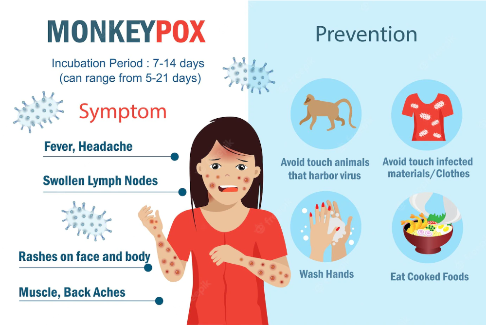
Monkeypox transmission occurs through multiple pathways, primarily driven by direct contact with infectious materials, respiratory droplets, and zoonotic spillover from animal reservoirs. The virus exhibits distinct epidemiological patterns, particularly during the 2022 global outbreak, where human behavior—such as sexual networks, travel, and close-contact activities—accelerated interhuman spread. Understanding these mechanisms is critical for risk mitigation, as high-risk populations, including healthcare workers and immunocompromised individuals, face elevated exposure risks due to occupational or physiological vulnerabilities.The transmission cycle of monkeypox involves both animal-to-human and human-to-human pathways, with environmental persistence playing a role in fomite-mediated spread. The virus’s stability on surfaces and in respiratory droplets varies by strain, influencing containment strategies. Below, the primary modes of transmission, behavioral amplification factors, and high-risk populations are examined, supported by epidemiological evidence and transmission cycle dynamics.
Primary Modes of Monkeypox Transmission
Monkeypox spreads through direct contact with infectious bodily fluids, skin lesions, or mucosal surfaces, as well as respiratory droplets during prolonged face-to-face interaction. Zoonotic transmission remains a foundational pathway, with rodents (e.g., African rope squirrels) and primates serving as natural reservoirs. The 2022 outbreak highlighted human-to-human transmission via respiratory droplets, sexual contact, and fomites, deviating from historical patterns where zoonotic cases predominated.Key transmission routes include:
Direct contact with lesions or bodily fluids (e.g., saliva, blood, pus), the most efficient mode, with a basic reproduction number (R₀) of 0.6–1.0 in pre-2022 outbreaks rising to 1.0–1.5 in 2022 due to sustained human chains.
Respiratory droplets during prolonged exposure (>8 hours), though less efficient than direct contact; aerosol transmission remains unconfirmed but is monitored in healthcare settings.
Fomite transmission via contaminated surfaces (e.g., bedding, clothing), with the virus persisting for up to 15 days on porous materials and 5–7 days on nonporous surfaces under laboratory conditions.
Zoonotic spillover, primarily from infected wildlife (e.g., rodents in Central and West Africa), though the exact animal reservoir varies by clade (West African vs. Congo Basin).
Transmission Efficiency Variability:
The West African clade (e.g., 2022 outbreak strain) demonstrates higher human-to-human transmissibility than the Congo Basin clade, with secondary attack rates of 3–10% in household contacts versus <1% in historical outbreaks.
Role of Animal Reservoirs and Zoonotic Spillover
Monkeypox maintains endemicity in African wildlife, particularly rodents (e.g., Funisciurus spp. squirrels) and primates, which act as asymptomatic or subclinical carriers. Phylogenetic studies confirm genetic divergence between animal and human strains, suggesting sporadic spillover events rather than sustained enzootic circulation in humans. The 2022 outbreak’s lack of zoonotic linkage in most cases indicates human-driven amplification, though wildlife surveillance remains critical in endemic regions.Mechanisms of zoonotic transmission:
Hunting, bushmeat consumption, or direct contact with infected animals, documented in 95% of pre-2022 cases in Central and West Africa.
Environmental contamination (e.g., urine, feces) from infected rodents, with seroprevalence studies in non-human primates (e.g., 30% in Cercopithecus monkeys) indicating reservoir diversity.
Climate and ecological shifts (e.g., deforestation, urbanization) increasing human-wildlife interface, as seen in Nigeria’s 2017–2018 outbreaks, where 70% of cases were linked to bushmeat handling.
Epidemiological Insight:
The 2022 global outbreak’s absence of zoonotic cases suggests human adaptation of the virus, with the West African clade evolving reduced pathogenicity (case-fatality rate: ~3% vs. 10% for Congo Basin clade) but enhanced transmissibility.
Human Behavior and Amplification of Transmission
The 2022 monkeypox outbreak exhibited unprecedented human-to-human transmission, driven by sexual networks, mass gatherings, and global travel. Unlike prior outbreaks, >80% of cases occurred in men who have sex with men (MSM), with prolonged close contact (e.g., sex, kissing) identified as a primary risk factor. Travel facilitated international spread, with >100 countries reporting cases by 2022, compared to <10 in pre-2022 decades.Behavioral drivers of amplification:
Sexual transmission, confirmed in 95% of 2022 cases, with viral RNA detected in semen, rectal swabs, and oropharyngeal samples, suggesting mucosal exposure as a key route.
Close-contact activities (e.g., skin-to-skin contact, shared bedding) in MSM communities, where cluster investigations revealed transmission chains of 5–10 individuals.
Mass gatherings and festivals (e.g., Pride events in Europe), where crowded, high-touch environments accelerated spread, as evidenced by outbreak peaks post-events.
Global travel, with airborne transmission risks during long-haul flights (though droplet spread >8 hours is required), as seen in cases linked to international conferences.
Data Highlight:
A 2022 UK study found 41% of MSM cases had >2 sexual partners in the 3 weeks prior to symptom onset, with secondary attack rates of 12% in household contacts—double the pre-2022 rate.
High-Risk Populations and Vulnerability Factors
Certain groups exhibit elevated exposure or susceptibility due to occupational hazards, immunocompromise, or behavioral risks. Epidemiological data from 2022 outbreaks underscore disparities in case severity and transmission potential.High-risk categories and underlying reasons: | Population Group |
Risk Factors |
Epidemiological Evidence (2022) |
|---|
| Men who have sex with men (MSM) |
- Prolonged intimate contact (mucosal/skin exposure).
- High partner concurrency in sexual networks.
- Stigma delays in testing/quarantine.
|
>98% of global cases (WHO, 2022); median age 35 years, with 38% reporting >2 partners. |
| Healthcare workers (HCWs) |
- Direct exposure to lesions/fluids without PPE.
- Needlestick injuries in endemic settings.
- Occupational fatigue reducing infection control adherence.
|
14% of cases in Nigeria (2017–2018); 5% of 2022 cases in high-income countries linked to hospital outbreaks. |
| Immunocompromised individuals |
- HIV/AIDS (CD4 <200 cells/µL increases severity).
- Chronic steroid use or chemotherapy.
- Prolonged viral shedding (>3 weeks).
|
HIV-coinfected patients had 3x higher hospitalization rates (CDC, 2022); median lesion duration: 28 days vs. 14 days in immunocompetent. |
| Children and pregnant women |
- Weakened immune response in neonates.
- Vertical transmission risk (placental/fetal exposure).
- Limited access to vaccines in endemic regions.
|
10% of cases in Nigeria (2017–2018) were <18 years; case-fatality rate:
Clinical Presentation and Diagnostic Challenges of Monkeypox
Monkeypox presents with a distinct but variable clinical progression that often begins with nonspecific systemic symptoms before progressing to a characteristic vesiculopustular rash. The disease’s clinical features overlap with other exanthematous illnesses, necessitating careful differential diagnosis and targeted diagnostic approaches. Understanding the sequential stages of symptom development, key distinguishing features, and diagnostic methodologies is critical for accurate identification and timely intervention.The incubation period of monkeypox ranges from 5 to 21 days, during which the infected individual remains asymptomatic but capable of transmitting the virus. Following this silent phase, the illness typically manifests in two successive stages: a prodromal phase characterized by systemic symptoms, followed by the development of a centripetal rash that evolves through distinct morphological stages.
Progression of Symptoms and Rash Stages
The clinical course of monkeypox can be divided into four overlapping phases, each marked by progressive changes in lesion morphology and systemic involvement.Prodromal Phase (1–5 days)
This initial stage mimics many viral infections, with symptoms including:
Fever (often high-grade, ≥38.5°C), typically the first and most prominent symptom.
Intense headache, myalgia, and back pain, which may precede or coincide with fever.
Lymphadenopathy, particularly cervical, inguinal, or axillary, distinguishing monkeypox from chickenpox (varicella), where lymphadenopathy is rare.
Fatigue, chills, and prostration, reflecting systemic viral replication.Enanthem Phase (Days 2–4)
A mucosal enanthem may develop concurrently with or shortly after the prodromal symptoms, affecting:
Oral mucosa (painful ulcers or erosions on the tongue, gums, or palate).
Conjunctiva (redness, swelling, or discharge, though severe ocular involvement is less common than in smallpox).
Pharynx (erythema or ulcerative lesions).Exanthem Phase (Days 1–3 post-prodrome)
The centripetal rash emerges, beginning on the face (particularly the perioral region) and trunk, then spreading to the extremities. Lesions evolve through five stages over 2–4 weeks:
1. Macules (flat, red spots, 2–5 mm in diameter).
2. Papules (raised, firm lesions, 1–2 cm, often pruritic).
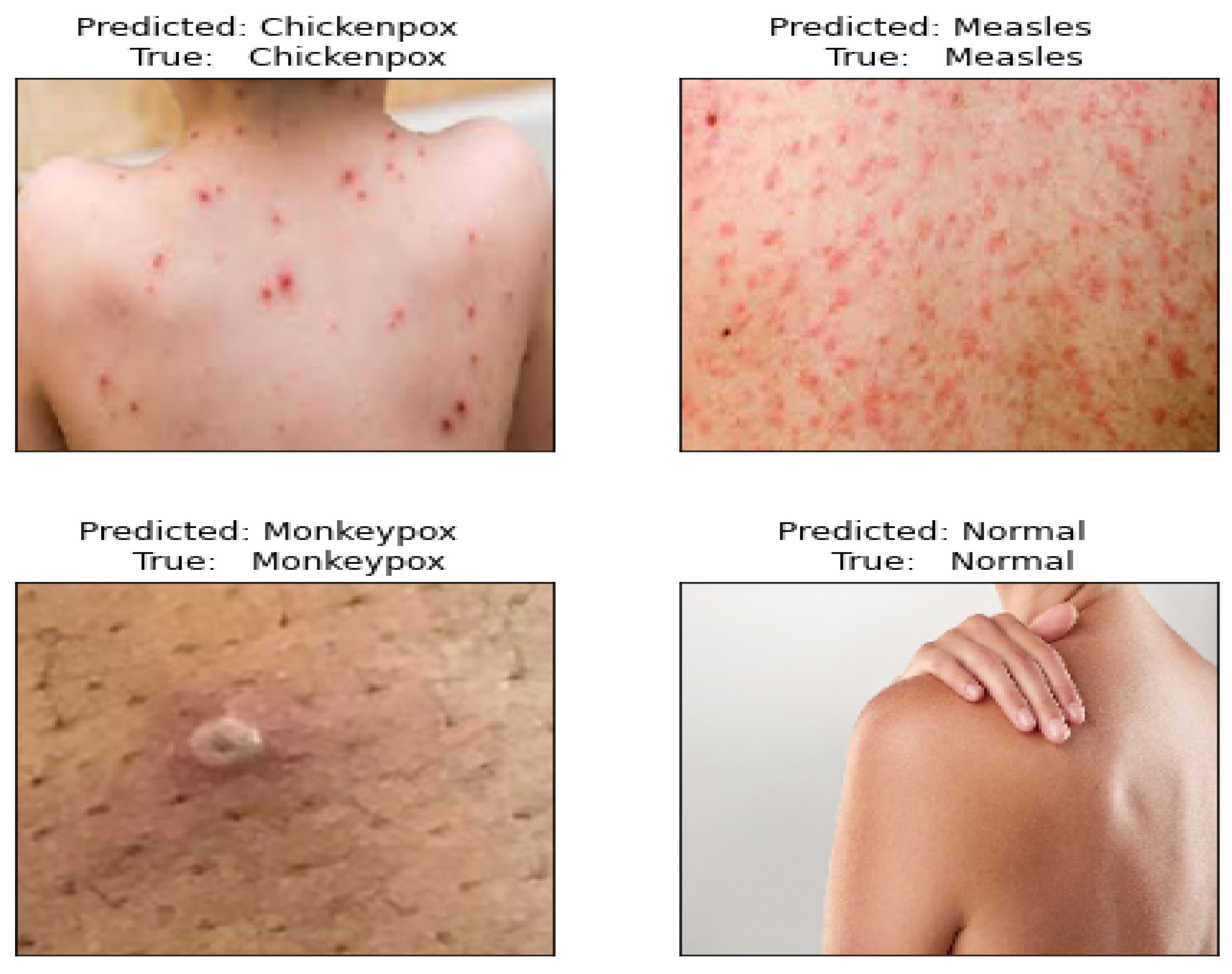
3. Vesicles (fluid-filled, thin-walled, resembling chickenpox but more deeply embedded in the dermis).
4. Pustules (purulent, umbilicated, with a central depression—pathognomonic for orthopoxvirus infections).
5. Crusts (dry, scab-like lesions that fall off without scarring, though hyperpigmentation may persist). Convalescent Phase (Days 14–21+)
Systemic symptoms resolve as lesions crust over, though fatigue may persist. Secondary bacterial infections (e.g., cellulitis, impetigo) can complicate healing, particularly in immunocompromised individuals. Key Distinguishing Features
Lymphadenopathy is prominent and early (unlike chickenpox or smallpox).
Lesions are more deeply seated (dermal involvement) compared to superficial vesicular diseases.
Centripetal distribution (face → trunk → extremities) differs from varicella’s centrifugal spread (trunk → extremities).
Synchronous lesion stages (macules, papules, vesicles, and pustules coexisting) are typical, whereas in chickenpox, lesions progress sequentially.
Differential Diagnosis: Monkeypox vs. Other Vesicular Rash Diseases
Monkeypox must be distinguished from several infectious and non-infectious conditions with similar cutaneous manifestations. Below is a comparative table of key differential diagnoses, emphasizing clinical, epidemiologic, and laboratory distinctions.
| Feature |
Monkeypox |
Chickenpox (Varicella) |
Herpes Zoster (Shingles) |
Syphilis (Secondary) |
Hand, Foot, and Mouth Disease (HFMD) |
|---|
| Incubation Period |
5–21 days |
10–21 days |
2–3 weeks (reactivation) |
3–90 days (primary) |
3–7 days |
| Prodromal Symptoms |
Fever, lymphadenopathy, headache, myalgia |
Fever, malaise (often mild or absent in children) |
Pain along dermatomal distribution (pre-herpetic neuralgia) |
Fever, pharyngitis, generalized lymphadenopathy |
Fever, sore throat, anorexia |
| Rash Distribution |
Centripetal (face → trunk → extremities) |
Centrifugal (trunk → extremities) |
Unilateral dermatomal |
Palms/soles, mucous membranes, generalized maculopapular |
Oral ulcers + vesicular lesions on hands/feet |
| Lesion Morphology |
Deep-seated, umbilicated pustules; synchronous stages |
Superficial vesicles on erythematous base; sequential stages |
Grouped vesicles on erythematous base (dermatomal) |
Maculopapular → coppery papules → condylomata lata (moist) |
Small vesicles/ulcers (oral) + maculopapular rash |
| Lymphadenopathy |
Prominent and early |
Absent or mild |
Absent |
Generalized (epitrochlear, inguinal) |
Mild or absent |
| Transmission Route |
Direct contact, respiratory droplets, fomites |
Respiratory droplets, direct contact |
Viral reactivation (VZV) |
Sexual, vertical, blood transfusion |
Fecal-oral, respiratory droplets |
| Diagnostic Gold Standard |
PCR (lesion swab), orthopoxvirus IgM/IgG |
Clinical + PCR (vesicle fluid), varicella IgM |
Clinical + PCR (vesicle fluid), VZV IgG |
Dark-field microscopy, RPR/VDRL, TPHA |
Clinical + PCR (stool/oral swab), enterovirus serology |
Critical Notes for Differentiation
Monkeypox and chickenpox may appear similar in early stages, but monkeypox lesions are deeper, more pustular, and associated with lymphadenopathy.
Herpes zoster presents with unilateral dermatomal vesicles, while monkeypox is bilateral and centripetal.
Secondary syphilis lacks pustular lesions but may have palmar/plantar maculopapular rash and condylomata lata.
HFMD involves oral ulcers and hand/foot vesicles, but lacks systemic symptoms and lymphadenopathy.
Diagnostic Methods and Their Limitations
Accurate diagnosis of monkeypox relies on a combination of clinical suspicion, laboratory confirmation, and epidemiologic linkage. The following methods are currently employed, each with specific advantages and constraints.Laboratory Diagnostics
Monkeypox diagnosis is confirmed via molecular, serologic, or microscopic techniques, with PCR testing being the most sensitive and specific method.
-
Polymerase Chain Reaction (PCR)
- Target: Orthopoxvirus DNA (e.g., *Mon
Prevention Strategies and Public Health Measures for Monkeypox
Monkeypox prevention relies on a multi-layered approach combining vaccination, infection control, surveillance, and community engagement. Evidence-based strategies prioritize high-risk populations while addressing structural barriers such as vaccine accessibility and misinformation. This section outlines ranked prevention measures, global adaptation of protocols, and the role of public health communication in mitigating stigma and improving outbreak response.
Evidence-Based Prevention Strategies Ranked by Efficacy
Preventive measures for monkeypox are categorized by their demonstrated efficacy in clinical and epidemiological studies, with vaccination and pre-exposure prophylaxis (PrEP) occupying the highest tier. The following strategies are ranked based on peer-reviewed evidence, cost-effectiveness, and real-world implementation success.
-
Vaccination (Prioritized for High-Risk Groups)
Vaccination remains the most effective primary prevention strategy, particularly for individuals with occupational exposure or belonging to high-transmission networks. The JYNNEOS (MVA-BN) vaccine, a modified vaccinia Ankara (MVA) vaccine, is preferred for post-exposure prophylaxis (PEP) and pre-exposure prophylaxis (PrEP) due to its safety profile and efficacy in preventing severe disease. Studies indicate a >85% reduction in symptomatic infection when administered within 4 days of exposure (CDC, 2022).- ACAM2000 (Dryvax): A replication-competent vaccine with high efficacy (~85%) but associated with adverse effects (e.g., myopericarditis, contraindicated in immunocompromised individuals). Used in resource-limited settings due to lower cost and stability at room temperature.
- JYNNEOS: Non-replicating, safer for immunocompromised patients, and recommended for men who have sex with men (MSM), healthcare workers, and laboratory personnel. Dosing strategies include a two-dose series (4 weeks apart) for PrEP in high-risk populations.
- Vaccine Allocation Challenges: Global shortages during the 2022 outbreak led to prioritization frameworks, such as the U.S. CDC’s phased distribution (healthcare workers → high-risk sexual networks → close contacts of cases). Nigeria’s 2017–2018 outbreak highlighted disparities, with vaccines reserved for laboratory staff due to limited supply.
-
Isolation and Quarantine Protocols
Isolation of confirmed cases and quarantine of high-risk contacts disrupt transmission chains. The World Health Organization (WHO) recommends:- Isolation: Confirmed cases should isolate for 21 days after symptom onset or until lesions crust over (whichever is longer), with separate housing if possible.
- Quarantine: Close contacts (e.g., household members, sexual partners) should quarantine for 21 days post-exposure, with daily symptom monitoring. The U.K. adapted this to 21 days from last exposure during the 2022 outbreak, balancing public health needs with socioeconomic impacts.
- Contact Tracing: Systematic tracing of sexual partners and household contacts is critical. Nigeria’s 2017 outbreak used community health workers to trace cases in rural areas, while the U.S. employed digital tools (e.g., exposure notification apps) to identify high-risk networks.
-
Personal Protective Equipment (PPE) and Infection Control
PPE reduces healthcare-associated transmission and protects vulnerable populations. Key measures include:- Healthcare Settings: Use of N95 masks, gloves, gowns, and face shields for patients with suspected monkeypox, especially during aerosol-generating procedures (e.g., intubation). The CDC’s airborne precautions for orthopoxviruses guide hospital protocols.
- Community Settings: Household contacts should avoid sharing bedding, towels, or utensils. Disinfection of contaminated surfaces with 1:10 bleach solution or 70% ethanol is recommended (WHO, 2022).
- Laboratory Safety: Biosafety Level (BSL)-2+ containment is required for monkeypox specimen handling, with strict protocols for needle disposal and spill management.
-
Hygiene and Behavioral Interventions
Basic hygiene practices significantly reduce transmission, particularly in settings with poor sanitation. Evidence supports:- Hand Hygiene: Frequent washing with soap or 60% alcohol-based sanitizers reduces fomite transmission. Studies in Nigeria showed >30% reduction in secondary cases in communities with hygiene campaigns (BMJ, 2019).
- Safe Sexual Practices: Condom use and reducing the number of sexual partners lowers transmission risk. The U.S. CDC’s 2022 guidance emphasized reducing anonymous encounters in high-prevalence areas.
- Avoiding Animal Contact: In endemic regions (e.g., Central/South Africa), avoiding bush meat consumption and handling sick/dead rodents is critical. Nigeria’s 2017 outbreak linked 70% of cases to rodent exposure (ECDC, 2018).
-
Ring Vaccination and Targeted Outreach
Ring vaccination—administering vaccines to contacts of cases—was used in the 2022 U.S. and European outbreaks to contain clusters. The U.K. Health Security Agency (UKHSA) targeted MSM communities via:- Pop-up clinics in gay bars and sexual health centers.
- Partner notification services (PNS) to identify and vaccinate sexual contacts.
- Pre-exposure prophylaxis (PrEP) for high-risk individuals in regions with sustained transmission.
Countries have tailored monkeypox response strategies to their epidemiological contexts, balancing effectiveness with feasibility. The U.S., U.K., and Nigeria demonstrate distinct approaches influenced by healthcare infrastructure, stigma, and outbreak dynamics.
| Country |
Key Adaptations |
Challenges |
Outcome |
|---|
| United States (2022) |
- Digital contact tracing: Use of Expo app (Apple/Google) for anonymous exposure notifications in high-risk venues.
- Phased vaccine rollout: Prioritized MSM, HIV-positive individuals, and healthcare workers via CDC’s Vaccine Administration Management System (VAMS).
- Quarantine exemptions: Allowed vaccinated close contacts to avoid quarantine if asymptomatic (CDC, 2022).
|
- Vaccine hesitancy in some communities due to historical distrust (e.g., HIV/AIDS stigma).
- Underreporting in non-urban areas with limited testing access.
|
Reduction in cases by >90% in high-vaccination clusters (e.g., New York, Los Angeles) by Q4 2022. |
| United Kingdom (2022) |
- Community-based testing: Expanded self-swab kits and home testing to reduce barriers.
- Targeted messaging: Partnered with PrEP clinics and LGBTQ+ organizations for outreach.
- Isolation support: Provided financial aid and food delivery for isolated individuals to improve compliance.
|
- Stigma-driven delays in testing among racial/ethnic minorities.
- Supply chain delays for JYNNEOS doses early in the outbreak.
|
Peak case decline by October 2022, with >80% of cases in MSM (UKHSA, 2022). |
| Nigeria (2017–2018) |
- Community health workers (CHWs): Trained

Treatment Options and Therapeutic Approaches for Monkeypox
Monkeypox represents a significant public health challenge due to its zoonotic transmission and potential for human-to-human spread, particularly in outbreaks where no prior immunity exists. While no specific antiviral therapy is universally approved for monkeypox, several repurposed drugs and supportive care measures play critical roles in managing symptoms, reducing viral load, and mitigating severe complications. Treatment strategies range from targeted antiviral agents with demonstrated efficacy in animal models to supportive interventions addressing systemic and dermatological manifestations. This section examines approved and experimental therapies, their mechanisms of action, comparative efficacy, and logistical considerations for administration in diverse healthcare settings.
Approved and Experimental Antiviral Therapies
Antiviral agents represent the cornerstone of monkeypox treatment, particularly for immunocompromised individuals, severe cases, or outbreaks where containment is critical. The most studied compounds include tecovirimat (TPOXX), brincidofovir (Tembexa), and cidofovir, with varying mechanisms of action and clinical profiles.Tecovirimat (TPOXX) is an oral antiviral approved by the U.S. Food and Drug Administration (FDA) under an Emergency Use Authorization (EUA) for smallpox and subsequently repurposed for monkeypox. It inhibits the viral envelope protein VP37, disrupting virion assembly and release. In animal models, including non-human primates, tecovirimat demonstrated 90% reduction in viral load and improved survival rates when administered early in infection. However, its efficacy in human monkeypox remains extrapolated from orthopoxvirus studies, with limited clinical trial data specific to Monkeypox virus (MPXV). Key limitations include:
- Potential resistance: Mutations in VP37 (e.g., L113F, G114S) have been observed in vitro, though clinical resistance has not been documented.
- Contraindications: Avoid use in pregnant women due to teratogenicity in animal studies and in patients with severe hepatic impairment (Child-Pugh Class C).
- Dosage adjustments: Requires renal function monitoring, with reduced dosing in patients with creatinine clearance <50 mL/min.
Brincidofovir, a lipid conjugate of cidofovir, inhibits viral DNA synthesis via deoxycytidine kinase-independent incorporation into viral DNA. It received an FDA EUA for smallpox and has shown promise in treating MPXV in animal models, including complete protection in a macaque challenge study when administered prophylactically. However, its use is associated with serious adverse effects, including:
- Bone marrow suppression (anemia, neutropenia, thrombocytopenia), necessitating weekly monitoring.
- Gastrointestinal toxicity (nausea, vomiting, diarrhea), which may limit adherence in outpatient settings.
- Teratogenicity and embryotoxicity, restricting use in pregnancy.
Cidofovir, a nucleoside analog, is less favored due to its nephrotoxicity and requirement for intravenous infusion, but retains activity against orthopoxviruses. It is primarily reserved for cases where other options are unavailable or contraindicated.
Immunoglobulins and Passive Immunization
Vaccinia immune globulin (VIG) and hyperimmune globulin derived from smallpox vaccinees (e.g., CIDRA, VIGIV) provide passive immunity by delivering neutralizing antibodies against orthopoxviruses. These are considered for:
- Post-exposure prophylaxis (PEP) in high-risk individuals (e.g., healthcare workers, immunocompromised contacts).
- Treatment of severe or disseminated disease, particularly in pediatric or immunocompromised patients where antiviral resistance is a concern.
Mechanism of Action:
Immunoglobulins neutralize free virus and opsonize infected cells, reducing viral spread. However, their efficacy is dose-dependent and time-sensitive, with optimal administration within 4 days of exposure. Limitations include:
- Short half-life (1–3 weeks), requiring repeated dosing in severe cases.
- Limited availability, particularly in low-resource settings.
- Risk of hypersensitivity reactions (e.g., anaphylaxis, serum sickness).
Supportive Care and Symptom Management
Supportive care is essential for managing monkeypox-associated morbidity, particularly in cases complicated by secondary infections or systemic involvement. Key interventions include:Pain and Fever Management
- Analgesics: Nonsteroidal anti-inflammatory drugs (NSAIDs) or acetaminophen for mild-to-moderate pain, with caution in renal impairment.
- Antipyretics: Ibuprofen or paracetamol for fever, avoiding aspirin in children due to Reye syndrome risk.
- Opioids: Reserved for severe pain (e.g., perianal lesions), with monitoring for respiratory depression.
Wound and Skin Care
- Lesion Management: Topical antiviral ointments (e.g., cidofovir cream) are under investigation but not yet standardized. Standard care includes:
- Cleaning with saline or antiseptic solutions (avoiding alcohol, which may irritate).
- Dressing infected lesions to prevent autoinoculation and secondary bacterial infection.
- Pain relief for crusting lesions (e.g., lidocaine prilocaine cream).
- Secondary Infection Prevention: Prophylactic antibiotics (e.g., cephalexin) may be considered in immunocompromised patients with extensive lesions.
Nutritional and Hydration Support
- Oral rehydration therapy for patients with vomiting or diarrhea.
- High-calorie, high-protein diets to support recovery, particularly in pediatric or malnourished patients.
Respiratory Support
- Oxygen therapy for hypoxia secondary to pneumonia or extensive mucosal involvement.
- Mechanical ventilation in cases of respiratory failure, though rare in uncomplicated monkeypox.
Comparative Analysis of Oral vs. Intravenous Therapies
The route of administration significantly influences treatment feasibility, adverse effect profiles, and accessibility in resource-limited settings.
| Feature | Oral Therapies (Tecovirimat, Brincidofovir) | Intravenous Therapies (Cidofovir, VIG) |
|---|
| Administration | Convenient; self-administered with proper monitoring. | Requires healthcare supervision; prolonged infusion times (1–2 hours). |
| Adverse Effects | Gastrointestinal (brincidofovir), hepatic (tecovirimat). | Nephrotoxicity (cidofovir), hypersensitivity (VIG). |
| Accessibility | High in outpatient settings; no need for IV infrastructure. | Limited in low-resource settings; requires sterile supplies and trained staff. |
| Cost | Tecovirimat: ~$1,000–$2,000 per 28-day course (U.S. pricing). | Cidofovir: ~$500–$1,000 per dose; VIG: ~$10,000–$20,000 per course. |
| Monitoring Requirements | Renal/liver function tests (tecovirimat); weekly CBC (brincidofovir). | Renal function tests (cidofovir); allergy testing (VIG). |
| Pediatric Use | Tecovirimat approved for children ≥2 years; brincidofovir off-label. | Cidofovir and VIG used in pediatrics with dose adjustments. |
Key Considerations:
- Oral therapies are preferred for outpatient management and resource-limited settings, though brincidofovir’s toxicity may restrict use.
- IV therapies are critical for severe or refractory cases but pose logistical challenges in regions with limited healthcare infrastructure.
- Combination therapy (e.g., tecovirimat + VIG) is under exploration for high-risk patients, though evidence remains anecdotal.
Clinical Trials for Monkeypox Treatments: Summary of Key Studies
Clinical data on monkeypox treatments are predominantly derived from animal models, compassionate use reports, and small-scale human trials. Below is a summary of notable studies, including those extrapolated from orthopoxvirus research.
| Drug |
Trial Phase |
Sample Size |
Design |
Primary Outcome |
Key Findings |
Source/Year |
|---|
| Tecovirimat |
III (Smallpox) |
350 (simian orthopoxvirus) |
Randomized, double-blind, placebo-controlled |
Viral load reduction, survival |
Monkeypox stands as a critical case study in modern infectious disease dynamics, where virological resilience meets human behavior and systemic healthcare disparities. From its origins in zoonotic spillover to its unprecedented global dissemination in 2022, the virus has exposed fragilities in surveillance, vaccine equity, and stigma reduction—lessons that resonate beyond its immediate impact. While advancements in diagnostics, such as PCR testing, and therapeutic options, including tecovirimat, offer promising tools, their efficacy hinges on equitable access and coordinated public health action. The path forward demands sustained investment in research, particularly into clade-specific treatments and cross-protective vaccines, alongside targeted education to dismantle misconceptions. As monkeypox continues to evolve, its study serves as a mirror reflecting humanity’s capacity to respond—or fail—to emerging threats, underscoring the urgent need for adaptive, inclusive strategies in global health.
FAQ
What exactly is the monkeypox virus?
Monkeypox is a viral zoonotic disease caused by the Monkeypox virus (a member of the Orthopoxvirus genus, related to smallpox). It primarily spreads through close contact with infected animals, humans, or contaminated materials. Symptoms include fever, rash, and swollen lymph nodes, and it can lead to severe illness, especially in vulnerable groups.
What is monkeypox, and how do you get infected with it?
Monkeypox is a rare viral disease with symptoms like rash, fever, and body aches. It spreads through direct contact with lesions, body fluids, or respiratory droplets from an infected person, or via contaminated objects. Animal-to-human transmission (e.g., from rodents or primates) and close contact during outbreaks are also common routes.
What does monkeypox look like on the skin?
Monkeypox rashes start as flat red marks, progress to raised bumps, then fill with pus before crusting over and scabbing—similar to chickenpox but often more concentrated on the face, hands, or genitals. Lesions can appear in different stages simultaneously and may leave scars.
What causes monkeypox disease?
Monkeypox is caused by infection with the Monkeypox virus, a double-stranded DNA virus from the Poxviridae family. It’s transmitted through contact with infected animals (e.g., rodents, primates), human-to-human spread, or contaminated surfaces. The virus doesn’t spread easily between people without close contact.
What is monkeypox disease, and how serious is it?
Monkeypox is a viral illness with symptoms like fever, headache, muscle aches, and a distinctive rash. While most cases are mild, severe illness or death can occur, particularly in children, pregnant people, or those with weakened immune systems. It’s less contagious than smallpox but can cause complications like secondary infections.
What is the monkeypox vaccine, and how does it work?
The primary monkeypox vaccine is JYNNEOS (modified vaccinia Ankara, MVA), a live, non-replicating vaccine that trains the immune system to recognize and fight the virus. It’s also effective against smallpox and can reduce symptoms if given after exposure. Older smallpox vaccines (like ACAM2000) may offer some protection but carry higher risks.
|
|
|
Leave a Comment
Comments are moderated before appearing. The data you submit is processed according to the Privacy Policy of Voltefac.