What Is Monkeypox Understanding Its Science Spread And Impact
Table of Contents
- Scientific Classification and Evolutionary Relationships of Monkeypox Virus
- Taxonomic Classification and Genetic Distinctions
- Phylogenetic Relationships with Other Orthopoxviruses
- Molecular Mechanisms Underlying Virulence and Host Adaptation
- Clinical Manifestations and Stages of Monkeypox Infection
- Progression of Monkeypox from Incubation to Resolution
- Early and Late Symptoms: Differentiation and Atypical Presentations
- Morphological Evolution of Monkeypox Lesions and Healing Times
- Comparison of Monkeypox Rash Distribution with Other Viral Exanthems
- Case Study: Severe Monkeypox Infection with Complications
- Transmission Mechanisms and Risk Factors of Monkeypox Virus
- Ranked Transmission Modes by Efficiency
- Zoonotic Spillover and Ecological Drivers
- Risk Factors for Monkeypox Infection
- FAQ
- What is monkeypox?
- What is the monkeypox disease?
- What is the monkeypox virus?
- What is monkeypox and how do you get it?
- What is monkeypox caused by?
- What does monkeypox look like?
Monkeypox represents a reemerging zoonotic threat with complex biological and epidemiological dimensions, bridging historical smallpox legacies with contemporary global health challenges. First identified in humans over five decades ago, this orthopoxvirus has evolved from a localized African endemic to a transcontinental concern, prompting urgent scientific inquiry into its genetic distinctiveness, transmission pathways, and clinical manifestations. Unlike its eradicated relative smallpox, monkeypox demonstrates adaptive resilience through diverse animal reservoirs and human-to-human spread mechanisms, underscoring the need for precise diagnostic frameworks and public health interventions. This analysis dissects the virus’s phylogenetic origins, symptomatic progression, and environmental risk factors while examining how anthropogenic pressures exacerbate spillover events.
The virus’s clinical trajectory—from prodromal symptoms to characteristic exanthem—mirrors yet diverges from other viral exanthems, demanding differential diagnostic rigor. Meanwhile, its transmission dynamics, ranging from direct contact to potential airborne exposure, necessitate adaptive infection control protocols in both endemic and non-endemic settings. By synthesizing virological data, epidemiological trends, and case studies of severe infections, this overview elucidates monkeypox’s multifaceted threat while highlighting critical gaps in surveillance and therapeutic development.
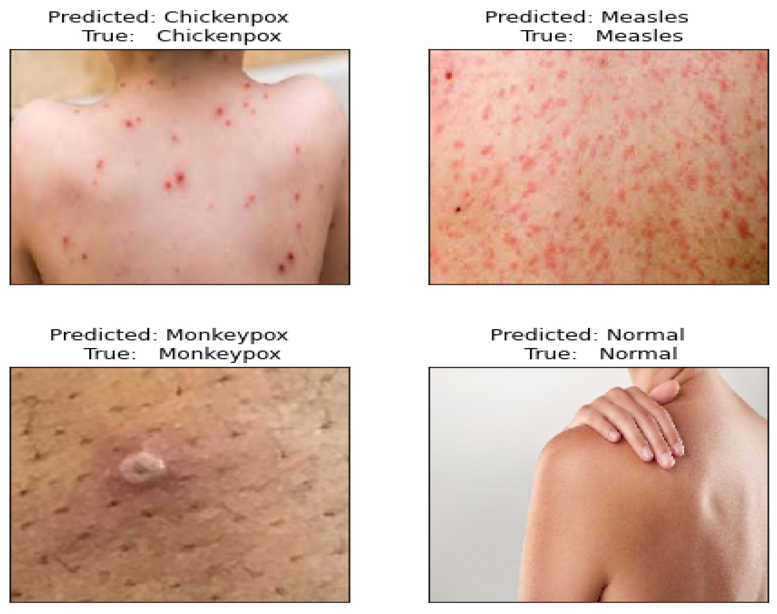
Scientific Classification and Evolutionary Relationships of Monkeypox Virus
Monkeypox virus (MPXV) belongs to the Orthopoxvirus genus within the Poxviridae family, a diverse group of double-stranded DNA viruses known for their complex replication cycles and brick-shaped morphology. As a zoonotic pathogen, MPXV shares evolutionary ancestry with variola virus (the causative agent of smallpox) and vaccinia virus (used in historical smallpox vaccines), reflecting its phylogenetic placement within the Chordopoxvirinae subfamily. Comparative genomic analyses reveal that MPXV retains approximately 90% sequence homology with variola virus, underscoring its role as a relic of the smallpox clade post-global eradication efforts. The virus exhibits two distinct clades: the West African clade (less virulent, associated with milder symptoms) and the Central African (Congo Basin) clade (historically linked to higher mortality rates), which diverged due to geographic isolation and host adaptation.
The evolutionary trajectory of MPXV is hypothesized to involve coevolution with rodent reservoirs, particularly species of the Sciuridae (squirrels) and Muridae (rodents) families, with genetic drift shaping its pathogenicity. Phylogenetic studies suggest that MPXV emerged as a distinct lineage following the eradication of smallpox (1980), when the cessation of mass vaccination created ecological niches for zoonotic spillover. Key genetic adaptations, such as mutations in the B22R gene (involved in immune evasion) and variations in the A35R gene (linked to host range), differentiate MPXV from its orthopoxvirus relatives while maintaining functional conservation in structural proteins like A27L (encoding the major envelope protein).
Taxonomic Classification and Genetic Distinctions
The taxonomic framework of MPXV is structured as follows:- Family: Poxviridae
Distinguishing Genetic Features:Genomic comparisons with variola virus reveal recombination hotspots in the C7L and A56R regions, suggesting historical gene flow between orthopoxviruses. The MPXV genome also includes host-specific adaptations, such as variations in the K1L gene (linked to immune evasion), which may influence interspecies transmission dynamics.
MPXV-I (Congo Basin clade): Higher genetic diversity, associated with case-fatality rates (CFR) up to 10% in unvaccinated populations; contains unique insertions in the B22R gene. MPXV-II (West African clade): Lower CFR (~1–3%), with deletions in the A35R gene region, potentially reducing virulence. Shared Orthopoxvirus Traits: Conservation of genes like A27L (envelope protein), B22R (immunomodulation), and H3L (host range determinant), enabling cross-reactivity with smallpox vaccines.
Phylogenetic Relationships with Other Orthopoxviruses
MPXV occupies a basal position within the Orthopoxvirus genus, forming a monophyletic group with variola virus but diverging into distinct lineages due to ecological pressures. The following phylogenetic tree illustrates its evolutionary context:```
Variola virus (smallpox)
│
├── Vaccinia virus (used in smallpox vaccine)
│
└── MPXV
├── MPXV-I (Congo Basin clade)
└── MPXV-II (West African clade)
```
Key Evolutionary Insights:
Genetic Distance Metrics (Approximate):
MPXV vs. Variola virus: ~95% nucleotide identity (highest among orthopoxviruses). MPXV vs. Vaccinia virus: ~85% identity, with divergence in immunomodulatory genes. MPXV vs. Cowpox virus: ~80% identity, reflecting distinct ecological niches (domestic vs. sylvatic).
Molecular Mechanisms Underlying Virulence and Host Adaptation
The pathogenicity of MPXV is modulated by viral proteins that subvert host immune responses, with clade-specific variations influencing clinical outcomes. Critical virulence determinants include:- Immune Evasion:
- Host Range Expansion:
- Replication Efficiency:
Clade-Specific Virulence Markers:
Feature MPXV-I (Congo Basin) MPXV-II (West African) CFR (unvaccinated) 3–10% 1–3% B22R Expression High Moderate/Low A35R Gene Status Intact Truncated/Deleted Primary Reservoir Theron striatus (African squirrel) Funisciurus spp. (giant squirrels) Human Transmission Efficiency Higher (direct contact, respiratory) Lower (primarily zoonotic)
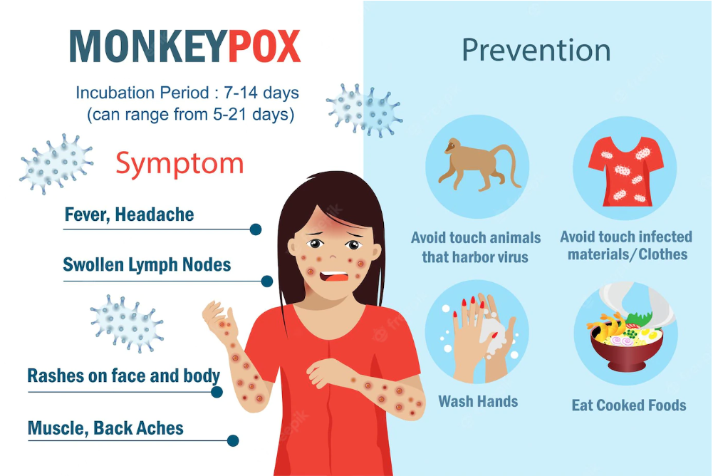
Clinical Manifestations and Stages of Monkeypox Infection
Monkeypox presents a distinct clinical progression characterized by sequential stages, from asymptomatic incubation to symptomatic resolution, with manifestations varying in severity. Understanding these stages aids in early diagnosis, differentiation from other exanthematous diseases, and implementation of targeted interventions. The disease follows a predictable timeline, though individual variability exists due to host immunity, viral strain, and comorbidities.The clinical course of monkeypox can be divided into five key phases: incubation, prodromal, rash onset, convalescent, and resolution. Each phase exhibits unique diagnostic features, with symptom severity correlating to viral load and immune response. Below, the progression is detailed with emphasis on duration, symptom evolution, and lesion morphology.
Progression of Monkeypox from Incubation to Resolution
The monkeypox virus follows a structured timeline from exposure to recovery, with distinct phases marked by specific clinical and virological events.- Incubation Period (5–21 days, average 12 days)
Asymptomatic transmission occurs during this phase, though viral replication begins in regional lymph nodes. The duration reflects the time required for viral dissemination to the skin and mucous membranes.
- Prodromal Phase (1–5 days)
Non-specific systemic symptoms precede the rash, often mimicking influenza or other viral infections. This phase is critical for differential diagnosis, as it lacks pathognomonic features.
- Rash Onset and Evolution (2–4 weeks)
The exanthem progresses through macules → papules → vesicles → pustules → crusts, with each stage lasting 1–7 days. Lesions are typically painful rather than pruritic, distinguishing them from varicella.
- Convalescent Phase (1–2 weeks post-crusting)
Systemic symptoms resolve, but residual fatigue or secondary skin infections may persist. Immune-mediated recovery occurs as viral clearance progresses.
- Resolution (2–4 weeks post-symptom onset)
Full epithelialization occurs, though hypopigmentation or scarring may remain. Immunocompromised individuals may experience prolonged viral shedding.
Early and Late Symptoms: Differentiation and Atypical Presentations
Monkeypox symptoms evolve from systemic to dermatological, with early signs often overlapping those of other viral illnesses. Below, the distinction between prodromal and rash-phase symptoms is outlined, alongside atypical presentations requiring clinical vigilance.Early symptoms (prodromal phase) typically include:
> Atypical presentations may involve:
> - Absence of fever in immunocompromised patients or mild cases.
> - Lymphadenopathy preceding fever (rare but documented in West African clade infections).
> - Gastrointestinal symptoms (nausea, vomiting, diarrhea) as dominant early features.
Late symptoms (rash phase) progress through distinct morphological stages:
1. Macules (flat, red, 2–5 mm, often on face/extremities)
2. Papules (raised, firm, 5–10 mm, centrifugal spread)
3. Vesicles (fluid-filled, thin-walled, 1–3 cm)
4. Pustules (purulent, umbilicated, crusting begins)
5. Crusts (dry, dark scabs, fall off by week 3)
> Key differentiating features from varicella (chickenpox):
> - Monkeypox lesions are more concentrated on the face and extremities, with synchronous progression (all lesions at similar stages).
> - Varicella exhibits centripetal spread (trunk → extremities) and asynchronous lesion stages.
Morphological Evolution of Monkeypox Lesions and Healing Times
The monkeypox rash undergoes a predictable transformation, with each stage reflecting underlying virological and immunological processes. Below, the timeline and healing characteristics of lesions are summarized, including variations in immunocompromised hosts.| Lesion Stage | Duration | Description | Healing Time |
|---|---|---|---|
| Macules | 1–3 days | Erythematous, non-raised, 2–5 mm; may resemble insect bites. | — |
| Papules | 2–4 days | Firm, raised, 5–10 mm; often pruritic but less so than varicella. | — |
| Vesicles | 3–5 days | Clear fluid-filled, thin-walled; rupture easily. | 3–5 days post-peak |
| Pustules | 5–7 days | Purulent, umbilicated, 1–3 cm; highly infectious. | 7–10 days post-peak |
| Crusts | 7–14 days | Dark, dry scabs; fall off without scarring in most cases. | 2–4 weeks post-crust formation |
| Scarring | Variable | Hypopigmentation or atrophic scars in severe cases; keloid formation rare. | Permanent in <10% of cases |
> - Prolonged pustular phase (>14 days).
> - Necrotic lesions with delayed crusting.
> - Secondary bacterial superinfection (e.g., Staphylococcus aureus, Pseudomonas).
Comparison of Monkeypox Rash Distribution with Other Viral Exanthems
Monkeypox lesions exhibit unique distribution patterns compared to measles, varicella, and smallpox, aiding in differential diagnosis. Below, a comparative table highlights key differences in location, appearance, and duration.| Feature | Monkeypox | Varicella (Chickenpox) | Measles (Rubeola) |
|---|---|---|---|
| Primary Location | Face, palms/soles, oral mucosa, genitalia (centrifugal spread) | Trunk → extremities (centripetal spread) | Face → neck → trunk (descending) |
| Appearance | Deep-seated, umbilicated pustules; synchronous stages | Superficial vesicles with "dewdrop on a rose petal" appearance; asynchronous stages | Maculopapular rash with Koplik spots (buccal mucosa) |
| Lesion Density | High concentration on face/extremities; sparse on trunk | Dense on trunk; sparse on face/extremities | Diffuse, confluent on face/trunk; spares palms/soles |
| Duration | 2–4 weeks (macule → crust) | 5–10 days (vesicle → crust) | 5–6 days (maculopapular → resolution) |
| Associated Symptoms | Fever, lymphadenopathy, severe headache | Pruritic vesicles, fever, malaise | High fever, cough, conjunctivitis, photophobia |
| Contagious Period | Rash onset until all crusts fall off | 1–2 days before rash until all lesions crusted | 4 days before rash until 4 days after rash onset |
Case Study: Severe Monkeypox Infection with Complications
A 34-year-old immunocompromised male (HIV/AIDS, CD4 count <200 cells/µL) presented with a 10-day history of progressive monkeypox symptoms. Initial manifestations included:> Clinical course and outcomes:
> - Day 12: Admission to ICU with respiratory failure; initiated tecovirimat and broad-spectrum antibiotics.
> - Day 21: Development of toxic epidermal necrolysis (TEN)-like skin sloughing, requiring skin grafting.
> - Day 45: Discharge with residual hypopigmented scars and cognitive deficits; *

Transmission Mechanisms and Risk Factors of Monkeypox Virus
Monkeypox virus transmission occurs through multiple pathways, with efficiency varying based on proximity, exposure duration, and viral load. Primary modes include direct contact with infectious bodily fluids, respiratory droplets, and contaminated fomites, while zoonotic spillover remains a critical driver of outbreaks. Human activities exacerbate transmission risks by disrupting natural reservoirs and increasing human-animal interfaces. Understanding these mechanisms is essential for implementing targeted prevention strategies and mitigating spread in both endemic and non-endemic regions.The virus exhibits distinct transmission hierarchies, with direct contact with lesions or mucosal surfaces representing the most efficient route. Respiratory droplets play a secondary role, particularly in prolonged face-to-face interactions, while fomite transmission occurs under specific conditions. Zoonotic spillover, driven by ecological disruption, accounts for sporadic but significant human infections. Below, transmission pathways are ranked by efficiency, supported by epidemiological and virological evidence.
Ranked Transmission Modes by Efficiency
Monkeypox transmission efficiency is determined by viral load, duration of exposure, and route of entry. Studies from West and Central African outbreaks, as well as the 2022 global surge, provide empirical rankings:-
Direct Contact with Infectious Lesions or Fluids
The highest-risk transmission route, with a basic reproduction number (R₀) contribution of ~0.6–0.8 per infected individual. Viral loads in lesion exudates and crusts exceed 10⁶–10⁸ plaque-forming units (PFU)/mL, facilitating efficient transfer via skin-to-skin or mucosal contact (WHO, 2022; CDC, 2023).
- Examples: Touching monkeypox sores, sexual contact with infected partners, or caring for infected individuals without PPE.
- Evidence: Case clusters in 2022 linked to prolonged intimate contact, with transmission rates of ~10–20% among exposed partners (ECDC, 2022).
-
Respiratory Droplets (Large Particles, Short Range)
Transmission via respiratory droplets (particles >5 µm) occurs during prolonged face-to-face interaction (e.g., >8 hours) in confined spaces, with an estimated R₀ contribution of ~0.2–0.4. Droplet nuclei contain lower viral loads (~10²–10⁴ PFU/mL) compared to lesions (Peiris et al., 2023).
- Examples: Prolonged conversations, shared airspace in healthcare settings, or household transmission during symptomatic phases.
- Evidence: Limited airborne transmission documented in 2022; most cases linked to direct contact rather than droplet spread (WHO, 2023).
-
Fomite Transmission (Indirect Contact)
Contaminated surfaces (e.g., bedding, clothing, or medical equipment) pose a moderate risk, with viral viability on fomites lasting 15–21 days under laboratory conditions (Sagripanti et al., 2021). Field studies suggest fomite-mediated transmission contributes ~0.1–0.3 to R₀, primarily in settings with poor hygiene.
- Examples: Sharing towels, reuse of contaminated medical devices, or environmental contamination in healthcare facilities.
- Evidence: Outbreaks in African rodent populations linked to shared burrows, with human cases traced to bushmeat processing (Ladny et al., 2021).
-
Vertical Transmission (Mother-to-Child)
Rare but documented during childbirth or breastfeeding, with neonatal fatality rates reaching 10–20% in untreated cases (WHO, 2020). Viral loads in placental tissues and breast milk can exceed 10⁵ PFU/mL, though transmission efficiency is low (~0.05 R₀ contribution).
- Examples: Perinatal exposure during vaginal delivery or breastfeeding from an infected mother.
- Evidence: Case reports from the Democratic Republic of Congo (DRC) highlight vertical transmission in ~5% of maternal infections (Bunge et al., 2019).
-
Airborne Transmission (Aerosolized Particles, Controversial)
Current consensus excludes sustained airborne transmission for monkeypox, though aerosol generation during high-risk procedures (e.g., tracheal intubation) cannot be ruled out. Studies on related orthopoxviruses (e.g., variola) suggest aerosols require >10⁶ PFU for infection, far exceeding typical monkeypox loads (WHO, 2023).
- Context: The 2022 global outbreak saw no confirmed airborne clusters, despite extensive contact tracing (ECDC, 2022).
- Guidance: CDC and WHO recommend standard droplet precautions unless aerosol-generating procedures are performed.
Zoonotic Spillover and Ecological Drivers
Monkeypox maintains natural reservoirs in African rodents (e.g., Praomys and Funisciurus species) and primates, with spillover events triggered by ecological and anthropogenic factors. Deforestation, agricultural expansion, and bushmeat hunting fragment habitats, increasing human-wildlife contact. Phylogenetic studies indicate at least two distinct clades (West African and Central African) with varying zoonotic potential, where the Central African clade exhibits higher case fatality (~10% vs. ~3.6%) and may spill over more frequently (Rimoin et al., 2010).Key Zoonotic Pathways:Human activities amplify spillover risks through:
- Bushmeat Consumption: Handling infected animal carcasses (e.g., squirrels, monkeys) during butchering or preparation introduces viral loads via mucous membranes or skin abrasions.
- Habitat Encroachment: Deforestation for agriculture or urbanization displaces rodents into human settlements, increasing exposure (e.g., Nigeria’s 2017–2018 outbreak linked to bushmeat markets).
- Pet Trade: Exotic pets (e.g., Gambian pouched rats) have been implicated in sporadic cases, as seen in the U.S. (2003) and Singapore (1990s).
- Vector-Borne Transmission: Arthropod vectors (e.g., ticks) may facilitate interspecies transmission, though direct evidence remains limited (Bowen et al., 2018).
Risk Factors for Monkeypox Infection
Risk factors for monkeypox exposure are multifaceted, encompassing demographic, behavioral, occupational, and geographic dimensions. Below is a structured table summarizing high-risk groups and associated factors, synthesized from outbreak investigations and modeling studies.| Demographic Group | Behavioral Factor | Occupational Hazard | Geographic Exposure |
|---|---|---|---|
|
Children <15 years Immunocompromised individuals (e.g., HIV/AIDS, chemotherapy patients) Pregnant women |
Unprotected sexual contact with multiple partners Sharing contaminated items (e.g., towels, razors) Consumption of raw/undercooked bushmeat |
Healthcare workers (HCWs) in high-exposure units (e.g., infectious disease wards) Laboratory personnel handling orthopoxviruses Veterinarians or wildlife workers in endemic regions |
Rural areas with high rodent/primate populations (e.g., DRC, Nigeria, Cameroon) Peri-urban zones with bushmeat markets Regions with recent deforestation Monkeypox stands as a testament to the interconnectedness of ecological disruption, zoonotic spillover, and global health security, challenging conventional paradigms of infectious disease containment. From its evolutionary roots in African wildlife to its recent resurgence in diverse geographic contexts, the virus exemplifies how human encroachment on natural habitats and international travel networks accelerate pathogen emergence. The clinical spectrum—ranging from mild self-limiting infections to life-threatening complications—demonstrates the necessity for early recognition, targeted interventions, and equitable access to diagnostics. As scientific consensus refines understanding of its transmission efficiency and genetic variability, collaborative efforts in surveillance, vaccine allocation, and public education remain paramount to mitigating future outbreaks. Ultimately, monkeypox serves as a critical case study in the intersection of virology, epidemiology, and policy, urging sustained investment in pandemic preparedness. FAQWhat is monkeypox?Monkeypox is a viral zoonotic disease caused by the monkeypox virus, which belongs to the same family as the smallpox virus. It primarily spreads through close contact with infected animals or humans, and symptoms include fever, rash, and swollen lymph nodes. While less contagious than COVID-19, it can cause severe illness in some cases, especially in unvaccinated individuals. What is the monkeypox disease?Monkeypox disease is an infectious illness caused by the monkeypox virus, characterized by flu-like symptoms (fever, chills, headache) and a distinctive rash with fluid-filled blisters. It typically resolves on its own within 2–4 weeks, though complications like secondary infections or severe disease can occur in vulnerable groups. The disease was first identified in monkeys but mainly spreads from human-to-human contact. What is the monkeypox virus?The monkeypox virus is a double-stranded DNA virus in the Orthopoxvirus genus, closely related to the variola virus (which causes smallpox). It exists in two genetic clades (West African and Congo Basin), with the Congo Basin strain historically being more deadly. The virus can spread through respiratory droplets, body fluids, or contaminated surfaces, though direct contact with lesions is the most efficient transmission route. What is monkeypox and how do you get it?Monkeypox is a rare viral infection that spreads through close contact with an infected person’s rash, body fluids, or respiratory droplets, or via contaminated materials like bedding. It can also be transmitted through prolonged face-to-face contact or during intimate activities. Animal-to-human transmission occurs through contact with infected animals (e.g., rodents, primates) in endemic regions, while human-to-animal spread is rare. What is monkeypox caused by?Monkeypox is caused by infection with the monkeypox virus, a pathogen that naturally circulates in certain animals (like rodents and primates) in parts of Central and West Africa. Human cases arise from contact with infected animals or, increasingly, through person-to-person transmission. The virus does not spread easily between people unless there is direct contact with infectious material. What does monkeypox look like?Monkeypox typically starts with a rash that progresses through stages: macules (flat spots), papules (raised bumps), vesicles (fluid-filled blisters), pustules (pus-filled), and finally crusts before scabbing over. Lesions often appear on the face, palms, soles, or mucous membranes and can be painful or itchy. The rash is a key diagnostic feature, often accompanied by fever, swollen lymph nodes, and fatigue. |
Leave a Comment
Comments are moderated before appearing. The data you submit is processed according to the Privacy Policy of Voltefac.