What Is Shin Splints Understanding Pain Prevention And Recovery
Table of Contents
- Definition and Basic Overview of Shin Splints
- Anatomical Location and Key Structures Involved
- Biomechanical Development of Shin Splints: Step-by-Step Pathophysiology
- Differential Diagnosis: Shin Splints vs. Other Lower-Leg Injuries
- Symptoms and Diagnostic Indicators of Shin Splints
- Progression of Symptoms from Early-Stage Discomfort to Severe Pain
- Diagnostic Process and Exclusionary Criteria
- Red Flags Indicating Serious Injury Requiring Immediate Medical Attention
- Symptom Variation by Activity Level and Functional Limitations
- Causes and Contributing Factors of Shin Splints
- Biomechanical Causes and Repetitive Stress Mechanisms
- Extrinsic Factors: Training Errors and Environmental Influences
- Intrinsic Factors: Anatomical and Physiological Vulnerabilities
- Flowchart: Chain Reaction from Poor Training Habits to Shin Splint Development
- Sport-Specific Contributions to Shin Splints
- Prevention Strategies and Best Practices for Shin Splints
- Modifying Training Routines to Prevent Shin Splints
- Role of Footwear in Prevention
- Warm-Up and Cool-Down Exercises for Shin and Lower-Leg Protection
- Strength and Mobility Exercises to Fortify Lower-Leg Structures
- Treatment and Recovery Protocols for Shin Splints
- Phased Recovery Plan Including RICE and Activity Modification
- Physical Therapy Interventions for Accelerated Healing
- Pain Management: OTC Medications vs. Specialist Interventions
- Long-Term Management and Return-to-Sport Protocols
- FAQ
- What does the pain from shin splints feel like?
- What causes shin splints?
- What exactly are shin splints?
- What is a shin splints injury?
- What is shin splints medically?
- What are shin splints and how do they happen?
Shin splints, or medial tibial stress syndrome, represent a common yet often misunderstood condition affecting athletes and active individuals alike, characterized by persistent pain along the inner shinbone. Unlike stress fractures or compartment syndrome, this overuse injury stems from repetitive microtrauma to the tibia’s soft tissues, exacerbated by biomechanical inefficiencies, improper training loads, or suboptimal footwear. While frequently dismissed as mere discomfort, shin splints can significantly impair mobility and performance if left unaddressed, underscoring the need for precise diagnosis and proactive management.
The condition manifests through a spectrum of symptoms, from dull aches during physical activity to debilitating pain that disrupts daily routines, particularly in high-impact sports such as running, soccer, or ballet. Distinguishing shin splints from other lower-leg injuries requires a nuanced understanding of its anatomical origins, progression patterns, and risk modifiers. This overview explores the biomechanical triggers, diagnostic markers, and evidence-based strategies to prevent, treat, and recover from this pervasive yet manageable condition.
Definition and Basic Overview of Shin Splints
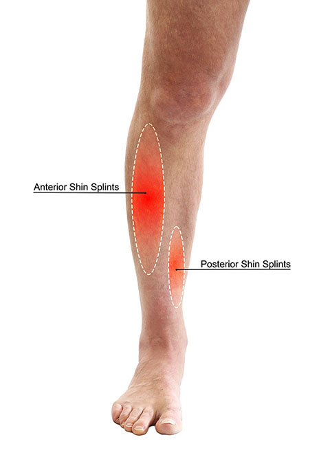
Medial Tibial Stress Syndrome (MTSS), commonly referred to as shin splints, is an overuse injury characterized by pain along the inner shinbone (tibia), typically affecting runners, dancers, and military recruits. Unlike stress fractures or compartment syndrome, shin splints involve inflammation of the connective tissues—including the tibialis posterior, soleus, and flexor muscles—rather than bone or nerve damage. The condition arises from repetitive stress on the lower leg, often exacerbated by poor biomechanics, inadequate footwear, or sudden increases in training intensity.
The full medical name, Medial Tibial Stress Syndrome (MTSS), distinguishes it from lateral shin pain (affecting the outer tibia) and highlights its primary location along the posterior-medial border of the tibia. Misdiagnosis frequently occurs due to overlapping symptoms with stress fractures or chronic exertional compartment syndrome (CECS), necessitating a differential analysis based on anatomical involvement and diagnostic imaging.
Anatomical Location and Key Structures Involved
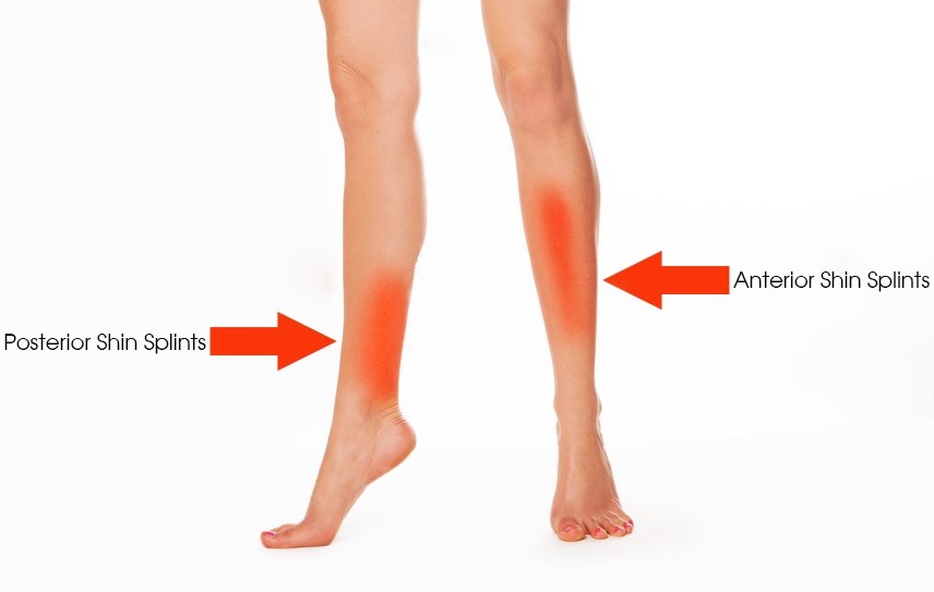
Shin splints primarily affect the posterior-medial tibia, where the tibialis posterior, soleus, and flexor digitorum longus muscles attach via their tendinous insertions. The interosseous membrane (connecting the tibia and fibula) and periosteum (fibrous tissue surrounding the bone) also play a role in pain transmission. Unlike lateral shin pain (often linked to the fibula or peroneal tendons), MTSS originates from soft-tissue irritation, not bone stress reactions.Key anatomical distinctions:
Biomechanical Development of Shin Splints: Step-by-Step Pathophysiology
The progression of shin splints follows a cumulative microtrauma model, where repetitive forces exceed tissue recovery capacity. Below is the sequential biomechanical breakdown:1. Increased Loading Forces
2. Muscle-Tendon Unit Fatigue
3. Connective Tissue Irritation
4. Chronic Adaptation Failure
Differential Diagnosis: Shin Splints vs. Other Lower-Leg Injuries
The following table compares shin splints (MTSS) with stress fractures, chronic exertional compartment syndrome (CECS), and tibial stress reactions to clarify diagnostic distinctions.| Feature | Shin Splints (MTSS) | Stress Fracture | CECS | Tibial Stress Reaction |
|---|---|---|---|---|
| Primary Location | Posterior-medial tibia (soft tissue) | Specific bone site (e.g., tibial shaft) | Anterior/lateral compartments (muscle/nerves) | Diffuse tibial bone (early stress fracture) |
| Pain Onset | Gradual, activity-related; resolves with rest | Sharp, localized; persists at rest (late stage) | Exercise-induced, resolves after 20–30 min | Dull ache during/after activity |
| Diagnostic Tools | Clinical exam; no imaging unless suspected fracture | X-ray (bone callus), MRI (edema) | Compartment pressure monitoring (>30 mmHg at rest) | MRI (bone marrow edema), bone scan |
| Risk Groups | Runners, dancers, military recruits; sudden training increases | Endurance athletes, osteopenic individuals | Athletes with tight calves, high VO₂ max demands | Novice athletes, females (higher risk due to Q-angle) |
| Key Symptom | Dull ache along medial tibia; tenderness to palpation of muscle attachments. |
Point tenderness over fracture site; night pain. |
Paresthesia (tingling) in foot/toes; swelling. |
Diffuse tibial pain; no specific trigger point. |
| Management Focus | Rest, ice, eccentric exercises, footwear correction | Immobilization, progressive loading, nutrition (calcium/vitamin D) | Fasciotomy (surgical release), activity modification | Relative rest, bone-loading modifications, bisphosphonates (severe cases) |
Symptoms and Diagnostic Indicators of Shin Splints
Shin splints, or medial tibial stress syndrome (MTSS), manifest through a spectrum of symptoms that evolve alongside the progression of the condition. Early-stage discomfort often goes unnoticed, particularly among individuals who engage in repetitive lower-leg activities such as running, jumping, or prolonged standing. As the condition advances, symptoms intensify, potentially leading to functional limitations and an increased risk of secondary injuries. Accurate diagnosis relies on a combination of clinical assessment, patient history, and exclusionary criteria to differentiate shin splints from other musculoskeletal pathologies. Healthcare professionals employ a structured diagnostic approach, incorporating physical examinations, imaging studies, and activity-specific symptom analysis to confirm the condition and guide treatment.The progression of shin splints symptoms is closely tied to biomechanical stress and tissue inflammation in the lower leg. Initial presentations typically include dull, aching pain along the inner (medial) edge of the shinbone (tibia), particularly during or after physical activity. Over time, symptoms may worsen, progressing to sharp, localized pain that persists even at rest, limits mobility, and disrupts daily activities. Athletes and active individuals often experience symptoms during high-impact exercises, while sedentary or less active individuals may notice discomfort during prolonged walking or standing. Understanding these patterns is critical for early intervention and preventing chronic complications.
Progression of Symptoms from Early-Stage Discomfort to Severe Pain
The symptomatic progression of shin splints follows a predictable trajectory, influenced by the duration and intensity of repetitive stress on the tibial periosteum, muscles, and connective tissues. Early-stage symptoms are typically activity-dependent, meaning pain emerges during exercise and subsides with rest. Common triggers include:As the condition advances, symptoms become more persistent and debilitating. The following stages outline the typical progression:
Early-Stage (Mild):
Dull ache or soreness along the inner shin, occurring only during physical activity (e.g., running, jumping). Pain resolves within minutes to hours post-exercise. No swelling or functional limitations observed.
Moderate-Stage (Intermediate):
Pain intensifies and may persist for hours after activity. Discomfort extends to low-impact activities (e.g., walking uphill or stair climbing). Possible mild swelling or tenderness upon palpation. Functional limitations begin to affect training or daily routines.
Late-Stage (Severe):Athletes often report symptoms that escalate with increased training load, while sedentary individuals may experience gradual onset due to sudden increases in activity (e.g., weekend warriors or new runners). The key distinction lies in the timing and triggers of pain, which healthcare professionals use to differentiate shin splints from other conditions like compartment syndrome or stress fractures.
Pain becomes constant, occurring both during and after activity, as well as at rest. Sharp, stabbing sensations may radiate along the shin. Swelling, bruising, or localized warmth may develop. Severe cases result in gait alterations (e.g., limping) or inability to bear weight. Risk of progression to stress fractures or chronic tendon pathology increases.
Diagnostic Process and Exclusionary Criteria
Diagnosing shin splints involves a multifactorial approach, combining patient history, physical examination, and diagnostic imaging to rule out more serious injuries. Healthcare professionals prioritize exclusionary criteria to ensure accurate identification, as shin splints share symptoms with conditions such as:The diagnostic process typically follows these steps:
1. Patient History and Symptom Analysis
2. Physical Examination
3. Imaging Studies
While shin splints are primarily a clinical diagnosis, imaging may be used to exclude other conditions:
Key Diagnostic Differentiators:Healthcare professionals rely on symptom patterns and exclusion of red flags to confirm MTSS. A delay in diagnosis can lead to chronic pain, muscle atrophy, or secondary injuries, underscoring the importance of early evaluation.
Shin splints: Diffuse pain along the medial tibia, worse with activity, no focal tenderness. Stress fracture: Sharp, localized pain with point tenderness; may worsen at night. Compartment syndrome: Pain out of proportion to activity, possible numbness/weakness; requires immediate intervention.
Red Flags Indicating Serious Injury Requiring Immediate Medical Attention
While shin splints are generally self-limiting with proper management, certain red flags signal potential complications or underlying conditions necessitating urgent care. The following checklist outlines warning signs that warrant immediate medical evaluation:Critical Red Flags for Serious Injury:
- Sudden onset of sharp, localized pain (suggestive of a stress fracture or tendon rupture).
- Swelling or bruising that does not resolve within 24–48 hours (possible compartment syndrome or vascular injury).
- Numbness, tingling, or weakness in the foot or lower leg (indicative of nerve compression or compartment syndrome).
- Pain that persists at rest or worsens at night (classic sign of a stress fracture).
- Fever or systemic symptoms (e.g., chills, fatigue) accompanying local pain (may indicate infection or systemic illness).
- Visible deformity or inability to bear weight (suggestive of a fracture, dislocation, or severe soft tissue injury).
- Pain that radiates down the leg or into the knee/ankle (potential nerve root involvement or referred pain from spinal issues).
Symptom Variation by Activity Level and Functional Limitations
Symptoms of shin splints vary significantly based on activity level, occupation, and biomechanical demands. Athletes and active individuals experience acute, activity-dependent pain, while sedentary or occupational workers may develop gradual, cumulative discomfort due to prolonged static loading. The following table maps symptom severity to functional limitations, providing a framework for clinical assessment and activity modification:| Symptom Severity | Activity Triggers | Functional Limitations | Athlete Impact | Sedentary/Occupational Impact | |||||||||||||||||||||||||||||||||||||||||||||||||||||||||||||||
|---|---|---|---|---|---|---|---|---|---|---|---|---|---|---|---|---|---|---|---|---|---|---|---|---|---|---|---|---|---|---|---|---|---|---|---|---|---|---|---|---|---|---|---|---|---|---|---|---|---|---|---|---|---|---|---|---|---|---|---|---|---|---|---|---|---|---|---|
| Mild (Early-Stage) | Running/jogging, jumping, prolonged walking | Pain resolves with rest; no activity restriction | Minimal impact; may continue training with caution | Discomfort during extended standing (e.g., retail workers, teachers) | |||||||||||||||||||||||||||||||||||||||||||||||||||||||||||||||
| Moderate (Intermediate)
Causes and Contributing Factors of Shin SplintsShin splints, or medial tibial stress syndrome (MTSS), arise from a complex interplay of biomechanical stressors, training errors, and individual anatomical variations. While repetitive impact forces on the lower leg are the primary trigger, the condition often develops due to cumulative microtrauma resulting from poor movement mechanics, inadequate recovery, or extrinsic factors such as footwear and training surfaces. Understanding these underlying causes is essential for both prevention and targeted intervention, as they vary significantly across sports and activities.The development of shin splints follows a predictable pattern where initial stress on the tibia and associated soft tissues exceeds the body’s adaptive capacity. This process is influenced by both intrinsic factors (e.g., muscle imbalances, structural abnormalities) and extrinsic factors (e.g., training load, equipment). Below, the biomechanical and environmental contributors are systematically analyzed, followed by a comparative assessment of how different sports exacerbate the condition. Biomechanical Causes and Repetitive Stress MechanismsThe primary biomechanical culprit in shin splints is excessive and repetitive loading on the tibia, particularly in the medial compartment where the posterior tibialis muscle attaches. This stress stems from three key mechanisms:1. Overuse of the Tibialis Posterior Muscle 2. Altered Gait Mechanics and Pronation 3. Muscle-Tendon Unit Dysfunction Key Biomechanical Formula: Extrinsic Factors: Training Errors and Environmental InfluencesExtrinsic factors often serve as the immediate triggers for shin splint onset, particularly when combined with intrinsic vulnerabilities. These include:
Intrinsic Factors: Anatomical and Physiological VulnerabilitiesIndividual anatomical traits predispose certain athletes to shin splints by altering force distribution. These include:
Flowchart: Chain Reaction from Poor Training Habits to Shin Splint DevelopmentStep 1: Training Error → Sudden increase in distance/intensity, inadequate recovery, or poor footwear selection. Step 2: Biomechanical Compensation → Overpronation, reduced shock absorption, or muscle fatigue (e.g., tibialis posterior dysfunction). Step 3: Microtrauma Accumulation → Repetitive stress on the tibia’s periosteum and connective tissues, exceeding tissue tolerance. Step 4: Inflammatory Response → Localized pain, swelling, and reduced performance, progressing to chronic inflammation if unaddressed. Step 5: Structural Adaptation Failure → Persistent microtears lead to fibrosis, weakening the muscle-tendon-bone junction and increasing injury recurrence. Sport-Specific Contributions to Shin SplintsDifferent sports impose unique movement patterns that heighten shin splint risk. Below is a comparative analysis:
|
Leave a Comment
Comments are moderated before appearing. The data you submit is processed according to the Privacy Policy of Voltefac.